Types of Joints: Classification and Examples
By Dr. Zubair Khalid, DVM, MS, PhD ·
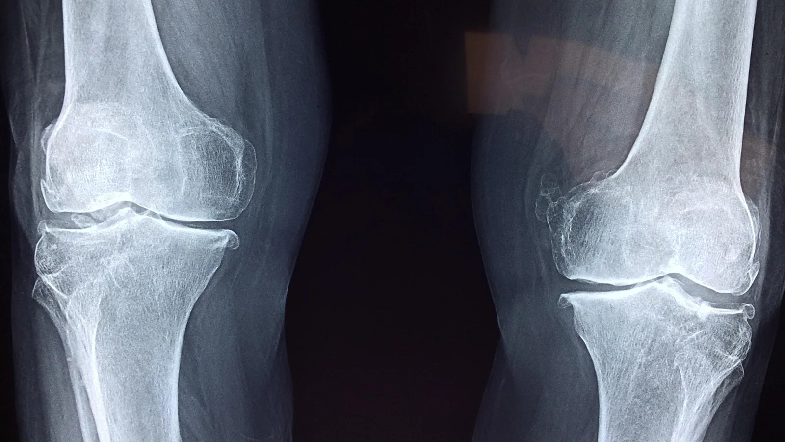
A joint is any union between two or more bones, whether that union is a rigid seam of connective tissue, a pad of cartilage, or a fluid-filled cavity that permits free movement. Veterinary anatomy classifies every joint twice: once by the tissue that holds the bones together (structural classification) and once by how much movement the joint allows (functional classification).
These two schemes run in parallel, and the fastest way to lose marks on an anatomy exam is to treat them as one system. A joint can be described structurally as fibrous and functionally as synarthrotic in the same sentence, and both statements are correct because they answer different questions. This article separates the schemes, gives a clear veterinary example for each class, and shows how the same joint type looks different across species, from the carnivore stifle to the equine fetlock.
This article is educational and is not a substitute for veterinary diagnosis or treatment.
Why Joint Classification Matters in Veterinary Practice
Joint type determines what a joint can do and what can go wrong with it. A synovial joint with a wide cavity and hyaline cartilage can develop septic arthritis, osteochondrosis, or a cruciate ligament rupture. A cartilaginous joint with a fibrocartilaginous disc can herniate, as in the intervertebral disc of the dog. A fibrous joint such as a skull suture is designed to fuse and rarely becomes a clinical problem in a healthy animal.
When a veterinarian reads a radiograph, performs arthrocentesis, or plans a surgical stabilization, the classification of the joint in front of them sets the rules. You cannot aspirate synovial fluid from a suture. You cannot expect a synchondrosis to remodel like a stifle. Knowing the types of joints, and knowing which type you are dealing with, is the first step in every orthopedic decision.
The Two Parallel Classification Schemes
Structural classification asks one question: what tissue connects the bones? The answer places the joint into one of three groups, fibrous, cartilaginous, or synovial.
Functional classification asks a different question: how much movement does the joint permit? The answer places the joint into one of three groups, synarthrosis (immovable), amphiarthrosis (slightly movable), or diarthrosis (freely movable).
Anatomists use both schemes because each one predicts something the other does not. Structural class tells you what the joint is made of and therefore how it heals, how it responds to infection, and how it appears on imaging. Functional class tells you what the joint does mechanically and therefore how much load it transmits and how much range of motion it permits.
Structural Classes
- Fibrous joints: bones united by dense connective tissue rich in collagen, with no joint cavity.
- Cartilaginous joints: bones united by cartilage, either hyaline cartilage or fibrocartilage, with no joint cavity.
- Synovial joints: bones separated by a fluid-filled cavity, lined by a synovial membrane and capped with articular (hyaline) cartilage.
Functional Classes
- Synarthrosis: essentially immovable.
- Amphiarthrosis: slightly movable.
- Diarthrosis: freely movable.
The mapping between the two schemes is close but not perfectly rigid. Fibrous joints are synarthrotic. Cartilaginous joints are usually amphiarthrotic. Synovial joints are diarthrotic. The canine sacroiliac joint shows why the mapping is a guide rather than a law: it is a combined joint with a caudoventral diarthrosis and a craniodorsal symphysis in the same structure [1].
Summary Comparison Table
| Structural class | Functional class | Tissue between bones | Veterinary example |
|---|---|---|---|
| Fibrous (suture) | Synarthrosis | Dense fibrous connective tissue | Skull sutures of the dog |
| Fibrous (gomphosis) | Synarthrosis | Periodontal ligament | Tooth socket in the horse |
| Fibrous (syndesmosis) | Amphiarthrosis | Ligament or interosseous membrane | Distal tibiofibular union |
| Cartilaginous (synchondrosis) | Synarthrosis to amphiarthrosis | Hyaline cartilage | Costochondral junction of the rib |
| Cartilaginous (symphysis) | Amphiarthrosis | Fibrocartilage | Intervertebral disc of the dog |
| Synovial (simple) | Diarthrosis | Synovial fluid and hyaline cartilage | Canine stifle |
| Synovial (compound) | Diarthrosis | Synovial fluid, hyaline cartilage, menisci | Equine fetlock |
| Synovial (combined) | Diarthrosis plus symphysis | Synovial fluid plus fibrocartilage | Canine sacroiliac joint [1] |
Fibrous Joints
Fibrous joints unite bones with dense connective tissue and have no joint cavity. Collagen fibers run from one bone into the other, so the two bones behave as a single mechanical unit. Movement is minimal or absent, which is why fibrous joints are synarthrotic.
Sutures
A suture is a fibrous joint found only between the flat bones of the skull. The bones are separated by a thin layer of fibrous tissue that ossifies with age in many species, converting the suture into a synostosis. In the dog and cat, most skull sutures close early in life. In the horse and ruminant, some sutures remain visible for years. Sutures absorb and distribute the forces of mastication and protect the brain by allowing controlled deformation of the skull during impact.
Gomphoses
A gomphosis is a fibrous joint in which a peg-like structure sits in a socket, held by a periodontal ligament. The only true gomphoses in domestic mammals are the tooth roots in their alveoli. The periodontal ligament is a specialized fibrous connection that permits microscopic movement under load, which is essential for normal chewing and for the proprioceptive feedback that tells an animal how hard it is biting.
Syndesmoses
A syndesmosis is a fibrous joint in which the connecting tissue is a ligament or an interosseous membrane rather than a suture line. The union between the distal tibia and fibula is a syndesmosis, as is the union between the shafts of the radius and ulna in species where the two bones remain separate. Syndesmoses allow slightly more movement than sutures, so they are often classified functionally as amphiarthroses.
Cartilaginous Joints
Cartilaginous joints unite bones with cartilage and have no joint cavity. The cartilage may be hyaline, as in a synchondrosis, or fibrocartilage, as in a symphysis. Both subtypes are avascular, which limits their healing capacity and makes them vulnerable to degenerative change.
Synchondroses
A synchondrosis is a cartilaginous joint in which hyaline cartilage connects the bones. The classic veterinary example is the costochondral junction, where the bony rib meets the costal cartilage, and the costosternal junctions where the costal cartilages meet the sternum. Many synchondroses are temporary. The growth plates (physes) of long bones are synchondroses that ossify when skeletal maturity is reached. A persistent or abnormal physis is a common source of lameness in young, large-breed dogs.
Symphyses
A symphysis is a cartilaginous joint in which fibrocartilage connects the bones, often with a disc or pad of fibrocartilage between them. The intervertebral disc is the most important symphysis in veterinary anatomy. Each disc has an outer annulus fibrosus of fibrocartilage and an inner nucleus pulposus. The disc permits slight movement between adjacent vertebrae and absorbs compressive load. In chondrodystrophic breeds such as the Dachshund, the nucleus pulposus calcifies early and can herniate into the spinal canal, producing the classic Hansen type I disc extrusion.
The pelvic symphysis is another symphysis, uniting the two ossa coxae ventrally. It is amphiarthrotic in the immature animal and becomes progressively more rigid with age, though it retains some flexibility that is important during parturition in the mare and cow.
Synovial Joints
Synovial joints are the most complex and the most clinically important joint type. They are diarthrotic, meaning they permit free movement, and they share a set of defining features.
Defining Features
- A joint capsule, a sleeve of fibrous tissue that encloses the joint and is continuous with the periosteum of the articulating bones.
- A synovial membrane, the inner layer of the capsule, which produces synovial fluid.
- Synovial fluid, a viscous fluid that lubricates the articular surfaces, nourishes the cartilage, and carries immune cells and debris.
- Articular (hyaline) cartilage, a smooth layer covering the ends of the bones, which reduces friction and distributes load.
- A joint cavity, the space between the articulating surfaces, filled with synovial fluid.
The synovium is not a passive lining. It clears intra-articular damage-related material, limits the duration of inflammation, maintains stromal homeostasis, and regulates the entry of peripheral immune cells [2]. When those functions fail, the synovium can become an inflammatory niche that sustains low-grade joint inflammation and contributes to cartilage degeneration [2].
Synovial Joint Subtypes
- Simple synovial joint: two bones, one joint cavity. Example: the canine stifle.
- Compound synovial joint: more than two bones in one capsule, often with menisci. Example: the equine fetlock (metacarpophalangeal joint), which includes the third metacarpal, the proximal phalanx, and the proximal sesamoid bones.
- Combined synovial joint: a joint in which a synovial cavity and a fibrous or cartilaginous component act together. Example: the canine sacroiliac joint, which combines a caudoventral diarthrosis with a craniodorsal symphysis [1].
Synovial Joint Shapes and Movements
Synovial joints are further divided by the shape of their articular surfaces, which predicts the movement they allow.
- Hinge (ginglymus): movement in one plane. The equine fetlock and the canine elbow are hinge joints.
- Pivot (trochoid): rotation around a single axis. The atlantoaxial joint, which allows the head to rotate, is a pivot joint.
- Ball and socket (spheroidal): movement in multiple planes. The hip (coxofemoral) and shoulder joints are ball and socket joints.
- Condylar: movement in two planes, usually flexion and extension plus some rotation. The stifle is a condylar joint.
- Plane (gliding): flat surfaces that slide over one another. The facet joints between adjacent vertebrae are plane synovial joints [3].
- Saddle: movement in two planes with opposition. The carpometacarpal joint of the thumb in humans is the classic example, and the analogous joints in the digit of the dog show similar mechanics.
Structural and Functional Classification Compared
The table above shows the mapping. The clinical point is that the two schemes answer different questions and both are needed.
A fibrous joint is synarthrotic because dense connective tissue does not stretch. A cartilaginous joint is amphiarthrotic because cartilage deforms under load but does not slide. A synovial joint is diarthrotic because the articular surfaces are separated by fluid and can slide freely.
The canine sacroiliac joint is the clearest veterinary illustration of why the two schemes must be kept separate. Structurally it is a combined joint, part synovial and part fibrous. Functionally it is a diarthrosis in its caudoventral portion and a symphysis in its craniodorsal portion [1]. A student who tries to force it into a single structural box will miss the anatomy and the clinical implications for lumbosacral pain.
How Joint Type Is Observed and Tested
Gross Anatomy and Dissection
The first test is direct observation. A joint with a visible cavity, a capsule, and smooth articular cartilage is synovial. A joint with no cavity and cartilage between the bones is cartilaginous. A joint with no cavity and fibrous tissue between the bones is fibrous. Dissection of the canine stifle reveals the joint capsule, the cruciate ligaments, the menisci, and the articular cartilage of the femoral condyles and tibial plateau.
Imaging
Radiography shows the joint space, which is the radiolucent gap between the articular surfaces. In a synovial joint, the joint space is visible because the cartilage and fluid are not radiopaque. In a fibrous or cartilaginous joint, the joint space is narrow or absent. Ultrasonography can show synovial membrane thickening, which is a marker of synovitis. In an equine model of acute synovitis, horses treated with high-molecular-weight hyaluronic acid showed lower synovial membrane thickening and reduced lameness scores compared with controls [4].
Arthrocentesis
Arthrocentesis is the aspiration of synovial fluid from a joint cavity. It is only possible in synovial joints because only synovial joints have a cavity and fluid. Synovial fluid analysis includes total protein, white blood cell count, and cytology. In an equine LPS model of joint inflammation, a dose of 0.0625 ng LPS produced a statistically significant increase in synovial total protein and white blood cell concentrations from baseline at 8 hours after injection [5]. That result is used as a benchmark for what inflammation looks like in synovial fluid.
Histology
Histology distinguishes the tissue types directly. Fibrous joints show dense regular connective tissue. Cartilaginous joints show hyaline cartilage or fibrocartilage. Synovial joints show a synovial membrane with intimal and subintimal layers, and articular cartilage with its characteristic chondrocyte columns. The canine sacroiliac joint was characterized histologically as predominantly dense connective tissue with fibrocartilaginous components, which is what supported its classification as a combined joint [1].
Comparative Species Notes
The Carnivore Stifle
The canine and feline stifle is a simple condylar synovial joint. It has a joint capsule, synovial fluid, hyaline articular cartilage, two menisci, and the cranial and caudal cruciate ligaments. The stifle is diarthrotic and permits flexion and extension plus a small amount of rotation when the joint is flexed. In the dog, the cranial cruciate ligament is the most commonly ruptured ligament in the pelvic limb, and rupture leads to instability, meniscal injury, and progressive osteoarthritis. The stifle is the joint most often used to teach synovial joint anatomy because all the defining features are visible on dissection.
The Equine Fetlock
The equine fetlock (metacarpophalangeal joint) is a compound synovial joint. It includes the third metacarpal bone, the proximal phalanx, and the proximal sesamoid bones, all within a single joint capsule. The fetlock is a hinge joint that allows flexion and extension in one plane. It is the joint most commonly affected by osteoarthritis in the performance horse, and it is the joint used in equine models of synovitis and intra-articular therapy [4][5]. The compound nature of the fetlock, with sesamoid bones inside the capsule, is a key difference from the simple carnivore stifle.
The Canine Sacroiliac Joint
The canine sacroiliac joint is a combined joint. Its caudoventral portion is a diarthrosis with a synovial surface, and its craniodorsal portion is a symphysis with fibrocartilage [1]. The synovial surface area is smaller than the extrasynovial surface area, and the iliac surface areas are significantly larger than the sacral surface areas [1]. This anatomy is relevant to the clinical evaluation of lameness and lumbosacral pain syndromes in dogs [1].
The Intervertebral Disc
The intervertebral disc is a symphysis in all domestic mammals, but the ratio of annulus fibrosus to nucleus pulposus and the composition of the nucleus vary by species and by breed. In the dog, the disc is the site of the most common spinal cord compression syndrome in chondrodystrophic breeds. In the horse, disc disease is rare but has been reported. The disc is avascular and receives nutrition by diffusion from the adjacent vertebral bodies and from the surrounding soft tissues.
Clinical Relevance, Limitations and Common Mistakes
Joint type drives clinical decisions. Synovial joints are the target of arthrocentesis, intra-articular medication, arthroscopy, and joint replacement. Cartilaginous joints are the target of disc surgery and of treatments for physitis in young animals. Fibrous joints are rarely surgical targets except in skull trauma or dental disease.
Synovial inflammation is a central mechanism in osteoarthritis. The synovium is an immune niche in which activated macrophages, fibroblast-like synoviocytes, mast cells, T cells, and damaged chondrocytes interact with nociceptors to shape pain [6]. Synovial immune control failure, in which debris clearance and inflammatory resolution do not occur in parallel, allows a pathological cellular network to persist and to form an inflammatory niche that feeds back into cartilage degeneration and subchondral bone remodeling [2]. This is why synovitis is treated as a disease process and not just a symptom.
The most common mistakes students make are worth naming directly.
- Treating structural and functional classification as the same scheme. They are not. A joint has one structural class and one functional class, and the two are described separately.
- Assuming all fibrous joints are immovable. Syndesmoses are fibrous but slightly movable, so they are amphiarthrotic.
- Assuming all cartilaginous joints are temporary. The intervertebral disc is a permanent symphysis.
- Forgetting that synovial joints have a cavity. The cavity is the defining feature and the reason arthrocentesis is possible.
- Confusing the joint capsule with the synovial membrane. The capsule is the outer fibrous sleeve. The synovial membrane is the inner lining that produces fluid.
- Using human-only examples. The stifle, fetlock, sacroiliac joint, and intervertebral disc are the examples that matter in veterinary practice.
Limitations: individual animals vary in joint anatomy and in disease presentation, and a veterinarian should evaluate any suspected joint problem in person. Imaging and synovial fluid analysis are needed to confirm a diagnosis.
Quick Review
- Structural classes are fibrous, cartilaginous, and synovial. Functional classes are synarthrosis, amphiarthrosis, and diarthrosis.
- Fibrous joints are synarthrotic and have no cavity. Skull sutures are the classic example.
- Cartilaginous joints are amphiarthrotic and have no cavity. The intervertebral disc is the classic example.
- Synovial joints are diarthrotic and have a capsule, synovial fluid, and hyaline cartilage. The stifle is the classic example.
- The canine sacroiliac joint is a combined joint with a diarthrosis and a symphysis in one structure [1].
- The carnivore stifle is a simple condylar synovial joint. The equine fetlock is a compound hinge synovial joint.
- Synovial fluid analysis is only possible in synovial joints because only they have a cavity.
Frequently Asked Questions
What are the three structural types of joints?
The three structural types are fibrous, cartilaginous, and synovial. They are defined by the tissue that connects the bones: dense connective tissue, cartilage, or a synovial cavity with fluid.
What are the three functional types of joints?
The three functional types are synarthrosis (immovable), amphiarthrosis (slightly movable), and diarthrosis (freely movable). They are defined by how much movement the joint allows.
Are all synovial joints diarthrotic?
Yes. Synovial joints are diarthrotic by definition because the fluid-filled cavity and smooth articular cartilage permit free movement. The amount of movement varies with joint shape, but all synovial joints are freely movable.
What is an example of a cartilaginous joint in a dog?
The intervertebral disc is the best example. It is a symphysis with an outer annulus fibrosus and an inner nucleus pulposus, and it permits slight movement between adjacent vertebrae.
How is the equine fetlock different from the canine stifle?
The fetlock is a compound synovial joint that includes the third metacarpal bone, the proximal phalanx, and the proximal sesamoid bones in one capsule. The stifle is a simple synovial joint with two main bones, the femur and tibia, plus the patella.
Why can synovial fluid only be sampled from synovial joints?
Synovial fluid is produced by the synovial membrane and contained within the joint cavity. Fibrous and cartilaginous joints have no cavity and no synovial membrane, so there is no fluid to aspirate.
Related Articles
- What Kind of Science Is Environmental Science? A Classification Guide
- Comprehensive Classification of Types of Chicken Parasites: Ectoparasites and Endoparasites
- Epigenetic Modification: Types, Mechanisms, and Examples
- Epigenetic Factors: Types, Mechanisms, and Examples
- What is a Stimulus in Biology? Definition, Types & Response Examples
- Competition in Biology: Definition, Types, and Examples
- Base Pair in DNA: Rules, Types and Examples
Sources
- The Canine Sacroiliac Joint: 1. Classification and Morphometrics.
- Synovial immune control failure in osteoarthritis: from maintenance of tissue homeostasis to inflammatory niche formation.
- PDGFRα signaling regulates cartilage and fibrous tissue differentiation during synovial joint development.
- Effects of Intra-Articular Administration of High-Molecular-Weight Linear Hyaluronic Acid on Synovial Fluid Characteristics and Joint Environment.
- Investigation into the Short-Term Effects of Intra-Articular Allogenic PRP in the Equine LPS Model of Joint Inflammation: A Randomised Control Trial.
- Neuroimmune crosstalk in osteoarthritis pain: from synovial inflammation to pain sensitization.