
Comprehensive Classification of Types of Chicken Parasites: Ectoparasites and Endoparasites
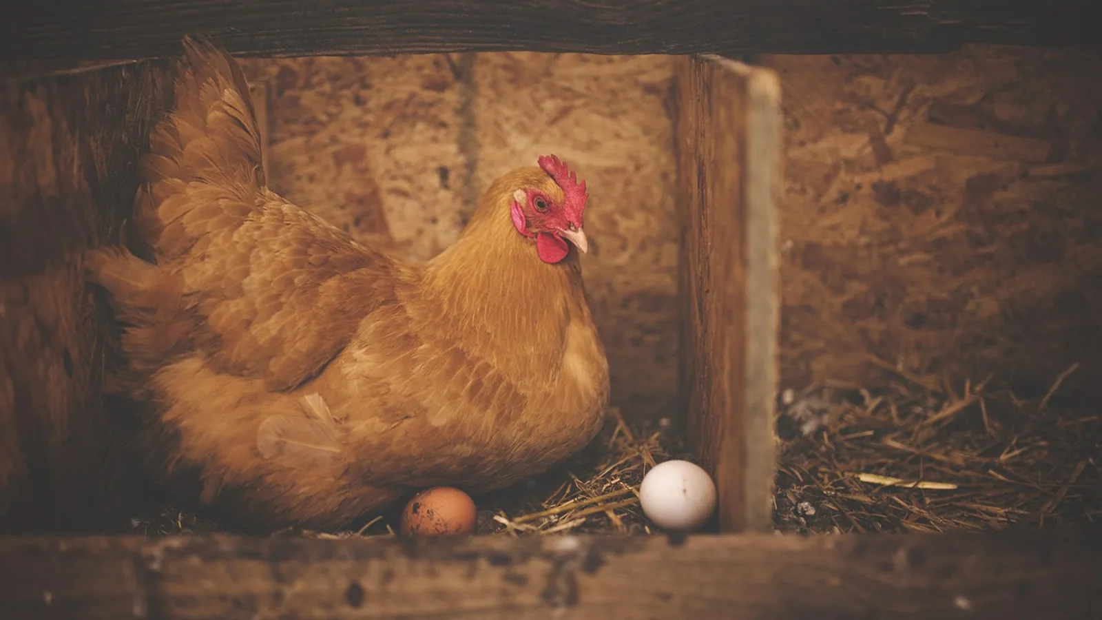
Chickens (Gallus gallus domesticus) are hosts to a diverse assemblage of parasitic organisms that cause substantial economic losses and welfare burdens in commercial and backyard production systems. These parasites are broadly classified into two ecological categories based on their anatomical niche: ectoparasites, which inhabit the external body surfaces, and endoparasites, which reside within internal organs, tissues, or lumina. This article provides a systematic classification of the major types of chicken parasites, emphasizing taxonomic grouping, life cycle biology, pathological mechanisms, and diagnostic principles. The classification framework presented here is essential for designing integrated control programs and for interpreting molecular and coprological surveillance data.
1. Classification Framework for Chicken Parasites
Parasites of chickens belong to the Kingdom Animalia (Phylum Arthropoda and Phylum Platyhelminthes, Nematoda, and Acanthocephala) and the Kingdom Protista (Phylum Apicomplexa and other protozoan phyla). A hierarchical classification based on location (ectoparasite versus endoparasite) and taxonomic phylum is standard in veterinary parasitology [1].
1.1 Ectoparasites
Ectoparasites are arthropods that feed on blood, feathers, skin, or tissue fluids from the external surface of the host. They are primarily represented by two classes within Phylum Arthropoda: Arachnida (mites and ticks) and Insecta (lice, fleas, and flies) [1, 2].
1.1.1 Mites (Subclass Acari)
Mites are the most economically significant ectoparasites of poultry. The major species include:
Dermanyssus gallinae (the poultry red mite). This species is a nocturnal blood-feeding mite that lives in cracks and crevices of poultry houses during the day and attacks chickens at night [3]. It causes anemia, reduced egg production, and is a vector for bacterial pathogens such as Erysipelothrix rhusiopathiae [4]. A detailed reference on its identification and control is provided in the article on Ectoparasites of Poultry: Dermanyssus gallinae, Ornithonyssus sylviarum, Knemidocoptes mutans, Knemidocoptes gallinae, and Argas persicus.
Ornithonyssus sylviarum (the northern fowl mite). This mite spends its entire life cycle on the host, feeding on blood and causing dermatitis, feather loss, and drop in egg production [3, 5].
Knemidocoptes mutans (the scaly leg mite). This burrowing mite resides in the epidermis of the legs and feet, causing hyperkeratosis, crust formation, and deformation known as scaly leg [6].
Knemidocoptes gallinae (the depluming mite). This mite burrows into the skin at the base of feathers, leading to intense pruritus, feather loss, and self-trauma [6].
1.1.2 Ticks (Subclass Ixodida)
Argas persicus (the fowl tick or tampan). This soft tick is an intermittent blood feeder that can cause anemia, paralysis due to salivary toxins, and transmission of Borrelia anserina, the agent of avian spirochetosis [7].
1.1.3 Lice (Order Phthiraptera)
Chewing lice (suborder Mallophaga) are host-specific, obligate ectoparasites that feed on feather barbules, skin scales, and blood from irritated skin. Common species include Menopon gallinae (the shaft louse), Menacanthus stramineus (the body louse), and Liperus caponis (the wing louse) [8]. Heavy infestations cause feather damage, weight loss, and reduced egg production [8, 9].
1.1.4 Fleas (Order Siphonaptera)
The sticktight flea (Echidnophaga gallinacea) is the most important flea species in chickens. Adults attach permanently to the skin around the comb, wattles, and eyes, feeding on blood and causing ulceration and anemia [2, 10].
1.1.5 Flies (Order Diptera)
Larvae of certain flies cause myiasis in chickens. The black blowfly (Phormia regina) and the green bottle fly (Lucilia sericata) can infest wounds or soiled feathers, leading to tissue destruction and secondary bacterial infection [11].
1.2 Endoparasites
Endoparasites are classified into helminths (nematodes, cestodes, trematodes) and protozoa (coccidia, flagellates, and blood parasites). These organisms inhabit the gastrointestinal tract, respiratory tract, liver, and other visceral organs.
1.2.1 Nematodes (Roundworms)
A comprehensive clinical reference for several key species is available in the article on Respiratory and Intestinal Nematodes of Poultry: Syngamus trachea, Ascaridia galli, Heterakis gallinarum, and Capillaria obsignata.
Ascaridia galli. This large roundworm inhabits the lumen of the small intestine. It has a direct life cycle via the fecal-oral route. Heavy burdens cause intestinal obstruction, weight loss, and reduced egg production [12].
Heterakis gallinarum. This cecal nematode is notable as the vector for Histomonas meleagridis, the causative agent of blackhead disease (histomonosis) in turkeys and occasionally in chickens [13].
Capillaria obsignata (the crop worm). This thin nematode infects the crop and small intestine, causing catarrhal inflammation, reduced feed conversion, and diarrhea [12].
Syngamus trachea (the gapeworm). This red nematode attaches to the tracheal mucosa, causing dyspnea, head shaking, and death from asphyxiation in heavy infestations [14].
Tetrameres fissispina and Tetrameres americana. These proventricular nematodes cause proventriculitis, glandular dilation, and secondary bacterial infections [15].
1.2.2 Cestodes (Tapeworms)
Cestodes require an intermediate host such as beetles, snails, or houseflies. Common species include:
Raillietina cesticillus, Raillietina tetragona, and Raillietina echinobothrida. These tapeworms attach to the small intestinal mucosa, causing catarrhal enteritis, nutrient malabsorption, and poor growth [16]. A detailed reference on Davainea proglottina is provided in the article on Davainea proglottina in Chickens: Microscopic Identification, Snail Intermediate Hosts, and Tapeworm Lifecycle Management.
Davainea proglottina. This small cestode has a very short proglottid chain. Its intermediate host is the slug or snail. Infections cause hemorrhagic enteritis and high mortality in young birds [17].
1.2.3 Trematodes (Flukes)
Postharmostomum gallinum and Echinostoma revolutum. These flukes reside in the ceca and small intestine, respectively. They require snail intermediate hosts and are less common in confined poultry systems [18].
1.2.4 Protozoa
Protozoan parasites are among the most pathogenic endoparasites in chickens.
Coccidia (Genus Eimeria). Seven species of Eimeria infect chickens. Each species is site-specific in the intestinal tract. The most pathogenic species include Eimeria tenella (ceca), Eimeria necatrix (midgut), and Eimeria maxima (midgut to lower small intestine). Infections cause enteritis, hemorrhagic diarrhea, and death [19]. A detailed discussion of predisposing factors is provided in the article on What Causes Coccidiosis in Chickens: Etiology, Transmission, and Predisposing Factors in Flock Management.
Histomonas meleagridis. This flagellated protozoan causes blackhead disease or histomonosis. It primarily affects turkeys but can cause morbidity in chickens. The organism is transmitted within the eggs of Heterakis gallinarum and directly via the fecal-oral route [13].
Trichomonas gallinarum and Tetratrichomonas gallinarum. These flagellates inhabit the ceca and upper intestine, causing typhlitis and diarrhea. They are transmitted by the fecal-oral route [20].
Spironucleus meleagridis (formerly Hexamita meleagridis). This flagellate is a significant pathogen in turkeys but also causes enteritis in chickens, especially in young birds [21].
Leucocytozoon caulleryi and Leucocytozoon sabrazesi. These blood protozoa are transmitted by blackflies (Simuliidae). They invade erythrocytes and leucocytes, causing anemia and organ enlargement [22]. A comprehensive reference is provided in the article on Leucocytozoonosis in Poultry: Leucocytozoon Transmission by Blackflies, Clinical Signs, and Integrated Control Strategies.
Plasmodium gallinaceum. This species causes avian malaria in chickens. It is transmitted by mosquitoes (Culicidae) and infects red blood cells, leading to anemia and spleenomegaly [23].
2. Diagnostic Overview
Accurate classification of parasite type is necessary for selecting diagnostic methods. Table 1 summarizes primary diagnostic techniques for each parasite group.
Table 1: Diagnostic approaches for major chicken parasite categories.
| Parasite Category | Common Genera | Primary Diagnostic Specimen | Diagnostic Methods |
|---|---|---|---|
| Mites (ectoparasite) | Dermanyssus, Ornithonyssus, Knemidocoptes | Skin scrapings, vent swabs, house crevice debris | Microscopic identification, morphological keys |
| Lice (ectoparasite) | Menopon, Menacanthus, Liperus | Feathers, skin | Visual inspection, stereomicroscopy |
| Nematodes (endoparasite) | Ascaridia, Heterakis, Capillaria, Syngamus | Feces, tracheal mucus | Fecal flotation (NaCl, ZnSO4), Baermann technique for Syngamus |
| Cestodes (endoparasite) | Raillietina, Davainea | Feces | Fecal flotation for proglottids, microscopic egg identification |
| Coccidia (endoparasite) | Eimeria | Feces | Oocyst flotation, sporulation to determine species |
| Blood protozoa | Leucocytozoon, Plasmodium | Blood smear, blood | Giemsa-stained thin and thick blood films, PCR |
3. Life Cycle Overview and Transmission Pathways
The transmission strategies of chicken parasites differ substantially between direct and indirect life cycles. Direct life cycles (e.g., Ascaridia galli, Eimeria spp.) involve fecal contamination of feed, water, or litter and do not require an intermediate host [12, 19]. Indirect life cycles (e.g., cestodes, Histomonas meleagridis) require an invertebrate intermediate host, complicating control [13, 16]. Ectoparasites such as D. gallinae exhibit a mixed feeding ecology; they reside off the host between blood meals but can be transmitted through contaminated equipment, clothing, and bird-to-bird contact [3].
The Mermaid diagram below illustrates a decision workflow for initial parasite classification based on clinical presentation and sample type.
graph TD
 A["Clinical Presentation: Pruritus, Feather Damage, Anemia"] --> B{External Examination}
 B --> C[Mobile arthropod on skin or feathers]
 B --> D[Burrowing or scaly lesions on legs/face]
 C --> E[Collect ectoparasite sample]
 E --> F["Microscopic ID: Mite, Louse, Tick, Flea"]
 D --> G[Skin scraping]
 G --> H[Knemidocoptes species]

 A2["Clinical Presentation: Diarrhea, Weight Loss, Dyspnea"] --> I{Fecal or Blood Sample}
 I --> J[Fecal flotation positive for oocysts]
 I --> K[Fecal flotation positive for eggs]
 I --> L[Blood smear positive for protozoa]
 J --> M["'Eimeria species (coccidia')"]
 K --> N[Intestinal nematode or cestode]
 L --> O[Leucocytozoon or Plasmodium]
4. Pathophysiology and Clinical Consequences
Parasitic infections in chickens produce a spectrum of pathological effects.
Ectoparasites cause blood loss leading to iron deficiency anemia, especially in heavy infestations of D. gallinae and A. persicus [3, 7]. Chronic mite infestation reduces eggshell quality and hen-day production [5]. Burrowing mites cause proliferative dermatitis and secondary bacterial infections [6]. Lice damage feather integrity, impairing thermoregulation and increasing feed intake requirements [8].
Endoparasites induce mechanical damage, nutrient competition, and immune suppression. Ascaridia galli infection reduces digestibility of proteins and fats [12]. Eimeria species cause direct destruction of intestinal epithelial cells, leading to osmotic diarrhea, protein-losing enteropathy, and hemorrhage [19]. In coccidiosis, the severity of disease varies by Eimeria species, with E. tenella causing fatal cecal hemorrhage and E. maxima inducing substantial weight loss [19].
Blood protozoa such as Leucocytozoon caulleryi and Plasmodium gallinaceum cause hemolytic anemia, organomegaly, and can be fatal in naive flocks [22, 23].
5. Epidemiological Factors
The prevalence of specific parasites depends on housing system, stocking density, biosecurity level, and climate. In battery cage systems, ectoparasite prevalence is lower than in free-range or litter-based systems due to reduced contact with contaminated substrate [3]. Conversely, coccidiosis is more prevalent in litter-based systems due to high oocyst concentrations in feces [19].
The introduction of new birds without quarantine often introduces ectoparasites and helminths to naive flocks. Wild birds and rodents serve as reservoir hosts for several parasites, including D. gallinae and Heterakis gallinarum [24].
6. Control Principles
Control strategies must be tailored to the parasite classification.
Ectoparasite control relies on environmental sanitation and acaricide application. For D. gallinae, acaricides must reach hiding places in cracks and crevices. Resistance to pyrethroids has been documented in some populations [25]. For lice and mites on the host, dust formulations containing permethrin or organophosphates are effective [8].
Endoparasite control uses anticoccidial agents (ionophores and synthetic compounds), anthelmintics (benzimidazoles, levamisole), and pasture management. Anthelmintic resistance in Ascaridia galli has been reported to benzimidazoles [26]. Vaccination is used for coccidiosis, with live attenuated Eimeria vaccines providing protective immunity [27].
A comprehensive discussion on antimicrobial resistance in the poultry production environment is provided in the article on Antimicrobial Resistance in Livestock-Associated Staphylococcus aureus: Genomic Epidemiology and One Health Implications.
7. Conclusion
Chicken parasites are taxonomically and ecologically diverse, falling into the major categories of ectoparasites (mites, ticks, lice, fleas, flies) and endoparasites (nematodes, cestodes, trematodes, protozoa). Accurate classification guides diagnostic testing, treatment selection, and preventive management. Integrated control programs must account for the life cycle specifics, host-parasite interactions, and environmental persistence of each parasite type. Continued surveillance for drug resistance and emerging parasites is essential for sustainable poultry production.
References
[1] Taylor, M. A., Coop, R. L., & Wall, R. L. (2015). Veterinary Parasitology (4th ed.). Wiley Blackwell.
[2] Soulsby, E. J. L. (1982). Helminths, Arthropods and Protozoa of Domesticated Animals (7th ed.). Baillière Tindall.
[3] Kettle, D. S. (1990). Medical and Veterinary Entomology. CAB International.
[4] Chirico, J., Eriksson, H., Fossum, O., & Jansson, D. (2003). The poultry red mite, Dermanyssus gallinae, a potential vector of Erysipelothrix rhusiopathiae causing erysipelas in hens. Medical and Veterinary Entomology, 17(2), 232-234.
[5] DeVaney, J. A. (1986). Effects of the northern fowl mite, Ornithonyssus sylviarum, on egg production and quality in chickens. Poultry Science, 65(5), 850-854.
[6] Hiepe, T., & Ribbeck, R. (1993). Veterinärmedizinische Arachno-Entomologie. Gustav Fischer Verlag.
[7] Mahmoud, M. S., & Abdel-Rahman, M. A. (2003). Tick paralysis caused by Argas persicus in poultry. Veterinary Parasitology, 113(3-4), 299-304.
[8] Axtell, R. C. (1999). Poultry integrated pest management: status and future. Integrated Pest Management Reviews, 4(1), 53-73.
[9] Trudeau, M. E., & Bennett, G. W. (1991). Effects of the chicken body louse, Menacanthus stramineus, on growth and reproduction of chickens. Poultry Science, 70(4), 805-810.
[10] Capinera, J. L. (2008). Encyclopedia of Entomology (2nd ed.). Springer.
[11] Hall, M. J. R., & Wall, R. (1995). Myiasis of humans and domestic animals. Advances in Parasitology, 35, 257-334.
[12] Ruff, M. D. (1998). Nematodes of the digestive tract. In Diseases of Poultry (10th ed., pp. 897-912). Iowa State University Press.
[13] McDougald, L. R. (2008). Histomonosis (blackhead) in poultry. In Diseases of Poultry (12th ed., pp. 1065-1072). Blackwell Publishing.
[14] Beasley, J. N., & Williams, J. E. (1970). Gapeworm infection in chickens. Avian Diseases, 14(1), 147-153.
[15] Permin, A., & Hansen, J. W. (1998). The prevalence and distribution of Tetrameres species in chickens. Veterinary Parasitology, 77(1), 47-55.
[16] Reed, W. M., & van Heerden, J. (2003). Cestode infections in poultry. Veterinary Clinics of North America: Food Animal Practice, 19(3), 671-684.
[17] Norton, C. C. (1966). Davainea proglottina in chickens: life cycle and pathology. Journal of Helminthology, 40(1-2), 95-106.
[18] Williams, I. C. (1960). The life cycle of Postharmostomum gallinum in the chicken. Parasitology, 50(3-4), 515-525.
[19] McDougald, L. R., & Fitz-Coy, S. H. (2008). Coccidiosis. In Diseases of Poultry (12th ed., pp. 1048-1064). Blackwell Publishing.
[20] Friedrich, R., & Huber, J. (1987). Pathogenicity of Trichomonas gallinarum in chickens. Avian Pathology, 16(2), 239-250.
[21] Barnes, H. J., & Gross, W. B. (1993). Spironucleosis in turkeys and chickens. In Diseases of Poultry (9th ed., pp. 771-775). Iowa State University Press.
[22] Morii, T., & Shinjo, T. (1972). Leucocytozoon caulleryi: pathology and diagnosis. National Institute of Animal Health Quarterly, 12(4), 177-185.
[23] Ball, G. H., & Chao, J. (1964). Avian malaria: life cycle and effects on the host. Journal of Protozoology, 11(3), 327-335.
[24] Duff, J. P., & Hudson, P. J. (1988). The role of wild birds in the epidemiology of Heterakis gallinarum. Parasitology, 96(1), 87-94.
[25] Fiddes, M. D., Le Gresley, S., Parsons, D. G., & Coles, G. C. (2005). Prevalence of the poultry red mite (Dermanyssus gallinae) in England. Veterinary Record, 157(8), 232-235.
[26] Shoop, W. L., & Haines, H. J. (1993). Benzimidazole resistance in Ascaridia galli of chickens. Poultry Science, 72(1), 121-124.
[27] Williams, R. B. (2002). Anticoccidial vaccines for broiler chickens: pathways to success. Avian Pathology, 31(4), 317-353. *** Disclaimer: This article is for educational and informational purposes only. It is not intended to substitute for professional veterinary advice, diagnosis, treatment, or regulatory guidance. Always consult a licensed veterinarian or qualified specialist regarding animal health, disease diagnosis, and therapeutic decisions.