Tibial Nerve: Course, Branches, and Function
By Dr. Zubair Khalid, DVM, MS, PhD ·
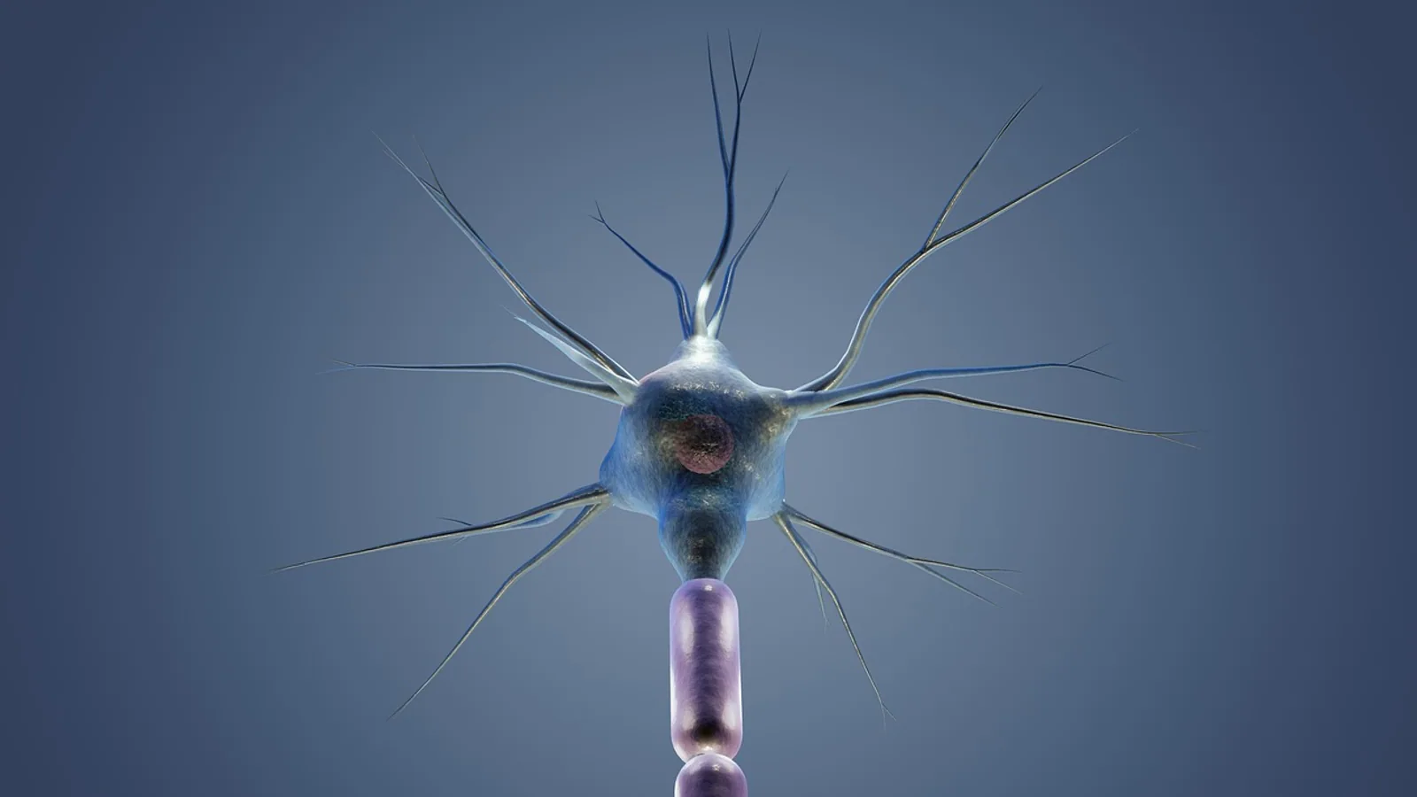
The nervus tibialis (n tibialis), or tibial nerve, is the larger of the two terminal branches of the sciatic nerve and the principal motor and sensory nerve of the caudal pelvic limb. It supplies the extensor muscles of the hock, the digital flexors, and the plantar skin, and it is the nerve most often implicated when a dog or cat suddenly cannot bear weight on a hind limb with a dropped hock.
This article traces the tibial nerve from its origin in the lumbosacral plexus through the caudal thigh, the popliteal fossa, and the tarsal canal, lists its named branches, and compares its anatomy and clinical behavior across dogs, cats, horses, and ruminants. This article is educational and is not a substitute for veterinary diagnosis or treatment.
Origin and Root Values
The tibial nerve arises from the lumbosacral plexus, a network formed by the ventral rami of the last lumbar and first sacral spinal nerves. In the dog, the sciatic nerve is formed mainly by contributions from L6, L7, and S1, with variable input from L5 and S2. The tibial nerve carries fibers from the same roots, predominantly L7 and S1 in the dog and cat.
Because the tibial and common fibular (peroneal) nerves travel together inside a common epineurial sheath for most of the thigh, they are best regarded as two functionally distinct nerves sharing one gross trunk. In the dog, this shared trunk is the sciatic nerve proper. In the horse, the same arrangement is present, and the two divisions separate only near the stifle.
A cadaveric case report in humans described a rare variation in which the common fibular nerve and tibial nerve exited the pelvis independently and converged about one inch inferior to the piriformis muscle to form a single sciatic trunk [1]. Equivalent variants are recognized in small animals and are one reason a nerve block intended for one division can produce unexpected results.
Course Through the Caudal Thigh
The tibial nerve descends in the caudal thigh between the biceps femoris and semitendinosus muscles, deep to the gluteal musculature. In the horse, this is the exact corridor used for a caudocranial midfemoral ultrasound-guided injection. A convex probe is placed perpendicular to the femur at the level of the distal end of the third trochanter, and the tibial and fibular nerves are located between the biceps femoris and semitendinosus. A 21-cm needle is inserted at roughly a 60-degree angle in a caudocranial direction toward the fascial plane. In a cadaveric dye-spreading study, both nerves were successfully stained in 51.85% of limbs (14 of 27), only the tibial nerve in 7.41% (2 of 27), and only the fibular nerve in 3.70% (1 of 27) [2]. That study is a reminder that even image-guided techniques do not guarantee complete coverage of both divisions from a single injection.
In the dog, the tibial nerve gives off muscular branches to the caudal thigh muscles, including the biceps femoris, semitendinosus, and semimembranosus, before reaching the stifle. These branches are short and arise from the caudal aspect of the parent trunk.
Popliteal Fossa and the Divergence from the Fibular Nerve
At the level of the stifle, the sciatic trunk divides into the tibial nerve and the common fibular nerve. The tibial nerve continues distally between the heads of the gastrocnemius muscle, running in the popliteal fossa. This is the region where the nerve is most accessible for direct palpation and where it is at risk during stifle surgery.
The common fibular nerve wraps laterally around the fibular head, where it is superficial and vulnerable. The tibial nerve stays medial and deep, running with the caudal tibial vessels. This anatomical separation matters clinically. A lesion at the fibular head produces a dropped hock with knuckling of the digits (the classic peroneal or fibular deficit), while a lesion of the tibial nerve produces a plantigrade stance with loss of digital flexion and loss of the withdrawal reflex.
Branches to the Extensor Muscles of the Hock
The tibial nerve supplies the main extensor of the hock, the gastrocnemius, through two or more muscular branches that enter the medial and lateral heads of the muscle. In the rat, a targeted muscle reinnervation model grafted the proximal tibial nerve into the gastrocnemius muscle and preserved muscle wet weight and reduced fibrosis compared with transection alone [3]. This confirms that the gastrocnemius is the principal target of the tibial nerve's motor output in the caudal limb.
The superficial digital flexor muscle also receives a branch from the tibial nerve. In the dog, this muscle flexes the digits and helps extend the hock through its calcaneal attachment. The deep digital flexor muscle, which includes the flexor digitorum lateralis, flexor digitorum medialis, and the interflexorius, is supplied by one or more branches of the tibial nerve.
In primates, the soleus muscle is innervated by two branches of the tibial nerve, a posterior branch supplying the major posterior part and an anterior branch supplying the anterior bipennate part [4]. The posterior branch was present in all nine primate species studied, while the anterior branch was present in only five. This comparative detail is useful because it shows that even within a single muscle, the tibial nerve can have variable intramuscular branching patterns that are not tied to the distribution of the other branches.
Tarsal Canal and Plantar Branches
The tibial nerve enters the tarsal canal (also called the tarsal tunnel) on the medial side of the hock, passing deep to the flexor retinaculum and between the tendons of the digital flexors. In a fetal cadaver study of 116 feet, the bifurcation of the tibial nerve was located within the tarsal tunnel in all cases, most frequently proximal to the medial malleolus-calcaneal axis [5]. The medial calcaneal nerve was classified into six types, with Type 2 being the most prevalent at 39.7% of specimens. An anastomosis between the medial and lateral plantar nerves was found in four patterns, with Type 1 occurring in 81.89% of cases.
In the dog, the tibial nerve divides into the medial and lateral plantar nerves within or just distal to the tarsal canal. These plantar nerves continue along the plantar metatarsus and give off the plantar digital nerves that supply the skin and joints of the digits. In the horse, the tibial nerve continues as the plantar nerves after a junction that is located at a maximum of 85 mm proximal to the proximal aspect of the calcaneal tubercle, and the medial cutaneous branch joins at a maximum of 150 mm proximal to the same landmark [6].
The equine tibial nerve has a mean thickness of 6 ± 1 mm, and the mean distance from the nerve to the cranial border of the superficial digital flexor is 11 ± 6 mm [6]. No anatomical variations of the tibial nerve were observed in that dissection study of 10 paired cadaver hindlimbs, which supports the generally recommended site for tibial nerve perineural injection at about 100 mm proximal to the calcaneal tubercle.
In the dromedary camel, the tibial nerve possesses many branches along its course to the plantar skin of the metatarsus and supplies the axial and abaxial plantar surfaces of the fourth digit, the interdigital surfaces, and the plantar-axial and plantar-abaxial surfaces of the third digit [7]. This pattern is broadly similar to other ungulates and is relevant for regional anesthesia of the distal limb.
Table: Branches, Targets, and Sensory Territory
| Branch | Target muscle or tissue | Sensory territory | Notes |
|---|---|---|---|
| Muscular branches in caudal thigh | Biceps femoris, semitendinosus, semimembranosus | None | Arise before the stifle |
| Gastrocnemius branches | Gastrocnemius (medial and lateral heads) | None | Main hock extensors |
| Superficial digital flexor branch | Superficial digital flexor | None | Flexes digits, assists hock extension |
| Deep digital flexor branches | Deep digital flexor group | None | Flexor digitorum lateralis, medialis, interflexorius |
| Medial calcaneal nerve | None | Medial and plantar heel skin | Six described types in fetal cadavers [5] |
| Medial plantar nerve | Flexor muscles and intrinsic foot muscles | Medial plantar skin and medial digits | Anastomoses with lateral plantar nerve in most feet [5] |
| Lateral plantar nerve | Intrinsic foot muscles | Lateral plantar skin and lateral digits | Continues as plantar digital nerves |
| Plantar metatarsal nerves | None | Plantar metatarsal skin | Give rise to plantar digital nerves |
| Medial cutaneous branch (horse) | None | Medial tarsal and metatarsal skin | Joins up to 150 mm proximal to calcaneal tubercle [6] |
Function: Motor and Sensory Roles
The tibial nerve is the principal flexor nerve of the pelvic limb. It drives digital flexion through the superficial and deep digital flexor muscles, and it drives hock extension through the gastrocnemius. In the standing animal, the gastrocnemius and the digital flexors work together to lock the hock and maintain the digitigrade or unguligrade stance.
Sensory function of the tibial nerve covers the plantar surface of the metatarsus and digits, plus the medial and plantar heel through the medial calcaneal nerve. In the horse, the medial cutaneous branch supplies the medial tarsal and metatarsal skin [6]. In the camel, the tibial nerve supplies the plantar skin of the metatarsus and the axial and abaxial surfaces of the third and fourth digits [7].
The tibial nerve also carries proprioceptive and reflex afferents. In a feline spinal locomotion model, low-intensity electrical stimulation of the distal tibial nerve or its medial plantar branch at levels activating large-diameter afferents produced no measurable effect on flexor or extensor activity during walking. Higher stimulus intensities that recruited smaller fibers increased swing height when delivered in late stance or early swing. Further increases that recruited noxious afferents terminated ongoing stance and produced a flexor withdrawal response of the paw [8]. This study shows that the tibial nerve carries multiple afferent populations with distinct functional roles in locomotion.
Comparative Anatomy: Dog, Cat, Horse, and Ruminant
Dog
In the dog, the tibial nerve is the larger terminal branch of the sciatic nerve. It supplies the gastrocnemius, superficial digital flexor, deep digital flexor, and the intrinsic muscles of the pes. It divides into medial and lateral plantar nerves at the tarsal canal. The dog's tibial nerve is the nerve most often affected by iatrogenic injury during stifle surgery, and it is the nerve used in experimental models of peripheral nerve repair [9].
A case report described an 8.5-year-old neutered male Dobermann with a malignant peripheral nerve sheath tumor of the tibial nerve that presented with non-weight-bearing left pelvic limb lameness and severe pain on hock flexion. CT and ultrasound showed a homogeneously thickened, contrast-enhancing segment of the left tibial nerve with muscle atrophy. Limb-sparing partial neurectomy with 2-cm margins achieved complete excision and no recurrence at 12 and 24 months [10]. This case shows that the tibial nerve can be the site of primary neoplasia in dogs, not just traumatic or iatrogenic injury.
A separate case report described a dog that developed a painful tibial nerve neuroma 10 days after iatrogenic injury during preparation of a reverse saphenous conduit flap. The dog had severe pain without nerve deficits. MRI showed an enlarged tibial nerve at the injury site. Surgical resection and autologous saphenous nerve graft transplantation produced immediate pain relief and full recovery by 6 months [11]. Neuroma formation should be considered when a dog develops severe pain and lameness after surgery near the tibial nerve, even shortly after the procedure.
Cat
In the cat, the tibial nerve has a similar course and branching pattern to the dog. The feline tibial nerve is used extensively in locomotion research because the cat's spinal cord can generate stepping movements after complete spinal transection. The distal tibial nerve and its medial plantar branch are the standard stimulation sites in these preparations [8]. Clinically, cats with tibial nerve injury show a dropped hock, loss of digital flexion, and a plantigrade stance, with loss of the plantar withdrawal reflex.
Horse
In the horse, the tibial nerve runs along the medial aspect of the tibia and joins the plantar nerves at a maximum of 85 mm proximal to the calcaneal tubercle [6]. The medial cutaneous branch joins at up to 150 mm proximal to the same landmark. The nerve is about 6 mm thick and lies about 11 mm from the cranial border of the superficial digital flexor. Perineural anesthesia of the tibial nerve is performed blindly or with ultrasound guidance, and the generally recommended site is about 100 mm proximal to the calcaneal tubercle [6].
A cadaveric study of a caudocranial midfemoral approach showed that a single ultrasound-guided injection can stain both the tibial and fibular nerves, but complete staining of both was achieved in only about half of the limbs [2]. This is a practical limitation for equine practitioners who want to block both divisions with one injection.
Ruminant
In ruminants, the tibial nerve follows the same general plan as in the horse, running caudomedially in the thigh and dividing into plantar nerves at the tarsus. The dromedary camel study provides detailed evidence that the tibial nerve supplies the plantar skin of the metatarsus and the axial and abaxial surfaces of the third and fourth digits [7]. In cattle and sheep, the tibial nerve is a target for regional anesthesia of the distal limb, and its plantar branches are the ones most often blocked for foot surgery.
Clinical Relevance, Limitations and Common Mistakes
Tibial nerve lesions in dogs and cats produce a characteristic triad: dropped hock, loss of digital flexion, and knuckling of the digits onto the dorsal surface. The limb may appear plantigrade because the gastrocnemius can no longer extend the hock. The withdrawal reflex is lost or reduced, and the plantar skin is analgesic.
The single most common mistake is confusing a tibial nerve deficit with a common fibular (peroneal) nerve deficit. Both can cause knuckling, but the mechanisms differ. A fibular nerve lesion causes loss of digital extension and loss of hock flexion, so the animal drags the dorsum of the paw and cannot flex the hock. A tibial nerve lesion causes loss of digital flexion and loss of hock extension, so the hock drops and the paw may knuckle because the flexors cannot correct the position. The two deficits can coexist if the lesion is proximal in the sciatic trunk.
A second common mistake is assuming that a dropped hock always means a tibial nerve lesion. Rupture of the gastrocnemius tendon, avulsion of the calcaneal tubercle, and Achilles mechanism injury can all produce a dropped hock without nerve involvement. Palpation of the tendon and assessment of the withdrawal reflex help separate the two.
A third pitfall is relying on a single reflex. The tibial nerve contributes to the withdrawal reflex, but so do the sciatic trunk and the spinal segments L6 to S1. A lesion that spares the tibial nerve but damages the sciatic trunk more proximally can still reduce the withdrawal reflex. Electrodiagnostic testing, including somatosensory evoked potentials, can localize the lesion more precisely. In dogs undergoing thoracolumbar hemilaminectomy, tibial nerve somatosensory evoked potentials recorded from the scalp showed that retraction of the multifidus muscles reduced amplitude and increased latency, while removal of disc material increased amplitude [12]. That study is a reminder that surgical manipulation alone can alter tibial nerve conduction without direct nerve injury.
A fourth pitfall is underestimating the risk of iatrogenic injury. Tibial nerve injury has been reported after preparation of a reverse saphenous conduit flap in a dog, and neuroma formation caused severe pain within 10 days [11]. In humans, tarsal tunnel syndrome from tibial nerve compression is well described, and electrodiagnostic testing found compression at the laciniate ligament or abductor hallucis in 59 of 91 patients, at the high ankle in 69 of 91, and at both sites in 51 of 91 [13]. The same principle applies in animals. Compression can occur at more than one site, and a single release may not resolve all symptoms.
Ultrasound-guided perineural injection of the tibial nerve with a sub-anesthetic dose of lidocaine improved symptoms and nerve conduction velocity in humans with tarsal tunnel syndrome [14]. This technique is not yet standard in veterinary practice, but it illustrates the principle that tibial nerve dysfunction can be diagnosed and monitored with targeted injections.
Individual animals vary, and a veterinarian should evaluate any suspected nerve deficit in person. The information here is a study framework, not a diagnostic protocol.
Frequently Asked Questions
What does the tibial nerve do?
The tibial nerve supplies the gastrocnemius, the digital flexor muscles, and the plantar skin of the metatarsus and digits. It is the main flexor nerve of the pelvic limb.
What happens if the tibial nerve is damaged in a dog?
A dog with tibial nerve damage typically has a dropped hock, loss of digital flexion, and knuckling of the digits. The withdrawal reflex is reduced or absent.
How do I tell a tibial nerve lesion from a peroneal nerve lesion?
A tibial nerve lesion causes a dropped hock and loss of digital flexion. A peroneal nerve lesion causes loss of digital extension and loss of hock flexion, with the animal dragging the dorsum of the paw.
Is the tibial nerve the same as the sciatic nerve?
No. The tibial nerve is one of the two terminal branches of the sciatic nerve. The other is the common fibular nerve.
What is the tarsal canal?
The tarsal canal is the passage on the medial side of the hock through which the tibial nerve and the flexor tendons run. The tibial nerve divides into the medial and lateral plantar nerves within or just distal to this canal.
Why is the tibial nerve important in horses?
In horses, the tibial nerve continues as the plantar nerves and supplies the plantar skin and deep structures of the distal limb. It is a target for perineural anesthesia, with the recommended injection site about 100 mm proximal to the calcaneal tubercle.
Can a tibial nerve injury heal?
Some tibial nerve injuries improve with time, especially if the nerve is bruised rather than cut. Complete transection usually requires surgical repair, and recovery depends on the distance the axons must regrow.
What tests are used to assess the tibial nerve?
Veterinarians assess the tibial nerve with the withdrawal reflex, gait observation, and palpation of the Achilles mechanism. Electrodiagnostic tests such as somatosensory evoked potentials can provide more precise information.
Related Articles
- Branches of Biology
- biology course
- Online regulatory affairs courses
- Veterinary CE for Integrative Medicine
- Pre-Veterinary Course Requirements
- Regulatory Writing Courses and Certifications
Sources
- Sciatic and Posterior Femoral Cutaneous Nerve Anomalies and Their Clinical Implications for Sciatica and Piriformis Syndrome: A Cadaveric Case Report.
- Equine cadaveric study suggests tibial and fibular nerve block is feasible with a single ultrasound-guided injection via a caudocranial midfemoral approach.
- Targeted muscle reinnervation surgery modulates apoptosis and synaptic plasticity to improve motor function following tibial nerve injury in rats.
- Evolutionary Implications of the Human Soleus Muscle Based on the Comparative Anatomy of Detailed Intramuscular Nerve Distribution Patterns in Primates.
- Branches of the tibial nerve in the foot of fetal cadavers.
- An Explorative Anatomical Study on Inter-Individual Variation of the Tibial Nerve and Landmarks for Perineural Anesthesia in Horses.
- Anatomical Study of the Nerve Supply of the Dromedary Camel (Camelus dromedarius) in the Distal Hindlimb with a Special Reference to the Cutaneous Innervation.
- Reflex and functional responses to distal tibial/medial plantar nerve electrical stimulation in the treadmill-locomoting spinal cat.
- Tibial Nerve Repair in a Dog Model: Effect of Local and Systemic Administration of Erythropoietin.
- Case Report: Partial neurectomy and limb-sparing treatment for SOX-10 expressing epithelioid malignant nerve sheath tumour of the tibial nerve.
- Management of an Early-Onset, Painful Tibial Nerve Neuroma Using an Autologous Nerve Graft.
- Somatosensory evoked potentials of the tibial nerve during the surgical decompression of thoracolumbar intervertebral disk herniation in dogs.
- An Investigation of Common Anatomical Sites of Tibial Nerve Compression in Persons With Clinical Findings of Tarsal Tunnel Syndrome.
- The role of ultrasound-guided perineural injection of the tibial nerve with a sub-anesthetic dosage of lidocaine for the diagnosis of tarsal tunnel syndrome.