Somatic System: Divisions and Key Functions
By Dr. Zubair Khalid, DVM, MS, PhD ·
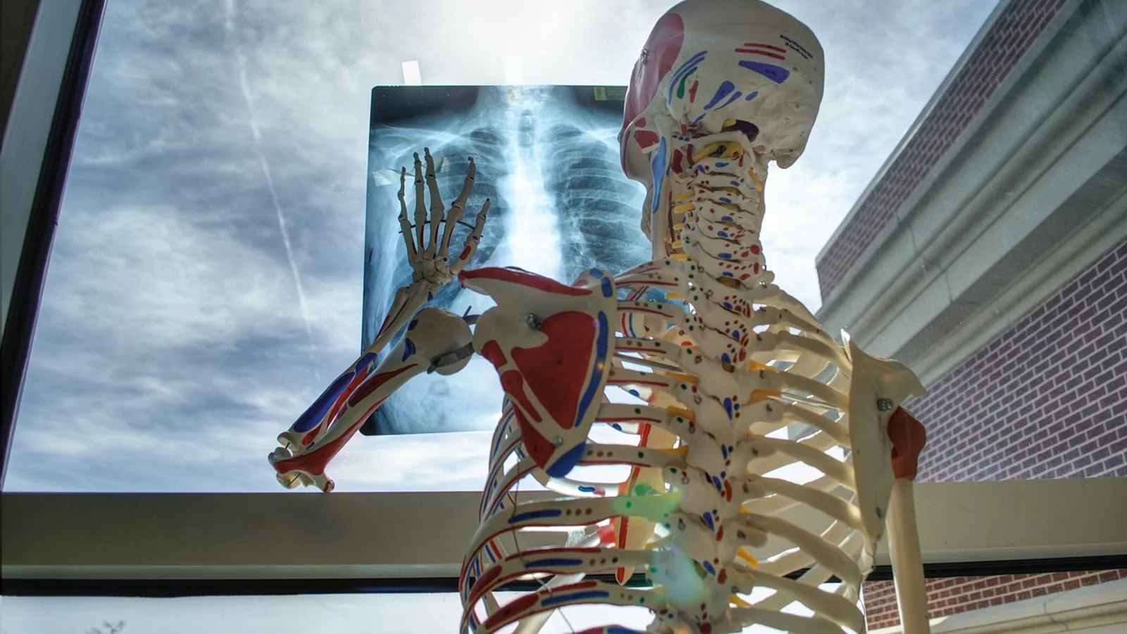
The somatic system is the part of the nervous system that carries conscious sensation from the body and produces voluntary movement by activating skeletal muscle. It is built from a sensory (afferent) division that brings information toward the central nervous system and a motor (efferent) division that sends commands out to muscle.
This system matters in every veterinary visit because it is what you test when you check a gait, a withdrawal reflex, a menace response, or a postural reaction. A dog that cannot bear weight, a cat with a dropped jaw, a horse with a wobbly hind end, all point to somatic pathways that are either working, injured, or blocked. Understanding the divisions keeps the examination logical instead of a random list of tests.
What Somatic Means in Anatomy
The word somatic comes from the Greek soma, meaning body. In anatomy it describes structures of the body wall and limbs, as opposed to visceral structures of the internal organs. Somatic muscle is skeletal muscle. Somatic sensation is touch, pressure, pain, temperature, and proprioception from skin, joints, and muscle. Visceral sensation and visceral muscle (cardiac and smooth muscle) belong to the autonomic system.
This is the first place students slip. Somatic does not mean "of the whole body" and it has nothing to do with somatic cells in genetics, where the term describes non-reproductive cells. In neuroanatomy, somatic means body-wall and voluntary.
A second useful distinction is that somatic and autonomic fibers often travel in the same nerve. The cranial tibial muscle of the pig, a classic skeletal muscle, receives somatic motor axons, sensory axons, and a substantial autonomic supply that most likely serves its blood vessels [1]. So a nerve is not purely somatic or purely autonomic. The classification applies to the individual fiber and its target.
The Two Divisions of the Somatic System
Sensory (Afferent) Division
Afferent means conducting inward. Somatic sensory neurons carry signals from receptors in skin, muscle, tendon, joint capsule, and periosteum toward the central nervous system. Their cell bodies sit in the dorsal root ganglia of spinal nerves and in the sensory ganglia of cranial nerves. These are pseudounipolar neurons, meaning they have a single process that splits into a peripheral branch and a central branch. In the pig cranial tibial model, sensory neurons measured 23 to 67 micrometers in diameter and were located in the ipsilateral L7 to S1 spinal ganglia [1].
Somatic sensory fibers are classified by size and myelination, which determines conduction speed. Large myelinated fibers carry proprioception and fine touch. Small myelinated and unmyelinated fibers carry pain and temperature. This matters clinically because a disease can hit one class before another. In transthyretin amyloidosis, thermal perception thresholds rise before many other signs, reflecting early loss of thin nerve fibers [2].
Motor (Efferent) Division
Efferent means conducting outward. Somatic motor neurons have their cell bodies in the ventral horn of the spinal cord gray matter or in the motor nuclei of the brainstem. Their axons leave the central nervous system and end on skeletal muscle fibers. Each axon is single, heavily myelinated, and fast conducting. In the pig, somatic motoneurons in the S1 ventral horn ranged from 33 to 102 micrometers in diameter, larger than the sensory and autonomic cells in the same study [1].
The somatic motor neuron is the final common pathway. Every voluntary movement, every reflex, and every postural adjustment converges on these cells. When a clinician tests a reflex, the goal is to confirm that this pathway is intact from receptor to muscle.
The Neuron Chain: Somatic Versus Autonomic
The cleanest way to separate the somatic system from the autonomic nervous system is to count neurons in the motor chain.
A somatic motor pathway uses one neuron. The cell body sits in the central nervous system. The axon travels uninterrupted to the target skeletal muscle and releases acetylcholine onto nicotinic receptors. There is no ganglion in the middle of the chain.
An autonomic motor pathway uses two neurons. The first (preganglionic) neuron sits in the central nervous system and synapses in an autonomic ganglion. The second (postganglionic) neuron then travels to the target, which is cardiac muscle, smooth muscle, or a gland. Autonomic ganglia are the relay stations that the somatic system does not have.
This structural difference has functional consequences. A single somatic axon can produce a fast, precise, all-or-nothing contraction. Autonomic output is slower, more diffuse, and modulated at the ganglion. The two systems also cooperate. In the rat, stimulating neurons in the midbrain ventral tegmental area increased both renal sympathetic nerve activity and hindlimb muscle vasodilation, showing that somatic and autonomic control are coordinated from shared brainstem and midbrain centers [3]. Descending neurons in the rostral ventromedial medulla innervate both sympathetic preganglionic neurons and motor regions, and their activity tracks muscle tone and sympathetic tone together across arousal states [4].
The Neuromuscular Junction
The neuromuscular junction is the synapse between a somatic motor axon terminal and a skeletal muscle fiber. It is the point where the nervous system converts an electrical command into a mechanical contraction.
The sequence runs in five steps.
- An action potential arrives at the axon terminal.
- Voltage-gated calcium channels open and calcium enters the terminal.
- Synaptic vesicles fuse with the presynaptic membrane and release acetylcholine into the synaptic cleft.
- Acetylcholine binds nicotinic acetylcholine receptors on the motor end plate of the muscle fiber.
- The muscle fiber depolarizes, calcium is released inside the fiber, and the fiber contracts.
Acetylcholinesterase in the cleft then breaks down acetylcholine so the signal stops.
The nicotinic receptor is the defining feature. Somatic motor output always ends on nicotinic receptors at the muscle. This is why succinylcholine, a nicotinic blocker, abolishes somatic muscle contraction. In a canine bladder reinnervation study, succinylcholine virtually eliminated bladder pressure increases produced by stimulating transferred somatic nerves, confirming that the reinnervated pathway retained the nicotinic mechanism of the original somatic supply [5]. In normal dogs, succinylcholine had no effect on nerve-evoked bladder pressure but strongly reduced urethral and anal sphincter pressure, because those sphincters are skeletal muscle under somatic control [5].
The junction is also a site of disease. Myasthenia gravis attacks the nicotinic receptor and produces exercise-induced weakness. Tick paralysis impairs acetylcholine release. Botulism blocks release. Each condition produces a different pattern of weakness, but all converge on the same junction.
Cranial and Spinal Nerve Components
Somatic fibers reach the body through two routes.
Spinal Nerves
Each spinal nerve forms from a dorsal root and a ventral root. The dorsal root carries somatic and visceral sensory fibers into the spinal cord. The ventral root carries somatic motor fibers out, along with autonomic preganglionic fibers in the thoracolumbar region. After the roots merge, the spinal nerve divides into a dorsal ramus that supplies the epaxial muscles and skin of the back, and a ventral ramus that supplies the limbs and body wall. The thoracic spinal nerves and epaxial muscles of Bothrops jararaca show the same ultrastructural organization of motor neurons, Schwann cell sheaths, and innervated skeletal fibers seen in mammals, which reflects deep evolutionary conservation of the somatic motor system across vertebrates [6].
Cranial Nerves
Cranial nerves carry somatic components in three functional columns.
General somatic efferent fibers innervate the extraocular muscles and the tongue. The oculomotor (CN III), trochlear (CN IV), and abducens (CN VI) nerves innervate the six eye muscles, while the hypoglossal (CN XII) nerve innervates the tongue. These neurons originate in the midbrain and hindbrain and project to specific muscles with precise laterality. The oculomotor neurons innervate the ipsilateral orbit except for the superior rectus and levator palpebrae, which are contralaterally innervated, and the trochlear neurons innervate the contralateral superior oblique [7]. This is why a lesion in one oculomotor nucleus can produce signs in both eyes.
General somatic afferent fibers carry touch, pain, and temperature from the face and oral cavity, mainly through the trigeminal nerve (CN V). Special somatic afferent fibers carry vision, hearing, and balance through CN II, CN VIII, and related pathways. The distinction between general and special somatic afferent is a functional classification, not a structural one, and it appears in the standard scheme of cranial nerve components.
The phrenic nerve is a good example of a somatic nerve with vital function. It arises from cervical spinal segments and innervates the diaphragm. Correct innervation depends on guidance molecules. When Npn-1 was conditionally ablated in somatic motor neurons, phrenic nerves defasciculated and ectopic innervated muscles formed in the diaphragm, showing that somatic motor axons actively shape the muscles they contact [8].
How the Somatic System Is Tested in Practice
A neurologic examination tests the somatic system in a fixed sequence.
- Postural reactions (knuckling, hopping, placing) test proprioceptive sensory input, motor output, and the connections between them.
- Spinal reflexes (patellar, withdrawal, perineal) test the local reflex arc. An absent reflex points to a lesion in the sensory or motor limb of that arc. An exaggerated reflex points to loss of descending inhibition.
- Cranial nerve examination tests the somatic components of CN III, IV, V, VI, VII, X, XI, and XII.
- Gait and muscle palpation assess bulk, tone, and symmetry.
Electrodiagnostics extend the examination. Electromyography records spontaneous muscle fiber activity and helps localize a lesion to the motor neuron, nerve, or muscle. Nerve conduction studies measure how fast a somatic nerve carries a signal. Quantitative sensory testing measures thermal and vibratory thresholds and can detect small fiber loss before routine examination finds it [2].
Skin and nerve biopsy can quantify somatic and autonomic fibers separately. In female cadavers, double immunofluorescent staining for beta III tubulin (a global axonal marker) and myelin basic protein (a myelinated nerve marker) allowed researchers to calculate autonomic density as tissue stained with beta III tubulin alone and somatic density as tissue stained with both markers [9]. Somatic density was highest in the external urethral sphincter, which is expected because that sphincter is skeletal muscle [9]. This technique is a research and diagnostic tool for separating the two systems in tissue.
Somatic and Autonomic Systems Work Together
The somatic system does not operate in isolation. Many behaviors require coordinated somatic and autonomic output.
Penile erection is one example. In the rat, reflex erection involves hemodynamic events under autonomic control and contractions of perineal muscles under somatic control, and the two are interconnected at the spinal level [10]. Blocking the perineal muscle contractions or interrupting afferent input from the penis changes the hemodynamic response, showing bidirectional interaction [10].
Respiration is another. The diaphragm is somatic muscle, but its activity is modulated by autonomic and brainstem circuits that also control heart rate and blood pressure. After cervical spinal cord injury, loss of descending inspiratory drive to the phrenic nucleus causes partial diaphragm paralysis, and recovery depends on plasticity of the surviving somatic motor system [11].
This coupling is why a patient with a somatic problem can show autonomic signs and vice versa. It is also why clinicians assess both systems when a neurologic or neuromuscular disease is suspected.
Quick-Reference Table
| Feature | Somatic Motor | Autonomic Motor |
|---|---|---|
| Neuron chain | One neuron | Two neurons |
| Ganglion | None | Present |
| Myelination | Heavily myelinated | Preganglionic myelinated, postganglionic usually unmyelinated |
| Neurotransmitter | Acetylcholine | Acetylcholine then norepinephrine or acetylcholine |
| Receptor on target | Nicotinic | Nicotinic then adrenergic or muscarinic |
| Target | Skeletal muscle | Cardiac muscle, smooth muscle, glands |
| Conscious control | Yes | No |
| Conduction speed | Fast | Slower |
| Effect | Precise, discrete contraction | Diffuse, sustained modulation |
Quick Review
- The somatic system has a sensory (afferent) division and a motor (efferent) division.
- Somatic motor pathways use one myelinated neuron that releases acetylcholine onto nicotinic receptors of skeletal muscle.
- Autonomic motor pathways use two neurons with a ganglion in between.
- Somatic sensation comes from skin, muscle, joint, and tendon. Visceral sensation is autonomic.
- Somatic fibers travel in spinal nerves (dorsal and ventral roots) and in cranial nerves (general and special somatic components).
- The neuromuscular junction is the nicotinic synapse where motor command becomes contraction.
- Somatic and autonomic systems are coordinated by shared brainstem and midbrain centers.
Clinical Relevance, Limitations and Common Mistakes
Somatic dysfunction is a daily problem in veterinary practice. A dog with intervertebral disc disease loses proprioception and motor function in the hind limbs because the spinal cord carries somatic sensory and motor tracts. A horse with laryngeal hemiplegia has atrophy of the cricoarytenoideus dorsalis, a skeletal muscle innervated by the recurrent laryngeal nerve, a branch of the vagus. A cat with a perineal urethrostomy may have somatic and autonomic nerve disruption in the surgical field, which is why nerve-sparing technique matters [9].
The most common mistakes students and clinicians make are worth naming.
First, treating the somatic system as only motor. The sensory division is half the system and is often the first to fail. Thermal and pain testing can detect small fiber loss before motor signs appear [2].
Second, assuming a nerve is purely somatic or purely autonomic. The cranial tibial muscle receives both, and the autonomic supply likely serves its vessels [1]. A nerve injury can therefore produce mixed signs.
Third, confusing somatic symptom disorder with a somatic nerve problem. Somatic symptom disorder is a psychiatric diagnosis involving excessive thoughts and behaviors about bodily symptoms, and it is studied with autonomic and stress measures, not with nerve conduction [12]. The word somatic appears in both contexts but means different things.
Fourth, forgetting that the neuromuscular junction is a target. Weakness that worsens with exercise and improves with rest suggests a junctional problem, not a nerve or muscle problem.
Fifth, missing the coordination between systems. A patient with somatic signs may also have autonomic instability, and treating only one system can leave the other untreated [3][4].
The limitation of any framework is that individual animals vary. A reflex that is normal in one dog may be absent in another with a different body condition or age. This article is educational and is not a substitute for veterinary diagnosis or treatment. A veterinarian who examines the patient in person is the only reliable source of a diagnosis.
Frequently Asked Questions
What is the somatic system in simple terms?
The somatic system is the part of the nervous system that lets an animal feel the body and move it voluntarily. It carries touch, pain, and position sense inward and sends movement commands outward to skeletal muscle.
How does the somatic motor pathway differ from the autonomic motor pathway?
The somatic pathway uses one myelinated neuron from the central nervous system to the muscle. The autonomic pathway uses two neurons with a ganglion between them.
What neurotransmitter and receptor does somatic motor use?
Somatic motor neurons release acetylcholine onto nicotinic receptors on the skeletal muscle motor end plate.
Why is the neuromuscular junction important in veterinary medicine?
It is the final step where nerve signal becomes muscle contraction. Diseases that block or destroy the nicotinic receptor cause weakness and are diagnosed by testing this junction.
Can a nerve carry both somatic and autonomic fibers?
Yes. Many nerves carry somatic motor, somatic sensory, and autonomic fibers, and the autonomic fibers often supply blood vessels within the muscle.
What happens when the somatic system is damaged?
Damage can cause loss of sensation, weakness, paralysis, or altered reflexes, depending on which part of the pathway is affected. The pattern of signs helps localize the lesion.
flowchart TD
    A[Stimulus at receptor] --> B[Somatic sensory neuron]
    B --> C[Dorsal root ganglion]
    C --> D[Spinal cord or brainstem]
    D --> E[Processing and integration]
    E --> F[Somatic motor neuron]
    F --> G[Myelinated axon]
    G --> H[Neuromuscular junction]
    H --> I[Nicotinic receptor]
    I --> J[Skeletal muscle contraction]
    E --> K[Autonomic pathway]
    K --> L[Two neuron chain with ganglion]
Related Articles
- Cell Division
- Transcription Bubble: Formation, Function, and Key Concepts
- Nucleotide and Nucleoside: Structure, Function, and Key Differences
- Coding vs Noncoding RNA: Key Differences and Functions
- Nucleotide Parts: Structure, Function, and Key Concepts
- Somatic Variant Calling Workflow: Key Differences from Germline and How to Optimize for Low-VAF Detection
- Somatic Nervous System: Structure and Function
- Mitotic vs Meiotic: Key Differences in Cell Division
- Somatic Nerves: What They Are and What They Do
Sources
- Localization of the autonomic, somatic and sensory neurons innervating the cranial tibial muscle of the pig.
- Comparison of quantitative sensory testing and heart rate variability in Swedish Val30Met ATTR.
- Coactivation of renal sympathetic neurons and somatic motor neurons by chemical stimulation of the midbrain ventral tegmental area.
- Spinal projecting neurons in rostral ventromedial medulla co-regulate motor and sympathetic tone.
- Neuromuscular nicotinic receptors mediate bladder contractions following bladder reinnervation with somatic to autonomic nerve transfer after decentralization by spinal root transection.
- Ultrastructure of the somatic motor system in Bothrops jararaca (Reptilia, Squamata, Viperidae).
- Evolution and development of extraocular motor neurons, nerves and muscles in vertebrates.
- The role of Sema3-Npn-1 signaling during diaphragm innervation and muscle development.
- Somatic and autonomic nerve density of the urethra, periurethral tissue, and anterior vaginal wall: an immunohistochemical study in adult female cadavers.
- Reflex erection in the rat: reciprocal interplay between hemodynamic and somatic events.
- The challenges of respiratory motor system recovery following cervical spinal cord injury.
- Does childhood trauma impact daily psychobiological stress in somatic symptom disorder? An ambulatory assessment study.