Pons Function: Roles in Respiration, Sleep, and Cranial Nerves
By Dr. Zubair Khalid, DVM, MS, PhD ·
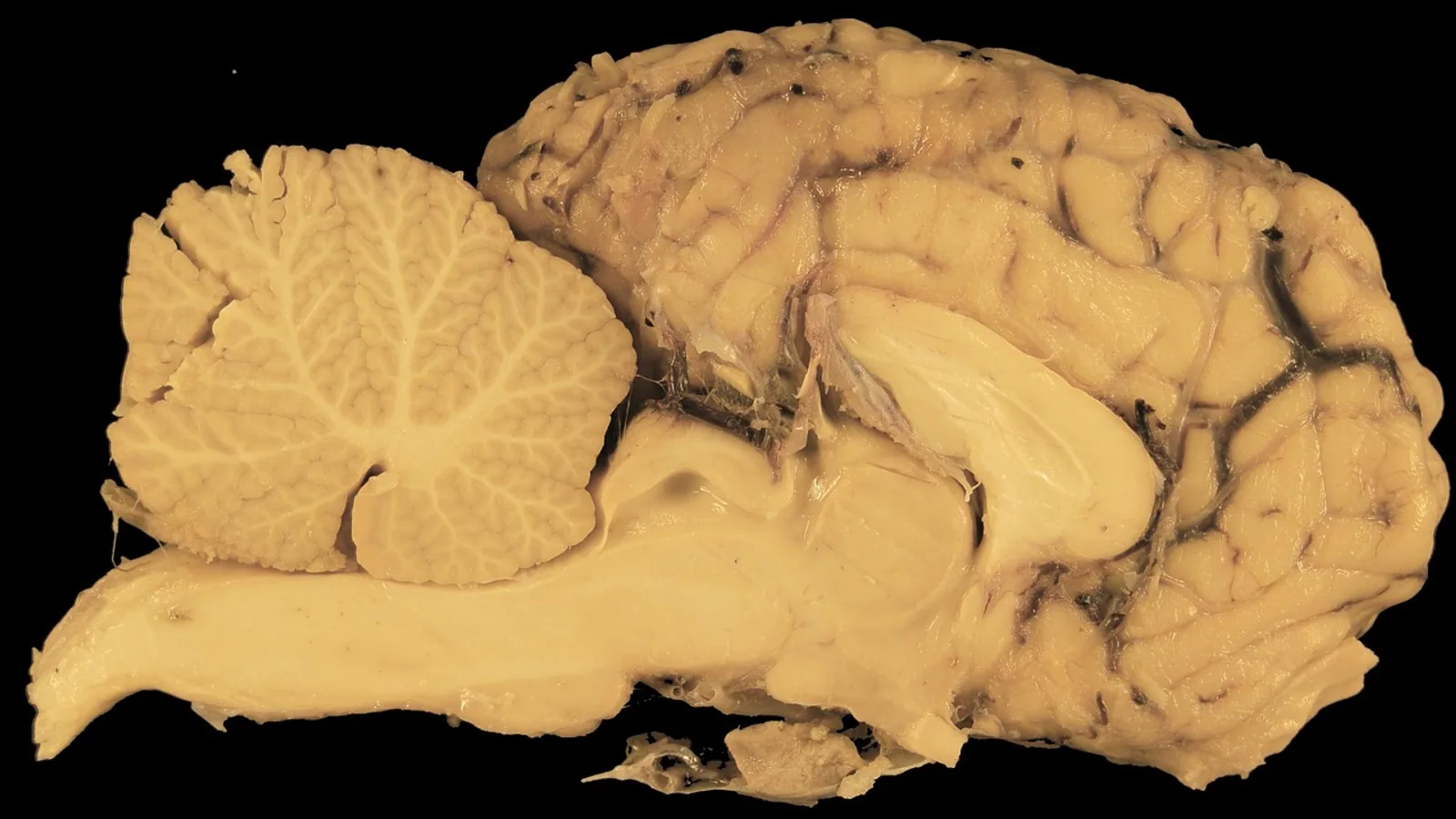
The pons is the middle segment of the brainstem, sitting between the midbrain above and the medulla oblongata below, and it serves as the main bridge carrying information between the cerebrum, the cerebellum, and the lower brainstem. Its core functions are respiratory rhythm modulation, rapid eye movement sleep onset, auditory processing, micturition control, and relaying the cranial nerve pathways for the trigeminal, abducens, facial, and vestibulocochlear nerves.
Understanding pons function matters in veterinary practice because even small pontine lesions can produce dramatic signs: sudden gait abnormalities in dogs, narcolepsy-cataplexy, vestibular disease, or life-threatening respiratory instability. This guide walks through each pontine function, maps nuclei to roles in a single reference table, and highlights species differences that change how these disorders present in dogs, cats, and horses.
What the Pons Is and Where It Sits
The brainstem connects the forebrain to the spinal cord and is divided caudo-rostrally into the medulla oblongata, the metencephalon (which includes the pons), and the mesencephalon [1]. The pons forms the middle of this three-part column. It sits ventral to the cerebellum and dorsal to the basilar artery in most domestic mammals, and it is the anatomical bridge between the cerebral hemispheres and the cerebellar hemispheres.
The brainstem is organized in three layers: a ventral basis, an intermediate tegmentum, and a dorsal tectum [1]. The pontine basis carries the massive corticopontine and pontocerebellar fiber systems that make the pons the largest relay station between the cortex and the cerebellum. The pontine tegmentum contains the cranial nerve nuclei and the reticular formation, the lattice of neurons and fibers that coordinates brainstem-based vital functions including arousal and consciousness [1].
The pons in the brain is not a passive cable. It contains its own nuclei, its own local circuits, and its own chemical signaling systems. That is why lesions here produce specific syndromes rather than a single generic brainstem picture.
Relay Between Cerebrum and Cerebellum
The largest single function of the pons is to relay information between the cerebrum and the cerebellum. Cortical motor and premotor areas send axons down to pontine nuclei in the basis pontis. Those pontine neurons then project across the midline into the contralateral cerebellar hemisphere through the middle cerebellar peduncle. The circuit runs in both directions, so the cerebellum can influence cortical motor planning and the cortex can update cerebellar predictions.
This relay is why the pons enlarged so dramatically during mammalian evolution. As human telencephalic volume increased, the pons, cerebellar hemispheres, cerebral crura, and pyramidal tracts all expanded together [1]. The same scaling principle applies across domestic species. Animals with more complex forelimb and jaw control, such as dogs and horses, have proportionally larger pontocerebellar systems than animals with simpler motor repertoires.
Clinically, this relay explains a well-known veterinary observation: pontine lesions in dogs cause gait abnormalities. Damage to the basis pontis or middle cerebellar peduncle interrupts the corticopontocerebellar loop, producing ataxia, wide-based stance, and dysmetria without necessarily causing obvious cranial nerve deficits. The gait problem is a relay problem, not a cerebellar problem in the primary sense.
Respiration and the Pneumotaxic Center
The pons contains the pneumotaxic center, a group of neurons in the rostral pontine tegmentum that modulates the rate and depth of breathing. The medulla generates the basic respiratory rhythm. The pons adjusts it. When the pneumotaxic center fires more actively, inspiration is cut short and respiratory rate rises. When it is suppressed, inspiratory time lengthens and tidal volume increases.
This pontine role is not isolated. The pons is part of a distributed respiratory network that includes the medulla, the hypothalamus, and the spinal cord. Hypothalamic nuclei such as the paraventricular nucleus, perifornical area, dorsomedial hypothalamus, and lateral and posterior hypothalamus have extensive interconnectivity with the pons, medulla, and spinal cord, and they modulate basal ventilation, responses to hypoxia and hypercapnia, and respiratory changes during sleep and exercise [2][3]. The pons is a mid-level node in that hierarchy, sitting between the automatic rhythm generators below and the behavioral and homeostatic drives above.
Pontine respiratory neurons are also vulnerable to injury. In a guinea pig model of recurrent apnea, animals subjected to repeated cycles of ventilatory arrest and recovery over three hours developed apoptotic neurons and glia in specific brainstem regions involved in respiratory control [4]. This finding supports the clinical concern that chronic sleep-disordered breathing can damage the very circuits that regulate breathing.
Sleep and REM Onset
The pons is the trigger zone for rapid eye movement sleep. Cholinergic neurons in the dorsal part of the rostral nucleus pontis oralis, a pontine tegmental nucleus, initiate the REM state when activated. In an in situ perfused rat brainstem preparation, microinjections of the cholinergic agonist carbachol (25 to 50 nanoliters of a 10 millimolar solution) into the pons triggered REM sleep-like episodes in 24 of 40 injection sites [5]. These episodes began roughly 151 seconds after injection, lasted about 20 minutes on average, and were most reliably elicited from the dorsal rostral nucleus pontis oralis [5].
The hallmark of those REM-like episodes was a specific pattern: depression of hypoglossal nerve activity with an increase in respiratory rate, while phrenic nerve activity changed little [5]. That pattern matches natural REM sleep, during which the tongue and pharyngeal muscles lose tone. In animals with compromised upper airway anatomy, this sleep-related hypotonia increases airway resistance or causes complete obstruction, which is why obstructive episodes are longest and most severe during REM sleep [5].
The pons also participates in arousal and consciousness through its reticular formation and its small aminergic and cholinergic projection nuclei, which influence mood, motivation, and cognition [1]. Pontine lesions can therefore produce hypersomnia, narcolepsy, or cataplexy. A four-year-old intact female cocker spaniel with brainstem meningoencephalitis affecting the mesencephalon, pons, and rostral medulla presented with acute progressive lethargy, hypersomnia, vestibular signs, and cataplexy [6]. The narcolepsy-cataplexy episodes in that dog were initially triggered by offering food and gradually resolved over three weeks with immunosuppressive treatment [6]. This case shows that symptomatic narcolepsy in dogs can be pontine in origin and can be reversible when the underlying inflammation is controlled.
Hearing and the Superior Olivary Complex
The pons houses the superior olivary complex, the first major binaural processing center in the auditory pathway. Sound information from the cochlear nuclei ascends to the superior olivary complex, where inputs from both ears are compared. This comparison is how mammals localize sound in space. The medial superior olive computes interaural time differences, and the lateral superior olive computes interaural level differences.
The superior olivary complex contains several named nuclei: the medial nucleus of the trapezoid body, the lateral nucleus of the trapezoid body, the lateral superior olive, and the superior paraolivary nucleus [7]. In the Etruscan shrew, a species that approximates ancestral mammalian conditions, these nuclei have been mapped in detail, and most labeled proteins show expression patterns comparable to rodents, although the superior paraolivary nucleus neurons are glycinergic rather than GABAergic and overall calcium-binding protein expression is low [7]. This kind of comparative work shows that the pontine auditory system is conserved in its overall plan but variable in its chemical details across species.
The vestibulocochlear nerve, cranial nerve VIII, enters the brainstem at the medullopontine sulcus after crossing the internal auditory canal and cerebellopontine angle cistern [8]. Its six nuclei are located in the lower pons [8]. This anatomical fact is why pontine lesions can cause hearing loss, vestibular signs, or both. In dogs and cats, brainstem auditory evoked response testing depends on the integrity of these pontine relays.
Cranial Nerve Pathways in the Pons
Four cranial nerves have their nuclei or principal pathways in the pons: the trigeminal (V), abducens (VI), facial (VII), and vestibulocochlear (VIII) nerves. The brainstem contains the motor and sensory nuclei of cranial nerves III through XII, arranged in a mediolateral direction, and unlike the spinal cord, their afferent and efferent fibers are not macroscopically separated [1].
Trigeminal Nerve (V)
The trigeminal nerve is the largest cranial nerve and carries sensation from the face and motor supply to the muscles of mastication. Its motor and sensory fibers have extra-axial and intra-axial courses that can be identified during brainstem mapping by recording responses from the masseter and tongue muscles [9]. The spinal trigeminal nucleus extends from the pons down into the medulla and upper spinal cord, and functional mapping in humans has shown a somatotopic arrangement of the three trigeminal branches along a perioral-to-periauricular axis, with overlap between dermatomes [10]. In veterinary patients, pontine trigeminal lesions can cause facial hypalgesia, absent jaw tone, or difficulty chewing.
Abducens Nerve (VI)
The abducens nerve supplies the lateral rectus muscle, which abducts the eye. Its nucleus sits in the caudal pons, and its fibers exit ventrally. The abducens nucleus also contains internuclear neurons that project to the contralateral medial rectus subnucleus of the oculomotor nerve, coordinating horizontal gaze. Electromyographic monitoring of the external rectus muscle is used during brainstem surgery to protect this nucleus [11].
Facial Nerve (VII)
The facial nerve has a complex course through the pons. Its motor nucleus lies in the ventrolateral pontine tegmentum, and its fibers loop around the abducens nucleus before exiting the brainstem. This loop creates the facial colliculus, a visible landmark on the floor of the fourth ventricle [11]. The facial nerve supplies the muscles of facial expression, and its parasympathetic fibers innervate the lacrimal and salivary glands. Pontine lesions affecting the facial nucleus or its intrapontine fibers cause facial paralysis, reduced tear production, and dry mouth. In SIDS research, increased expression of the beta-2 nicotinic acetylcholine receptor subunit has been found in the facial nucleus of infants who died of SIDS compared with controls, suggesting that cholinergic signaling in pontine nuclei is relevant to arousal and respiratory control [12].
Vestibulocochlear Nerve (VIII)
The vestibulocochlear nerve carries balance and hearing information into the lower pons. Its six nuclei include the cochlear nuclei, which process hearing, and the vestibular nuclei, which process balance [8]. The nerve enters at the medullopontine sulcus, and its canalicular and cisternal segments can be imaged with heavily T2-weighted MRI sequences such as FIESTA or CISS [8]. In dogs and cats, pontine vestibular disease produces head tilt, nystagmus, and ataxia, and it can be difficult to distinguish from peripheral vestibular disease without imaging.
The Pontine Micturition Center
The pontine micturition center, also called the Barrington nucleus, sits in the dorsolateral pontine tegmentum. It coordinates the switch between urine storage and voiding. When activated, it sends descending signals that relax the urethral sphincter and contract the detrusor muscle simultaneously. When damaged, the result is detrusor-sphincter dyssynergia, a condition in which the bladder contracts against a closed sphincter, leading to incomplete voiding and urinary retention.
In veterinary neurology, pontine micturition center dysfunction is most often seen with brainstem lesions from trauma, inflammation, or neoplasia. It is one of the reasons that animals with pontine disease need careful monitoring of bladder function, not just gait and cranial nerve status.
Function-by-Function Table: Pontine Nuclei and Their Roles
| Pontine nucleus or region | Primary function | Clinical relevance |
|---|---|---|
| Basis pontis and pontine nuclei | Relay between cerebrum and cerebellum | Gait abnormalities, ataxia, dysmetria in dogs |
| Pneumotaxic center (rostral pontine tegmentum) | Modulates respiratory rate and inspiratory time | Respiratory instability with pontine lesions |
| Nucleus pontis oralis (dorsal rostral part) | REM sleep onset via cholinergic activation | Narcolepsy, cataplexy, REM-related airway obstruction |
| Superior olivary complex | Binaural hearing and sound localization | Hearing deficits, abnormal brainstem auditory evoked responses |
| Trigeminal motor and sensory nuclei | Jaw movement, facial sensation | Facial hypalgesia, weak jaw tone |
| Abducens nucleus | Lateral rectus control, horizontal gaze | Inability to abduct the eye, gaze palsy |
| Facial nucleus | Facial expression, lacrimation, salivation | Facial paralysis, dry eye, dry mouth |
| Vestibulocochlear nuclei | Balance and hearing | Head tilt, nystagmus, deafness |
| Pontine micturition center | Coordination of bladder voiding | Urinary retention, detrusor-sphincter dyssynergia |
| Caudal pontine reticular nucleus | Acoustic startle reflex | Exaggerated or absent startle response |
The caudal pontine reticular nucleus deserves a note. It is the sensorimotor interface for the acoustic startle reaction, an archaic reflex that connects auditory inputs to motor output [13]. In gerbils, mice, and Etruscan shrews, the neurons of this nucleus form a continuum of soma sizes rather than distinct subpopulations, and Kv1.1 potassium channel expression increases continuously with soma size [13]. This suggests that the startle reflex is built from a graded population of neurons rather than discrete cell types.
How the Pontine Respiratory and Sleep Circuits Interact
The relationship between breathing and sleep is one of the most clinically important pontine functions. During REM sleep, the pons actively suppresses upper airway muscle tone while increasing respiratory rate [5]. This combination is protective in healthy animals because it prevents the airway from collapsing during the muscle relaxation of REM. But in animals with anatomical narrowing of the airway, such as brachycephalic dogs or horses with recurrent laryngeal neuropathy, the same pontine mechanism can tip the balance toward obstruction.
The pons does not work alone in this. Hypothalamic nuclei project to the pons and medulla and modulate respiration during awake and sleep states [2][3]. This means that a pontine lesion can unmask hypothalamic influences that were previously balanced, or vice versa. The respiratory network is a hierarchy, and the pons is a critical middle layer.
The following flow diagram traces the main pontine pathways from sensory input to clinical output.
flowchart TD
    A[Cerebral cortex] --> B[Pontine nuclei]
    B --> C[Cerebellum]
    C --> D[Motor coordination]
    E[Medulla rhythm generator] --> F[Pneumotaxic center]
    F --> G[Respiratory rate control]
    H[Cholinergic trigger] --> I[Nucleus pontis oralis]
    I --> J[REM sleep onset]
    K[Cochlear input] --> L[Superior olivary complex]
    L --> M[Sound localization]
    N[Cranial nerve nuclei] --> O[Trigeminal facial abducens vestibulocochlear]
    O --> P[Head and face function]
Comparative Species Differences
Pontine function is conserved across mammals, but the clinical expression of pontine disease varies by species.
In dogs, pontine lesions cause gait abnormalities as a prominent sign. The corticopontocerebellar relay is large and clinically eloquent in dogs, so even partial interruption produces visible ataxia and dysmetria. Dogs also develop symptomatic narcolepsy-cataplexy with pontine inflammation, as documented in a cocker spaniel with brainstem meningoencephalitis [6]. The cataplexy episodes in that dog were triggered by food and resolved with immunosuppression, which is a pattern veterinarians should recognize as potentially treatable [6].
In horses, laryngeal hemiplegia may involve the vagus nerve and its brainstem connections. The vagus nerve arises from the medulla, not the pons, but the two structures are functionally linked through the reticular formation and the respiratory central pattern generator. A horse with recurrent laryngeal neuropathy has a failure of abductor muscle function in the larynx, and the brainstem circuitry that coordinates laryngeal movement with breathing spans both the pons and the medulla. Pontine respiratory neurons influence the timing of laryngeal abduction during inspiration, so pontine dysfunction can contribute to upper airway obstruction in horses even when the primary lesion is in the recurrent laryngeal nerve.
In cats, pontine lesions often present with vestibular signs because the vestibular nuclei are large and the feline brainstem is compact. The same lesion that would cause gait abnormalities in a dog may cause a more obvious head tilt and nystagmus in a cat.
Across species, the pontine micturition center is a common target of brainstem disease. Any animal with a pontine lesion should have bladder function assessed, because urinary retention can develop silently and lead to secondary complications.
Clinical Relevance, Limitations and Common Mistakes
Pontine disease in veterinary patients is challenging because the signs are varied and the structure is small. A lesion in the pons can produce gait abnormalities, cranial nerve deficits, respiratory irregularity, sleep disorders, or bladder dysfunction, and the combination depends on which nuclei and tracts are involved.
The most common mistake is to attribute all vestibular signs to peripheral vestibular disease. Pontine vestibular nuclei lesions can mimic peripheral disease, and imaging is often needed to distinguish them. A second common mistake is to overlook sleep and respiratory signs in animals with brainstem disease. Narcolepsy-cataplexy can be mistaken for syncope or seizures, and REM-related airway obstruction can be mistaken for primary airway disease. A third mistake is to focus on gait and cranial nerves while ignoring bladder function, which can lead to unrecognized urinary retention.
Imaging the pons is difficult because of its compact nature and the proximity of critical structures. MRI is preferred over computed tomography for brainstem evaluation, and higher field strengths offer superior anatomic detail [14]. The compact nature of the brainstem poses challenges in distinguishing its nuclei on MRI, but adequate protocols including T1- and T2-weighted imaging, fluid-attenuated inversion recovery, susceptibility-weighted imaging, and diffusion-weighted imaging enable comprehensive evaluation [14]. Diffusion tensor imaging with fiber tractography provides additional qualitative and quantitative insight into white matter tracts [14].
Treatment depends on the underlying cause. Inflammatory lesions may respond to immunosuppression, as in the dog with meningoencephalitis of unknown origin [6]. Neoplastic lesions may require surgery, radiation, or palliative care. Neuropathic pain from pontine lesions can be severe and may require multimodal analgesia, including methadone as an add-on for refractory central pain [15].
This article is educational and is not a substitute for veterinary diagnosis or treatment.
Frequently Asked Questions
What does the pons do in a dog's brain?
The pons relays information between the cerebrum and cerebellum, modulates breathing rate, triggers REM sleep, processes hearing, coordinates bladder voiding, and hosts the nuclei for cranial nerves V, VI, VII, and VIII. Pontine lesions in dogs commonly cause gait abnormalities.
Can a dog recover from a pontine lesion?
Recovery depends on the cause. Inflammatory pontine lesions can resolve with immunosuppressive treatment, as seen in a dog with brainstem meningoencephalitis whose narcolepsy-cataplexy episodes resolved over three weeks. Structural lesions such as tumors may have a more guarded prognosis.
Why does the pons control breathing?
The pneumotaxic center in the rostral pons modulates the rate and depth of breathing generated by the medulla. It adjusts inspiratory time and respiratory rate in response to signals from the hypothalamus, cortex, and peripheral sensors.
What is the pneumotaxic center?
The pneumotaxic center is a group of neurons in the rostral pontine tegmentum that shortens inspiration and increases respiratory rate when active. It is one of several pontine regions that fine-tune the basic rhythm produced by the medulla.
How does the pons affect sleep?
Cholinergic neurons in the dorsal rostral nucleus pontis oralis trigger REM sleep. When activated, they produce REM-like episodes with upper airway muscle depression and increased respiratory rate, which is why obstructive sleep apnea worsens during REM.
Which cranial nerves pass through the pons?
The trigeminal, abducens, facial, and vestibulocochlear nerves have their nuclei or principal pathways in the pons. The vestibulocochlear nerve enters at the medullopontine sulcus and its six nuclei are located in the lower pons.
What is the pontine micturition center?
The pontine micturition center is a nucleus in the dorsolateral pontine tegmentum that coordinates bladder voiding by relaxing the urethral sphincter and contracting the detrusor muscle at the same time. Damage causes urinary retention and detrusor-sphincter dyssynergia.
Why do horses with laryngeal hemiplegia sometimes have brainstem involvement?
The vagus nerve arises from the medulla, but the brainstem circuitry that coordinates laryngeal movement with breathing spans both the pons and the medulla. Pontine respiratory neurons influence the timing of laryngeal abduction during inspiration, so pontine dysfunction can contribute to upper airway obstruction even when the primary lesion is in the recurrent laryngeal nerve.
Related Articles
- Bovine Neurological Anatomy: Cranial Nerves and Brainstem
- Equine Larynx Anatomy and Function in Respiration
- Respiration Biology
- Anticodon Sequence: Definition, Function, and Role in Translation
- 5 Prime Cap: Definition, Function, and Role in mRNA
- DNA Polymerase 1 2 3: Functions and Roles in Replication
- Cranial Nerve Mnemonic: Names, Numbers, and Functions
- Olfactory Nerve: Cranial Nerve I Pathway and Function
Sources
- Structural and functional anatomy of the brainstem.
- Modulation of respiration and hypothalamus.
- The role of the hypothalamus in modulation of respiration.
- Apnea produces neuronal degeneration in the pons and medulla of guinea pigs.
- REM sleep-like episodes of motoneuronal depression and respiratory rate increase are triggered by pontine carbachol microinjections in in situ perfused rat brainstem preparation.
- Symptomatic Narcolepsy/Cataplexy in a Dog with Brainstem Meningoencephalitis of Unknown Origin.
- Anatomy of superior olivary complex and lateral lemniscus in Etruscan shrew.
- The Vestibulocochlear Nerve: Anatomy and Pathology.
- A novel method of neurophysiological brainstem mapping in neurosurgery.
- Functional brainstem representations of the human trigeminal cervical complex.
- [[Monitoring of the Floor of the 4th Ventricle for Brainstem Surgery].](https://pubmed.ncbi.nlm.nih.gov/37211736/)
- Effects of cigarette smoke exposure on nicotinic acetylcholine receptor subunits α7 and β2 in the sudden infant death syndrome (SIDS) brainstem.
- In the Caudal Pontine Reticular Nucleus, Kv1.1 Expression Is Soma Size Dependent and Invariant During Postnatal Development.
- Computed tomography and magnetic resonance imaging of the brainstem.
- Successful pain control with add-on methadone for refractory neuropathic pain due to radiation necrosis in pontine metastatic lesion: a case report.