Olfactory Nerve: Cranial Nerve I Pathway and Function
By Dr. Zubair Khalid, DVM, MS, PhD ·
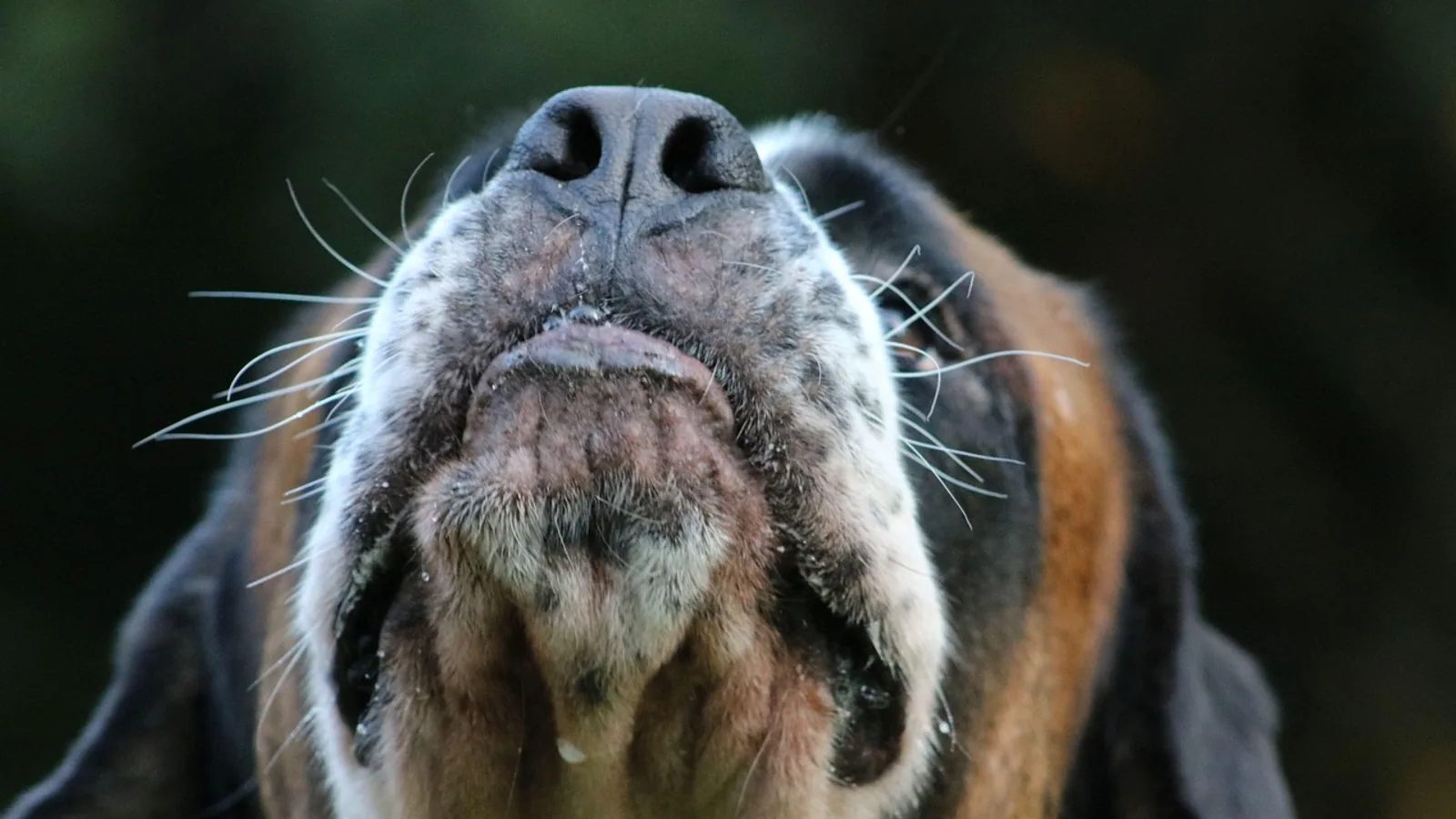
The olfactory nerve, cranial nerve I, is the first and shortest of the twelve cranial nerves and the only one that reaches the cerebral cortex without a relay in the thalamus. It carries smell information from olfactory receptor neurons in the nasal epithelium, through the perforations of the cribriform plate as the fila olfactoria, into the olfactory bulb, and then along the olfactory tract to the piriform cortex, amygdala, and entorhinal cortex.
This arrangement makes the olfactory nerve unusual in two ways. It is the only cranial nerve with direct cortical projection, and it is the only one whose primary sensory neurons are replaced continuously throughout life. For veterinary students, the pathway matters because it explains how a dog can detect a scent at parts per trillion, why nasal disease and head trauma can blunt smell, and why the olfactory brain sits so close to the limbic circuits that govern emotion and memory.
What the Olfactory Nerve Is and What It Is Not
The olfactory nerve is a special visceral afferent nerve. "Special" means it serves one dedicated sense rather than general sensation. "Visceral afferent" places it with the chemical senses that monitor the internal and external environment. Unlike the optic nerve, which is a true tract of the central nervous system wrapped in meninges, the olfactory nerve is a collection of peripheral axons that happen to synapse inside a cortical structure, the olfactory bulb.
That distinction has practical consequences. The olfactory nerve is unmyelinated and extremely thin, so it is fragile. Its axons cross the cribriform plate of the ethmoid bone through many small foramina, which is why blunt head trauma that shifts the brain relative to the skull can shear the fila olfactoria and produce permanent anosmia (loss of smell). The nerve also regenerates. Olfactory receptor neurons turn over roughly every 30 to 60 days, and their axons re-establish synapses in the olfactory bulb throughout adult life, a capacity matched by few other mammalian neurons.
The term "smell nerve" or "nerve for smell" refers to this same structure. There is no separate accessory olfactory nerve in the cranial nerve numbering, though many mammals have an accessory olfactory system, the vomeronasal organ and its nerve, which detects pheromones and projects to the accessory olfactory bulb. In dogs and cats the vomeronasal organ is well developed and opens into the incisive duct. In humans it is present during development but regresses and is generally considered nonfunctional.
The Pathway, Step by Step
The olfactory pathway runs in a fixed sequence from the nasal cavity to the cortex. Each step has a defined cell type and target, and each can be disrupted by different diseases.
Step 1: Olfactory Receptor Neurons in the Olfactory Epithelium
The first-order neurons are bipolar olfactory receptor neurons embedded in the olfactory epithelium, a specialized mucosa covering the ethmoturbinates and the caudal nasal septum. Each receptor neuron extends a single dendrite to the mucosal surface, where it ends in a knob bearing cilia. Odorant molecules dissolved in the mucus layer bind to G protein-coupled odorant receptors on those cilia.
Each receptor neuron expresses one odorant receptor gene, and all neurons expressing the same receptor converge on the same glomerulus in the olfactory bulb. This one receptor, one neuron, one glomerulus rule is the organizing principle of the system. It means the bulb contains a spatial map of receptor identity rather than a map of odor identity, and the brain must decode odor quality from the pattern of activated glomeruli.
The number of functional odorant receptor genes varies enormously across species, and this is the single biggest genetic difference between a dog's nose and a human's. Dogs carry a far larger repertoire of intact olfactory receptor genes than humans, which expands the range of volatile molecules their receptors can detect. The olfactory epithelium in dogs also covers a much larger surface area, folded over extensive turbinates that increase the contact between inhaled air and receptor neurons.
Step 2: Axons Through the Cribriform Plate as Fila Olfactoria
The unmyelinated axons of the receptor neurons collect into bundles called fila olfactoria. These bundles pass through the perforations of the cribriform plate of the ethmoid bone to enter the cranial cavity. The fila olfactoria are the actual olfactory nerve proper. They are surrounded by Schwann cells rather than oligodendrocytes, which is consistent with their peripheral identity.
Because the cribriform plate is rigid and the fila are delicate, this is the most mechanically vulnerable point in the pathway. Fractures of the cribriform plate from a fall, a vehicular injury, or a blow to the face can sever the fila and cause unilateral or bilateral anosmia. The same region is the route by which nasal tumors, fungal disease, and some infections can spread from the nasal cavity into the cranial vault.
Step 3: Synapse in the Olfactory Bulb Glomeruli
Inside the skull, the fila olfactoria terminate in the olfactory bulb, a laminated structure sitting on the cribriform plate beneath the frontal lobe. The bulb has several layers, and the first is the glomerular layer, where receptor axons synapse onto the dendrites of mitral cells and tufted cells.
A glomerulus is a spherical neuropil compartment roughly 100 to 200 micrometers across in rodents. Within it, thousands of receptor axons converge onto the apical dendrites of a small number of projection neurons. Each glomerulus receives input from one receptor type, and each mitral or tufted cell typically extends its primary dendrite into a single glomerulus. The result is a one-to-one labeled line from receptor type to projection neuron, modified by local inhibition.
That local inhibition is substantial. Periglomerular cells, GABAergic interneurons in the glomerular layer, provide inhibitory input to mitral and tufted cells and shape their responses. In awake mice, periglomerular cells show robust, odor-specific responses that differ between odorants, and their population changes with experience: repeated passive exposure reduces the number of odor-responsive cells, while active discrimination learning expands the responsive population and sharpens selectivity over training [1]. This is the cellular basis for the idea that smell is a learned sense, not a fixed one.
Step 4: Mitral and Tufted Cells and the Olfactory Tract
Mitral cells and tufted cells are the output neurons of the olfactory bulb. They are the second-order neurons of the pathway. Their axons form the olfactory tract, which runs caudally along the ventral surface of the frontal lobe.
Mitral and tufted cells are not interchangeable. They differ in soma size, laminar position, dendritic arborization, and projection targets. Tufted cells sit more superficially and tend to project to more anterior cortical targets, while mitral cells sit deeper in the mitral cell layer and project more broadly. Recent work has identified a further subdivision, superficial tufted cells, which have distinct voltage-gated conductances, fire at higher frequencies, and are modulated by insulin, with roughly 73 percent of superficial tufted cells co-expressing the Kv1.3 potassium channel and the insulin receptor [2].
The output of these cells is not a simple reflection of receptor input. Odor-evoked firing in mitral and tufted cells is relatively sparse in the awake animal, and the temporal pattern of spikes, particularly alignment with the respiratory cycle, carries much of the information about odor identity [3]. Decoding accuracy improves as more neurons are sampled and as information is integrated across multiple sniffs [3]. The bulb also implements a rapid temporal filter: mitral and tufted cells activated earliest in a sniff represent odor identity across concentrations, while cells connected to later-activated glomeruli are concentration dependent [4]. This is how an animal recognizes the same odor whether it is faint or strong.
Mitral and tufted cells are coupled to each other by gap junctions mediated by connexin 36. Eliminating connexin 36 in knockout mice reduces fine odor discrimination for similar odor pairs but not for dissimilar ones, and reduces mitral cell excitation in response to sensory afferent stimulation, most prominently in the late phase of the response [5][6]. The coupling therefore appears to sharpen discrimination between chemically similar odors rather than to support basic detection.
Step 5: Olfactory Tract to Piriform Cortex, Amygdala, and Entorhinal Cortex
The olfactory tract carries mitral and tufted cell axons to a set of cortical and limbic targets. The largest is the piriform cortex, the primary olfactory cortex, which occupies the ventral lateral surface of the temporal lobe in most mammals. The piriform cortex is the main site of odor quality discrimination and associative learning.
The amygdala receives direct olfactory input, particularly the cortical and medial nuclei. This projection is the anatomical basis for the strong emotional and autonomic responses to odors, including fear and avoidance. In mice, mitral and tufted cells in the dorsal olfactory bulb show three response patterns to a fear-inducing odorant: enhanced, suppressed, and nonresponsive. Suppressed cells encode active avoidance, while suppressed and enhanced cells together encode passive freezing [7]. Odor and emotion are wired together at the first cortical relay.
The entorhinal cortex receives olfactory input and serves as the gateway to the hippocampus. This is why odors are potent triggers of episodic memory, and why the olfactory system is one of the earliest affected in neurodegenerative disease.
Additional targets include the anterior olfactory nucleus, the olfactory tubercle, and the cortical nucleus of the amygdala. The anterior olfactory nucleus sits at the caudal end of the olfactory tract and projects back to the contralateral bulb through the anterior commissure, providing a route for comparing the two nostrils.
The Thalamic Exception
The olfactory system is the only sensory system that does not relay through the thalamus before reaching the cortex. Vision, hearing, touch, and taste all pass through a thalamic nucleus on the way to their primary cortical area. Smell does not. Mitral and tufted cell axons reach the piriform cortex directly.
This does not mean the olfactory system never uses the thalamus. The piriform cortex and other olfactory areas project to the mediodorsal thalamus, which then projects back to orbitofrontal cortex. That loop supports conscious odor identification, odor hedonics, and odor-guided decision making. But the primary pathway bypasses the thalamus entirely, which is why smells can evoke emotional and memory responses before a person or animal has consciously identified the odor.
Table of Relay Stations
| Order | Relay station | Principal cell type | Primary target | Key function |
|---|---|---|---|---|
| 1 | Olfactory epithelium | Olfactory receptor neuron | Olfactory bulb glomerulus | Detect odorants, transduce chemical signal to electrical signal |
| 2 | Olfactory bulb glomerular layer | Mitral cell, tufted cell | Olfactory tract | Receive receptor input, integrate with periglomerular inhibition |
| 3 | Olfactory bulb external plexiform layer | Granule cell (inhibitory) | Mitral and tufted cells | Dendrodendritic inhibition, sharpen temporal responses |
| 4 | Olfactory tract | Mitral and tufted cell axons | Piriform cortex, amygdala, entorhinal cortex | Deliver output to cortex and limbic system |
| 5 | Piriform cortex | Pyramidal neuron | Orbitofrontal cortex, thalamus, other cortices | Odor quality discrimination, associative learning |
| 6 | Amygdala | Cortical and medial nuclei | Hypothalamus, brainstem | Emotional and autonomic responses to odor |
| 7 | Entorhinal cortex | Layer II and III neurons | Hippocampus | Odor memory and spatial context |
A Step by Step View of the Pathway
The following flowchart shows the sequence from odorant molecule to cortical target.
flowchart TD
    A[Odorant molecule in air] --> B[Olfactory epithelium]
    B --> C[Olfactory receptor neuron]
    C --> D[Fila olfactoria]
    D --> E[Cribriform plate]
    E --> F[Olfactory bulb glomerulus]
    F --> G[Mitral and tufted cells]
    G --> H[Olfactory tract]
    H --> I[Piriform cortex]
    H --> J[Amygdala]
    H --> K[Entorhinal cortex]
    I --> L[Orbitofrontal cortex via thalamus]
Comparative Species Notes
The olfactory system varies more across mammals than any other sensory system. The differences are quantitative and qualitative, and they track the ecological importance of smell for each species.
Dogs Versus Humans
Dogs have a far larger repertoire of olfactory receptor genes than humans. The exact number of functional genes differs by breed and by the reference genome used, but the direction of the difference is consistent: dogs retain many more intact odorant receptor genes, which broadens the chemical space they can sample. Dogs also have a much larger olfactory epithelium, more extensive ethmoturbinates, and a larger olfactory bulb relative to total brain size.
The bulb size difference is the most visible anatomical marker. In humans, the olfactory bulb is a small structure at the base of the frontal lobe. In dogs, it is proportionally much larger, reflecting the greater number of receptor inputs and the greater computational demand of processing them. The olfactory bulb is also larger relative to brain size in cats, horses, and most livestock species than in humans.
These differences are not simply about sensitivity. A larger receptor repertoire and a larger bulb support finer discrimination between similar odors, which is the ability that matters for tracking, scent matching, and detecting specific volatile compounds. The connexin 36 work in mice shows that fine discrimination depends on precise circuit properties in the bulb, not just on receptor number [5][6].
Other Domestic Species
Cats have a well-developed olfactory system and a functional vomeronasal organ, used in the flehmen response to analyze pheromones. Horses have a large olfactory bulb and use smell extensively in social recognition and feed selection. Ruminants rely on smell for feed discrimination and for detecting predators, and their olfactory bulbs are proportionally large.
Birds have a smaller olfactory bulb than mammals of comparable body size in most species, but the relative size varies widely. Some seabirds and vultures have a well-developed olfactory system used for navigation and locating carrion. The olfactory nerve is present in all vertebrates.
What the Olfactory Bulb Does With the Signal
The olfactory bulb is not a passive relay. It transforms receptor input in several ways, and these transformations are the subject of most current research on the system.
The first transformation is concentration invariance. Receptor neurons respond monotonically to increasing odor concentration, but mitral and tufted cell responses can be non-monotonic, rising and then falling as concentration increases. This transformation is odor specific and depends on inhibitory circuits that scale with excitatory input [8]. The functional result is that the bulb can represent odor identity in a way that is relatively stable across the range of concentrations an animal encounters.
The second transformation is temporal filtering. Mitral and tufted cells activated earliest in a sniff carry identity information across concentrations, while later-activated cells carry concentration information [4]. The bulb uses the timing of the sniff cycle to separate what an odor is from how strong it is.
The third transformation is decorrelation. Odors that are chemically similar produce overlapping receptor activation patterns, and the bulb sharpens the difference between them. This depends on inhibition, including the connexin 36 gap junctions between mitral and tufted cells [5][6]. When that coupling is removed, mice lose the ability to discriminate similar odor pairs but retain the ability to discriminate dissimilar ones.
The fourth transformation is experience-dependent plasticity. The population of odor-responsive periglomerular cells changes with learning, becoming broader during difficult discrimination and then sharpening over training [1]. The bulb is not a fixed filter. It adapts to the odors that matter to the animal.
Activity-dependent gene transcription also shapes the bulb. Conditional deletion of the transcription factor NPAS4 in mitral and tufted cells alters the balance of excitation and inhibition, impairs temporally precise odor responses, and disrupts decoding of chemically similar aldehydes in cortex [9]. The bulb's wiring is refined by the animal's odor experience during development.
The Olfactory Nerve and the Rest of the Cranial Nerves
The olfactory nerve is numbered first among the cranial nerves, but it is unlike the others in several respects. It is the only one that carries a special sense without a thalamic relay. It is the only one whose first-order neurons are replaced throughout life. It is the only one whose axons are unmyelinated and whose cell bodies sit in the nasal epithelium rather than in a ganglion.
The other cranial nerves that serve the head and neck have their own distinct functions. The optic nerve carries vision, the trigeminal nerve carries facial sensation and motor supply to the muscles of mastication, the facial nerve carries taste from the anterior tongue and motor supply to the muscles of facial expression, and the vagus nerve carries parasympathetic and visceral afferent fibers. The olfactory nerve shares the nasal cavity with the trigeminal nerve, and the two are functionally distinct: the trigeminal nerve detects irritating or pungent chemicals, which is why ammonia and menthol are sensed even by people with anosmia.
Clinical Relevance, Limitations and Common Mistakes
The olfactory pathway is clinically relevant in several common scenarios. Head trauma can shear the fila olfactoria at the cribriform plate and cause anosmia. Nasal disease, including chronic rhinosinusitis, can reduce olfactory function by obstructing airflow or by inflaming the olfactory epithelium. In a rat model of eosinophilic chronic rhinosinusitis, reduced olfactory bulb volume was accompanied by thinning of the olfactory neuron layer and glomerular layer, increased microglia, elevated inflammatory cytokines, decreased glutamate, increased GABA, and a significant reduction in the spontaneous firing rate of mitral and tufted cells [10]. This shows that nasal inflammation can alter the bulb itself, not just the epithelium.
Systemic disease can also affect the pathway. In a mouse model of pancreatic cancer, degeneration of glomerular and mitral cells in the ventral olfactory bulb was associated with hyperosmia to food odors, and dopamine levels in the bulb were reduced [11]. This suggests that the olfactory bulb is sensitive to metabolic and systemic signals, not only to odors.
The olfactory bulb is also relatively resistant to ischemic injury. Mitral cells show lower susceptibility to excitotoxicity, efficient calcium homeostasis, high baseline antioxidant enzyme levels, and robust mitochondrial function, which together may explain their survival under ischemic stress [12]. This is unusual among neurons and is an active area of research.
Common mistakes in studying the pathway include treating the olfactory nerve as a single structure rather than a population of continuously replaced axons, assuming the bulb is a passive relay, and forgetting that the pathway bypasses the thalamus. Another common mistake is to confuse the olfactory nerve with the trigeminal nerve when testing nasal sensation. A final mistake is to assume that a large olfactory bulb automatically means a better sense of smell. Bulb size correlates with the number of receptor inputs, but the quality of olfactory performance depends on receptor repertoire, circuit properties, and learning.
Individual animals vary in olfactory ability, and any suspicion of olfactory dysfunction should be evaluated by a veterinarian. This article is educational and is not a substitute for veterinary diagnosis or treatment.
Frequently Asked Questions
What is the olfactory nerve?
The olfactory nerve is cranial nerve I, the nerve that carries smell information from the olfactory epithelium to the olfactory bulb.
Where does the olfactory nerve start and end?
It starts in olfactory receptor neurons in the nasal epithelium and ends in the olfactory bulb, where its axons synapse on mitral and tufted cells.
Why is the olfactory nerve the only cranial nerve without a thalamic relay?
Its axons project directly to the piriform cortex, amygdala, and entorhinal cortex, so smell reaches cortex without passing through the thalamus.
What are the fila olfactoria?
They are the bundles of unmyelinated olfactory receptor axons that pass through the cribriform plate to reach the olfactory bulb.
What do mitral and tufted cells do?
They are the output neurons of the olfactory bulb. They receive receptor input in glomeruli and send axons along the olfactory tract to olfactory cortex and limbic targets.
Do dogs really have a better sense of smell than humans?
Dogs have more olfactory receptor genes and a larger olfactory bulb relative to brain size, which supports finer odor discrimination.
Can the olfactory nerve regenerate?
Yes. Olfactory receptor neurons are replaced throughout life, and their axons re-establish synapses in the olfactory bulb.
What happens if the olfactory nerve is damaged?
Damage at the cribriform plate or in the nasal cavity can cause anosmia, a partial or complete loss of smell.
Related Articles
- Bovine Neurological Anatomy: Cranial Nerves and Brainstem
- MAPK Pathway: Mechanism, Function, and Clinical Relevance
- Wnt Signaling Pathway: Mechanisms, Functions, and Study Methods
- cAMP Signaling Pathway in Rat: Mechanisms and Functions
- Why Does My Ferret Smell, and How to Reduce It
- Why Does My Cat's Breath Smell?
- Glossopharyngeal Nerve: Anatomy, Function, and Pathway
- Optic Nerve and Chiasm: Anatomy and Pathway
Sources
- Experience-Dependent Plasticity of Periglomerular Cells in the Olfactory Bulb.
- The superficial tufted and mitral cell output neurons of the mouse olfactory bulb have dual roles in insulin sensing.
- Odor identity decoding by mitral/tufted cells in the olfactory bulb from large-scale pooled datasets.
- Rapid temporal processing in the olfactory bulb underlies concentration-invariant odor identification and signal decorrelation.
- Connexin 36-mediated gap junctions contribute to fine odour discrimination and late-phase excitation of mitral cells in the mouse olfactory bulb.
- Connexin 36-mediated gap junctions contribute to fine odor discrimination and excitation of mitral cells in the mouse olfactory bulb.
- Multimode neural population coding of diverse innate fear response by mitral and tufted cells.
- Evidence that interglomerular inhibition generates non-monotonic concentration-response relationships in mitral/tufted glomeruli in the mouse olfactory bulb.
- Deletion of NPAS4 in olfactory bulb principal neurons alters E/I balance and impairs decoding of chemically similar odour molecules.
- Neuroinflammation and neural activity in the olfactory bulb drives olfactory dysfunction in a rat model of eosinophilic chronic rhinosinusitis.
- Duloxetine improves hyperosmia in mice with pancreatic cancer by increasing dopamine levels in the olfactory bulb.
- Factors Contributing to Resistance to Ischemia-Reperfusion Injury in Olfactory Mitral Cells.