Optic Nerve and Chiasm: Anatomy and Pathway
By Dr. Zubair Khalid, DVM, MS, PhD ·
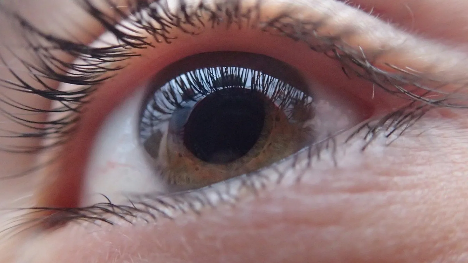
The optic nerve is the second cranial nerve (CN II), a central nervous system tract that carries axons from retinal ganglion cells to the brain, and the optic chiasm is the X-shaped midline structure where axons from the nasal retina of each eye cross to the opposite side. Together they form the first two segments of the visual pathway, converting light captured by photoreceptors into electrical signals that reach the visual cortex.
This pathway matters because its anatomy explains visual field deficits, guides neuro-ophthalmic imaging, and differs meaningfully across species. A horse with a lesion behind the chiasm loses vision in a different pattern than a dog with the same lesion, and understanding why requires knowing how many fibers cross in each species.
What the Optic Nerve Actually Is
A CNS Tract, Not a Peripheral Nerve
The optic nerve is a white matter tract of the central nervous system. This distinction is not semantic. Peripheral nerves are myelinated by Schwann cells and can regenerate after injury. The optic nerve is myelinated by oligodendrocytes and, in adult mammals, does not regenerate. Zebrafish are a notable exception: after optic nerve transection, adult zebrafish show retinal ganglion cell recovery by 2 weeks post-injury, remyelination by 4 to 5 weeks, and functional restoration of optokinetic response behavior [1].
The optic nerve develops from the neuroectodermal optic stalk, not from neural crest or mesoderm. Smarcc1-dependent chromatin remodeling in dorsal optic stalk progenitors drives the transition from a pigmented, retinal pigment epithelium-like state to astrocyte progenitors, and loss of Smarcc1 disrupts optic nerve head morphogenesis, causing glial lamina collapse and retinal ganglion cell degeneration [2]. The optic nerve head astrocytes that support retinal ganglion cell axons are therefore derived from the same embryonic tissue as the retina itself.
Retinal Ganglion Cells: The Output Neurons
Retinal ganglion cells (RGCs) are the projection neurons of the retina. They receive processed signals from photoreceptors via bipolar cells and from amacrine cells, then send axons through the optic nerve head into the optic nerve. RGCs are not a uniform population. They differ in morphology, function, and, critically for this topic, in the laterality of their projections.
Wang and colleagues purified ipsilaterally projecting RGCs from contralaterally projecting RGCs in embryonic mouse retina and found more than 300 genes differentially expressed between the two populations during the period of axonal outgrowth and decussation [3]. Among the confirmed differences, genes marking immaturity were expressed in postmitotic ipsilateral RGCs, and the complementary pair Igf1 and Igfbp5 was upregulated in contralateral and ipsilateral RGCs respectively [3]. This means the decision to cross or not cross is made molecularly, before the axon reaches the chiasm.
Optic Nerve Head
The optic nerve head is the site where unmyelinated RGC axons exit the globe. It is visible on fundoscopy as the optic disc. Myelination begins behind the lamina cribrosa, which is why the normal disc is pale but not white. In cynomolgus macaques, longer axial length is associated with larger Bruch's membrane opening distance, greater disc ovality, and a more nasal central retinal vein trunk position [4]. These parameters are measured clinically with spectral-domain optical coherence tomography and are relevant to species used in vision research.
The Optic Chiasm: Where Fibers Cross
Basic Architecture
The optic chiasm is located at the ventral surface of the diencephalon, above the sella turcica in humans and in a comparable position in domestic mammals. It is the structure where axons from the nasal retina of each eye cross the midline to join the contralateral optic tract, while axons from the temporal retina remain ipsilateral.
Isaac Newton proposed that optic nerve fibers cross at the chiasm based on his studies of light and vision, a hypothesis later confirmed anatomically and used to explain the classic patterns of visual field deficits [5]. The name chiasm comes from the Greek letter chi, reflecting the X-shaped gross appearance.
Internal Organization of the Chiasm
The chiasm is not a simple interchange. Horton and colleagues injected different fluorescent tracers into each eye of a rhesus monkey and examined serial coronal sections. They found a zone within the core of the anterior and mid portions of the optic chiasm that contained only crossing fibers, delineated by segregated, interwoven sheets of fibers from each eye [6]. This zone of decussation expanded as fibers coursed posteriorly toward the optic tracts. Eventually crossed fibers became completely intermingled with uncrossed fibers, and ocular separation was lost [6].
This internal segregation has clinical consequences. Sellar tumors compress the anterior chiasm, focusing force on the central compartment of crossing fibers, which explains why these tumors produce temporal visual field loss in each eye rather than a random pattern of deficits [6].
Molecular Control of Decussation
Decussation is not passive. In zebrafish, experimental induction of BMP4 at 15 hours post-fertilization results in complete ipsilateral projection of RGC axons and failure of commissural connections in the forebrain, partly through interaction with sonic hedgehog signaling and disrupted optic stalk morphogenesis [7]. Induction at 24 hours post-fertilization produces milder effects but still causes severe RGC projection pathologies, including fasciculation with the ipsilateral optic tract and innervation of one tectum by both eyes [7].
In humans, diffusion MRI can quantify the proportion of crossing fibers in the chiasm. Puzniak and colleagues used diffusion tensor imaging and constrained spherical deconvolution tractography in individuals with albinism and controls, detecting significant group differences in chiasmal crossing for both methods [8]. Estimates of crossing strength from diffusion MRI correlated significantly with fMRI estimates of misrouting in a subset of albinotic individuals [8]. This confirms that the proportion of crossing fibers is measurable in vivo and varies between individuals.
The Full Pathway: Retina to Visual Cortex


Step 1: Retina to Optic Nerve
RGC axons leave the retina at the optic disc, pass through the lamina cribrosa, and acquire myelin from oligodendrocytes. The optic nerve is organized topographically: fibers from the superior retina run superiorly, inferior fibers run inferiorly, and macular fibers occupy a central position. This topography is maintained throughout the pathway.
Step 2: Optic Nerve to Chiasm
The optic nerve travels posteriorly and medially to reach the chiasm. In humans, the nerve is approximately 4 to 5 cm long. In dogs, it is shorter. The intracranial portion lies above the diaphragma sellae and medial to the internal carotid artery.
Step 3: Chiasm
At the chiasm, fibers from the nasal retina cross to the opposite side. Fibers from the temporal retina remain on the same side. The result is that each optic tract contains fibers from the temporal retina of the ipsilateral eye and the nasal retina of the contralateral eye. This arrangement means each tract carries information from the contralateral visual field.
Step 4: Optic Tract to Lateral Geniculate Nucleus
The optic tract runs from the chiasm to the lateral geniculate nucleus (LGN) of the thalamus. The LGN is a laminated structure. In primates, the magnocellular layers receive input from large RGCs involved in motion detection, and the parvocellular layers receive input from smaller RGCs involved in fine detail and color. Each LGN layer receives input from only one eye, maintaining segregation.
The retinogeniculate visual pathway has four subdivisions: two decussating and two nondecussating pathways that cannot be identified on conventional structural MRI [9]. Diffusion MRI tractography can trace these subdivisions. He and colleagues compared four tractography methods and found that unscented Kalman filter with two-tensor modeling and constrained spherical deconvolution based probabilistic tracking produced the best reconstructions, with the probabilistic method better estimating the percentage of decussating fibers [9].
Step 5: Optic Radiation to Visual Cortex
From the LGN, axons project via the optic radiation (also called the geniculocalcarine tract) to the primary visual cortex in the occipital lobe. In humans, the optic radiation sweeps around the lateral ventricle. Meyer's loop carries fibers from the inferior retina and passes through the temporal lobe, which is why temporal lobe lesions can cause superior quadrantanopia.
Step 6: Visual Cortex
The primary visual cortex (Brodmann area 17, V1) is located in the occipital lobe. It receives input from the LGN in a retinotopic arrangement: adjacent points in the visual field map to adjacent points in the cortex. The cortex then processes and distributes visual information to extrastriate areas for motion, color, and object recognition.
Comparative Decussation and Visual Fields
Why Decussation Proportion Varies
The proportion of crossing fibers at the chiasm correlates with the degree of binocular overlap. In species with frontally placed eyes and large binocular fields, such as humans and cats, a substantial fraction of fibers remains ipsilateral. In species with laterally placed eyes and panoramic vision, such as horses and rabbits, almost all fibers cross.
This makes functional sense. The chiasm sorts fibers so that each hemisphere receives information from the contralateral visual field. In a lateral-eyed animal, the two eyes see almost entirely different parts of the world. Crossing most fibers ensures that each hemisphere gets a complete map of its contralateral field. In a frontal-eyed animal, the two eyes see largely the same field, and keeping some fibers ipsilateral allows for stereopsis.
Species Comparison Table
| Species | Eye Position | Binocular Field | Approximate Decussation | Functional Consequence |
|---|---|---|---|---|
| Human | Frontal | Large | Partial (nasal fibers cross, temporal fibers stay) | Stereopsis, depth perception |
| Dog | Frontal to intermediate | Moderate | Partial, more crossing than human | Some stereopsis, good motion detection |
| Cat | Frontal | Large | Partial, similar to human | Excellent stereopsis |
| Horse | Lateral | Small | High | Panoramic vision, limited stereopsis |
| Rabbit | Lateral | Very small | Very high | Near-complete panoramic vision |
The table values are qualitative because precise decussation percentages vary by individual and are difficult to measure in vivo in most species. The human data from diffusion MRI show that crossing proportion is measurable and differs between individuals, including in albinism [8].
Albinism and Misrouting
Albinism is associated with abnormally high proportions of crossing fibers at the chiasm. Puzniak and colleagues found that individuals with albinism had significantly more crossing streamlines than controls, and this finding correlated with fMRI estimates of misrouting [8]. This misrouting is thought to contribute to the visual deficits associated with albinism, including reduced stereopsis and abnormal optokinetic responses.
How the Pathway Is Studied
Diffusion MRI Tractography
Diffusion MRI measures the diffusion of water molecules in tissue. In white matter, diffusion is restricted by myelin and axonal membranes, so it is anisotropic (directionally dependent). Tractography algorithms use this anisotropy to reconstruct fiber pathways.
The retinogeniculate visual pathway is challenging to reconstruct because it contains both crossing and non-crossing fibers in close proximity. He and colleagues found that two-tensor unscented Kalman filter and probabilistic constrained spherical deconvolution methods produced the best results, with the probabilistic method better estimating decussation percentage [9].
Visual Evoked Potentials
Visual evoked potentials (VEPs) measure electrical activity in the visual cortex in response to visual stimuli. In a mouse model of optic nerve crush, flash VEP recordings were used to assess visual function after treatment with a DGAT1 inhibitor and CNTF [10]. This technique is used clinically in veterinary ophthalmology to assess retinal and optic nerve function in animals that cannot cooperate with behavioral testing.
Optical Coherence Tomography
Optical coherence tomography (OCT) measures retinal nerve fiber layer thickness and ganglion cell layer thickness. In recurrent optic neuritis, patients with preserved ganglion cell-inner plexiform layer thickness showed greater retinal nerve fiber layer swelling during acute attacks, while those with severe optic atrophy had limited swelling [11]. This principle applies to veterinary patients as well: a dog with pre-existing optic atrophy may show less disc edema during a new inflammatory episode than a dog with a healthy disc.
Fetal Ultrasound
In human fetuses, the optic chiasm can be measured by ultrasound. Wu and colleagues established normal width ranges for the decussation of the optic chiasm, optic nerves, and optic tracts from 19 to 40 weeks gestation, finding that widths increased linearly with gestational age [12]. Paladini and colleagues confirmed that the structure identified as the optic chiasm on ultrasound is indeed this anatomical structure by echoanatomic correlation in a neonatal specimen [13].
Clinical Relevance, Limitations and Common Mistakes
Clinical Relevance
Optic nerve and chiasm lesions produce characteristic visual field deficits. Because nasal fibers cross at the chiasm, a lesion compressing the chiasm from below (such as a pituitary mass) typically causes bitemporal hemianopia: loss of the temporal visual field in both eyes. A lesion behind the chiasm, such as an optic tract lesion, causes contralateral homonymous hemianopia: loss of the same side of the visual field in both eyes.
In veterinary medicine, optic nerve disease is common in dogs and cats. Optic neuritis, glaucoma, and neoplasia can all affect the optic nerve. Because the optic nerve is a CNS tract, it does not regenerate after injury in mammals. This is why optic nerve crush injuries in animal models cause permanent vision loss unless experimental treatments are used [10].
Neuromyelitis optica provides an interesting clinical example. Harvey and colleagues reported a patient with inflammation along the optic nerve, chiasm, and tract with no evidence of decussation of the inflammation [14]. The absence of decussation may relate to astrocyte localization within the chiasm, supporting the hypothesis that neuromyelitis optica is an astrocytopathy [14].
Limitations
Individual anatomy varies. The exact proportion of crossing fibers at the chiasm differs between individuals and species. Imaging findings must be interpreted in the context of the whole patient. This article is educational and is not a substitute for veterinary diagnosis or treatment.
Common Mistakes
Mistake 1: Calling the optic nerve a peripheral nerve. The optic nerve is a CNS tract myelinated by oligodendrocytes, not Schwann cells. This affects its regenerative capacity and its response to disease.
Mistake 2: Assuming all fibers cross at the chiasm. In humans and many domestic mammals, only nasal fibers cross. Temporal fibers remain ipsilateral.
Mistake 3: Confusing the optic tract with the optic nerve. The optic tract contains fibers from both eyes, while the optic nerve contains fibers from only one eye.
Mistake 4: Forgetting that decussation proportion varies by species. A horse has a different visual pathway organization than a dog, and this affects clinical presentation.
Mistake 5: Assuming the chiasm is a simple crossing point. The chiasm has internal organization, with a central compartment of crossing fibers that is vulnerable to compression [6].
Quick Review
- The optic nerve is a CNS tract, not a peripheral nerve, and is myelinated by oligodendrocytes.
- Retinal ganglion cell axons from the nasal retina cross at the chiasm, while temporal fibers remain ipsilateral in humans and many domestic mammals.
- The proportion of crossing fibers varies by species and correlates with binocular field size.
- The chiasm has a central compartment of crossing fibers that is vulnerable to compression by sellar masses [6].
- The pathway continues through the optic tract, lateral geniculate nucleus, optic radiation, and primary visual cortex.
- Diffusion MRI can quantify decussation and is used to study the retinogeniculate pathway [8][9].
- Optic nerve injury does not regenerate in adult mammals, though zebrafish show robust regeneration [1].
Frequently Asked Questions
What is the optic chiasm?
The optic chiasm is the X-shaped structure where axons from the nasal retina of each eye cross to the opposite side of the brain. It is located at the ventral surface of the diencephalon.
Do all optic nerve fibers cross at the chiasm?
No. In humans and many domestic mammals, only fibers from the nasal retina cross. Fibers from the temporal retina remain on the same side. In lateral-eyed animals like horses and rabbits, a higher proportion of fibers cross.
Why is the optic nerve considered part of the central nervous system?
The optic nerve is myelinated by oligodendrocytes, not Schwann cells, and is derived from neuroectoderm. This makes it a CNS tract rather than a peripheral nerve.
What happens if the optic chiasm is damaged?
Damage to the chiasm typically causes bitemporal hemianopia, which is loss of the temporal visual field in both eyes. This is because crossing nasal fibers are concentrated in the central chiasm.
Can the optic nerve regenerate after injury?
In adult mammals, no. The optic nerve does not regenerate after injury. Zebrafish can regenerate the optic nerve, with recovery of retinal ganglion cells by 2 weeks and remyelination by 4 to 5 weeks after transection [1].
How is the visual pathway imaged?
Diffusion MRI tractography can reconstruct the retinogeniculate pathway and estimate the proportion of crossing fibers [9]. Optical coherence tomography measures retinal nerve fiber layer and ganglion cell layer thickness, and visual evoked potentials assess cortical response to visual stimuli.
Related Articles
- Bovine Neurological Anatomy: Cranial Nerves and Brainstem
- Pug Health Problems: Pug Encephalitis Nerve Paralysis and Eye Proptosis
- Canine Thoracic Limb Nerves: Brachial Plexus and Innervation Patterns
- Equine Ocular Anatomy: Adnexa and Globe Structures
- Comparative Anatomy of the Ruminant and Equine Eye
- Ultrasound-Guided Celiac and Mesenteric Nerve Block in Dogs
- Olfactory Nerve: Cranial Nerve I Pathway and Function
- Glossopharyngeal Nerve: Anatomy, Function, and Pathway
Sources
- From injury to recovery: Spatiotemporal dynamics of the visual pathway during spontaneous structural and functional regeneration after optic nerve transection in zebrafish.
- Smarcc1 drives optic stalk patterning and optic nerve head astrocyte differentiation.
- Ipsilateral and Contralateral Retinal Ganglion Cells Express Distinct Genes during Decussation at the Optic Chiasm.
- Optic Nerve Head Parameters and Their Associations With Axial Length and Refractive Status in Cynomolgus Macaques.
- Isaac Newton's description of the optic chiasm.
- Decussating axons segregate within the anterior core of the primate optic chiasm.
- BMP Signaling Interferes with Optic Chiasm Formation and Retinal Ganglion Cell Pathfinding in Zebrafish.
- Quantifying nerve decussation abnormalities in the optic chiasm.
- Comparison of multiple tractography methods for reconstruction of the retinogeniculate visual pathway using diffusion MRI.
- Reduction of Lipid Droplets Modulates the Injury Response and Enhances CNTF-Induced Axon Regeneration Following Optic Nerve Crush.
- OCT-defined optic atrophy thresholds are associated with blunted optic nerve swelling in recurrent optic neuritis.
- Dimensions of the optic chiasm: quantitative ultrasound comparison between fetuses with anophthalmia/microphthalmia and normal fetuses.
- Assessment of fetal optic chiasm: an echoanatomic and reproducibility study.
- Absence of Decussation in Optic Pathway Inflammation in Neuromyelitis Optica and Its Implications for Astrocyte Localization.