Obturator Foramen: Anatomy, Borders, and Contents
By Dr. Zubair Khalid, DVM, MS, PhD ·
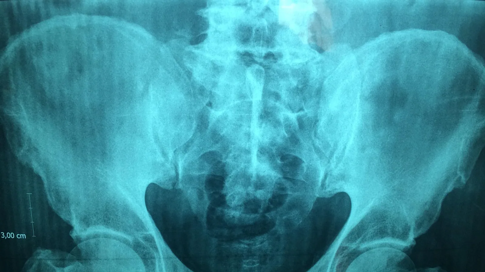
The obturator foramen is the large, mostly membrane-covered opening in the os coxae (hip bone) bounded by the pubis cranially and the ischium caudally. It transmits the obturator nerve, artery, and vein through a small gap at its craniodorsal margin called the obturator canal.
This single opening sits at the crossroads of pelvic biomechanics, comparative anatomy, and clinical neurology. It is the attachment site for the internal and external obturator muscles, the exit route for the entire obturator neurovascular bundle, and a frequent casualty of pelvic trauma. In dogs and cats it is a wide oval window that a finger can pass through. In horses and cattle it is comparatively smaller and more circular. In birds the picture changes again, because the foramen is closed by membrane and the obturator nerve finds a different exit. Understanding the ring, its borders, and what runs through it explains why obturator nerve entrapment produces a characteristic gait abnormality and why pelvic fractures near this site deserve careful neurologic assessment.
Why the Foramen Obturator Matters
The foramen obturator is one of the largest named openings in the mammalian skeleton. Every structure that leaves the pelvic cavity for the medial thigh must negotiate it or pass beside it. That makes the foramen a landmark for regional anesthesia, a hazard during pelvic surgery, and a diagnostic clue when a patient suddenly loses the ability to adduct a limb.
The opening is not a simple hole. It is a ring of bone with a membrane stretched across most of its area, leaving only a small tunnel for the neurovascular bundle. This arrangement gives the obturator muscles a broad surface for attachment while protecting the vessels and nerve that pass through the canal.
The Bony Ring: Pubis and Ischium
The os coxae develops from three centers of ossification: the ilium, the ischium, and the pubis. They meet at the acetabulum, the cup-shaped socket for the femoral head. The obturator foramen lies ventral to the acetabulum and is enclosed entirely by the pubis and ischium.
The pubis contributes the cranial border and the cranial part of the medial border. The ischium contributes the caudal border and the caudal part of the medial border. The two bones meet at the acetabular notch region laterally and at the pelvic symphysis medially. In the dog and cat the foramen is a large, oval or slightly triangular opening oriented with its long axis running craniocaudal. In the horse and ox the foramen is smaller relative to body size and tends toward a rounder or more oval shape.
The shape of the foramen varies with species, sex, and individual. In humans the classic teaching is that the foramen is oval in males and triangular in females, and quantitative Fourier analysis of human pelves supports a real but modest sex difference in outline shape [1]. The same principle applies in veterinary species, though the magnitude of dimorphism is smaller and less clinically useful. In dogs, the foramen is consistently large enough to admit a finger in medium and large breeds, which is why it is a convenient surgical corridor for certain procedures.
Borders of the Obturator Foramen
The foramen has four named borders. Each border is a bony ridge or surface that a student should be able to identify on a cleaned pelvis and on a radiograph.
Cranial Border
The cranial border is formed by the pubis, specifically the caudal surface of the pubic body and the adjacent part of the pubic ramus. It is a smooth, gently curved edge. The cranial border is the site where the obturator canal opens, because the canal is a groove between the pubis and the obturator membrane at the craniodorsal corner of the foramen.
Caudal Border
The caudal border is formed by the ischium, along the cranial edge of the ischial ramus. It is thicker and more robust than the cranial border in most species because it bears the attachment of the internal obturator muscle and part of the ischiocavernosus muscle. In the dog the caudal border is often slightly concave, following the curve of the ischial arch.
Medial Border
The medial border is the edge closest to the pelvic symphysis. It is formed by the pubis cranially and the ischium caudally, meeting at the junction of the two bones. In young animals this border is separated from its opposite side by the pelvic symphysis, a cartilaginous joint that ossifies with age. The medial border is the thinnest part of the ring and is a common site of fracture in the dog and cat.
Lateral Border
The lateral border is the edge closest to the acetabulum. It is formed by the pubis and ischium as they converge toward the acetabular notch. This border is thick and provides attachment for the obturator membrane and the obturator muscles. The lateral border is the part of the ring that a surgeon must avoid when placing implants, because the obturator vessels run just medial to it in some species.
The Obturator Membrane
The obturator membrane is a strong, fibrous sheet that covers almost the entire foramen. It attaches to the bony borders around the entire circumference except at the craniodorsal corner, where it leaves a gap. That gap is the obturator canal.
The membrane has two surfaces. The pelvic surface faces the pelvic cavity and gives origin to the internal obturator muscle. The extra-pelvic surface faces the medial thigh and gives origin to the external obturator muscle. The membrane is not a passive sheet. It transmits forces between the two obturator muscles and helps stabilize the hip during weight bearing.
In the dog and cat the membrane is thin enough to be translucent in fresh specimens. In the horse and ox it is thicker and more fibrous. In birds the membrane is complete and the foramen is fully closed, which is one of the key comparative differences in this region.
The Obturator Canal
The obturator canal is the neurovascular passage at the craniodorsal margin of the foramen. It is bounded cranially by the pubis, caudally by the obturator membrane, and laterally by the obturator internus muscle. The canal is short, typically less than a centimeter in dogs and cats, but it is the only route by which the obturator nerve, artery, and vein leave the pelvis.
The canal is not a rigid tube. Its walls are partly muscular and partly fibrous, which means its dimensions change with hip position and muscle tone. That flexibility is clinically relevant because it explains why obturator nerve entrapment can be positional rather than fixed.
In humans the obturator canal is on average 4.4 cm from the midpoint of the ischiopubic rami, and the trans-obturator sling passes on average 2.4 cm inferior-medial to the canal [2]. Those measurements come from human cadaveric dissection, but they illustrate a general principle: the canal is a small target surrounded by vulnerable structures.
Contents of the Obturator Canal
Three named structures pass through the obturator canal: the obturator nerve, the obturator artery, and the obturator vein. They travel together as a neurovascular bundle, but their branching patterns and relationships vary.
Obturator Nerve
The obturator nerve arises from the ventral divisions of the fourth, fifth, and sixth lumbar spinal nerves in the dog and cat. It descends along the medial surface of the ilium, enters the obturator canal, and divides into an anterior branch and a posterior branch. The anterior branch supplies the pectineus, adductor, and gracilis muscles. The posterior branch supplies the external obturator and sometimes the adductor magnus.
The point at which the nerve divides is variable. In a cadaveric study of 50 human lower limbs, the obturator nerve branched in the pelvic cavity in 56% of specimens, inside the obturator canal in 24%, and was undivided in 20% [3]. A separate study of 14 fresh-frozen specimens classified bifurcation as proximal to the foramen (type I, 14.3%), inside the foramen (type II, 64.3%), or distal to the foramen (type III, 21.4%) [4]. The practical takeaway is that the nerve may already be split before it reaches the canal, so a surgeon or anesthetist cannot assume a single trunk.
The nerve's dimensions are also relevant. In the same cadaveric series, the main trunk measured 108.26 ± 9.53 mm in length, 2.84 ± 0.88 mm in width, and 1.11 ± 0.35 mm in thickness. The anterior and posterior divisions were thinner, measuring 2.19 ± 0.82 mm and 0.99 ± 0.6 mm in width respectively [3]. These are human values, but they give a sense of scale for a structure that is small enough to be injured by a misplaced suture or a fracture fragment.
Obturator Artery
The obturator artery is a branch of the internal iliac artery in most domestic species. It runs through the canal and divides into an anterior branch and a posterior branch, which anastomose with the medial circumflex femoral artery and the caudal gluteal artery. In the dog the artery is small, typically less than 2 mm in diameter. In the horse and ox it is larger relative to body size.
The artery is the structure most at risk during trans-obturator procedures. In a study of 60 adults using CT angiography, the minimum distance from the medial cranial border of the obturator foramen to the obturator artery was 6.8 ± 1.9 mm on the left and 6.9 ± 1.6 mm on the right. The distance was greater than 3.2 mm in all patients but less than 5.0 mm in 23.3% of patients [5]. That means a cable passer or needle with a 5.0 mm outer diameter could contact the artery in nearly a quarter of patients. The same study defined an avascular "safe zone" within the foramen bounded by the artery and the pubis, with a mean area of 195.1 ± 63.3 mm² on the left and 190.3 ± 55.4 mm² on the right [5].
Obturator Vein
The obturator vein accompanies the artery and drains into the internal iliac vein. It is thin-walled and easily torn. In the trans-obturator sling study, the device passed on average 1.1 cm from the most medial branch of the obturator vessels [2]. The authors noted that the small caliber of the vessels and the confined space in which they would bleed make the consequences of injury uncertain, but the risk of injury is real.
Table: Borders and Contents of the Obturator Foramen
| Border or Region | Bony Contribution | Key Contents or Attachments |
|---|---|---|
| Cranial border | Pubis | Origin of obturator canal, pectineus attachment |
| Caudal border | Ischium | Internal obturator muscle attachment, ischiocavernosus |
| Medial border | Pubis and ischium | Pelvic symphysis, thinnest part of ring |
| Lateral border | Pubis and ischium near acetabulum | Obturator membrane attachment, obturator vessels nearby |
| Obturator canal | Pubis cranially, membrane caudally | Obturator nerve, artery, vein |
| Obturator membrane | Fibrous sheet across foramen | Origin of internal and external obturator muscles |
Species Differences in the Foramen Obturator
The size and shape of the foramen obturator vary widely across domestic species. These differences matter for surgery, radiology, and comparative anatomy.
Dogs and Cats
In dogs and cats the foramen is large and oval. It is oriented with the long axis running craniocaudal. The membrane is thin and the canal is short. The foramen is large enough in medium and large dogs to admit a finger, which is why it has been used as a surgical corridor for procedures such as vas deferens reconstruction and vascular bypass. In a canine study of triple pelvic osteotomy, reduction in tension of the external and internal obturator muscles was one of the most notable findings after surgery, along with atrophy of the middle gluteal muscle and entrapment of the sciatic nerve at the level of the ilial body [6]. That study did not specifically examine the obturator foramen, but it shows how pelvic surgery can alter the tension and function of the obturator muscles.
Horses and Cattle
In horses and cattle the foramen is relatively smaller and more circular. The membrane is thicker and more fibrous. The obturator nerve is larger in absolute terms because of the greater muscle mass it supplies, but the canal is proportionally narrower. This makes the nerve more vulnerable to compression if the foramen is fractured or if a hematoma forms within the canal.
Birds
In birds the obturator foramen is closed by membrane and the obturator nerve exits differently. The avian pelvis is fused to the synsacrum, and the foramen is not a route for a large neurovascular bundle in the same way as in mammals. The obturator nerve in birds typically exits through the foramen or through a separate opening, depending on the species. The key point for students is that the avian foramen is not a wide-open window. It is a membrane-covered space with a different functional anatomy.
Clinical Relevance, Limitations and Common Mistakes
The obturator foramen is a common site of pelvic fractures in dogs and cats. The medial border is thin and the ring is under stress during weight bearing, so a fall or a vehicular trauma can fracture the pubis or ischium near the foramen. Fractures here can impinge on the obturator canal and compress the obturator nerve.
Obturator nerve entrapment causes adductor weakness. The affected limb may splay outward when the animal stands, and the animal may have difficulty bringing the limb under the body. The gait abnormality is sometimes mistaken for a hip problem, but the neurologic examination will show normal hip range of motion and normal withdrawal reflex with weak adduction.
The nerve can also be injured during pelvic surgery. In a study of trans-obturator sling placement in human cadavers, the anterior and posterior divisions of the obturator nerve were on average 3.4 cm and 2.8 cm from the passed device, and the device passed on average 1.1 cm from the most medial branch of the obturator vessels [2]. The authors concluded that vascular and nerve structures are within 1 to 3 cm of the path of any device passed through the obturator foramen, so the risk of injury is inherent to the approach.
Exostoses, or bone spurs, can also form in the foramen. In a study of 221 human hip bones, exostoses were found in 21 (9.5%), with the right side affected twice as often as the left. They were more frequent in the medial area of the foramen and varied markedly in number and severity [7]. The authors noted that exostoses can cause nerve compression syndrome, local inflammatory reactions, and movement restrictions. The same principle applies in animals, though the prevalence is not well documented.
Congenital agenesis of the obturator foramen is extremely rare. In a study of 228 dried human hip bones, complete, incomplete, or mixed agenesis was observed in 15 of 114 pelvises (13.2%). Unilateral complete agenesis was identified in 3 right-sided and 4 left-sided hip bones, with an average bony plate thickness of 2.1 mm [8]. This is a human study, but it shows that the foramen can fail to form normally, which has implications for pelvic surgery and radiological interpretation.
The most common mistakes students make are confusing the foramen with the obturator canal, assuming the nerve always exits as a single trunk, and forgetting that the membrane covers most of the foramen. The foramen is the whole opening. The canal is the small passage at its craniodorsal corner. The membrane covers the rest.
This article is educational and is not a substitute for veterinary diagnosis or treatment.
Quick Review
- The obturator foramen is bounded by the pubis cranially and the ischium caudally.
- The obturator membrane covers most of the foramen, leaving the obturator canal open at the craniodorsal corner.
- The obturator nerve, artery, and vein pass through the canal.
- The nerve may branch before, inside, or after the canal, so its anatomy is variable.
- In dogs and cats the foramen is large and oval. In horses and cattle it is smaller and rounder. In birds it is closed by membrane.
- Pelvic fractures near the foramen can entrap the obturator nerve and cause adductor weakness.
- Exostoses and congenital agenesis are rare but documented variations that can complicate surgery.
Frequently Asked Questions
What is the foramen obturator?
The foramen obturator is the large opening in the hip bone formed by the pubis and ischium. It is covered by the obturator membrane except at the craniodorsal corner, where the obturator canal transmits the obturator nerve, artery, and vein.
What are the borders of the obturator foramen?
The cranial border is the pubis, the caudal border is the ischium, the medial border is the pubis and ischium near the pelvic symphysis, and the lateral border is the pubis and ischium near the acetabulum.
What structures pass through the obturator canal?
The obturator nerve, obturator artery, and obturator vein pass through the obturator canal. The nerve supplies the adductor muscles of the medial thigh.
How does the obturator foramen differ between dogs and horses?
In dogs and cats the foramen is large and oval. In horses and cattle it is relatively smaller and more circular. The membrane is also thicker in horses and cattle.
What happens if the obturator nerve is damaged?
Obturator nerve damage causes adductor weakness. The animal may splay the affected limb outward and have difficulty bringing it under the body. The gait abnormality can be mistaken for a hip problem.
Is the obturator foramen a common fracture site?
Yes. The medial border of the foramen is thin and under stress during weight bearing, so pelvic fractures commonly involve the pubis or ischium near the foramen. Fractures here can compress the obturator nerve.
Related Articles
- Cross-Border Regulatory Compliance
- Border Collie Lifespan Diet and Preventive Care Guide
- Border Collie Care, Training, Temperament, Health and Lifespan
- Qualitative Content Analysis: Methods and Applications
- Border Terrier Health Problems: Inherited Conditions and Preventive Screenings
- Border Collie Health Problems: Trapped Neutrophil Syndrome and MDR1
- Foramen Magnum: Anatomy, Contents, and Comparative Notes
- Jugular Foramen: Anatomy, Contents, and Clinical Notes
Sources
- Oval in males and triangular in females? A quantitative evaluation of sexual dimorphism in the human obturator foramen.
- Anatomy of the obturator region: relations to a trans-obturator sling.
- Morphology of the obturator nerve, an anatomical study with emphasis on its clinical implications.
- Anatomic Study of the Bifurcation of the Obturator Nerve: Application to More Precise Surgical/Procedural Localization.
- The Obturator Foramen "Safe Zone" for Anterior Pelvis Internal Fixation by Transobturator Cerclage.
- Effects of triple pelvic osteotomy on anatomic structures of the pelvic region in the dog.
- Exostoses in obturator foramen - an osteological study.
- Congenital Agenesis of the Obturator Foramen in an Asian Population and Its Clinical Significance.