Nucleus Tractus Solitarius: Anatomy and Function
By Dr. Zubair Khalid, DVM, MS, PhD ·

The nucleus tractus solitarius (NTS) is the principal brainstem relay for visceral afferent information carried by the vagus (cranial nerve X), glossopharyngeal (IX) and facial (VII) nerves. Every heartbeat, breath, swallow and gut contraction generates sensory traffic that terminates in this narrow column of gray matter in the dorsomedial medulla, where the signal is sorted, integrated and forwarded to the motor and autonomic nuclei that produce the appropriate reflex response.
This article covers the subdivisions of the NTS, the visceral afferent inputs it receives, its major efferent projections and the reflexes it governs. It is written for veterinary and biomedical students who need a working map of the nucleus, not a catalog of every peptide expressed within it.
Where the NTS Sits in the Brainstem
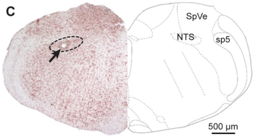

The NTS lies in the dorsomedial medulla oblongata, immediately beneath the floor of the fourth ventricle and just lateral to the dorsal motor nucleus of the vagus. The solitary tract, a bundle of primary afferent axons, runs longitudinally along the lateral edge of the nucleus. Afferents leave the tract and synapse onto NTS neurons along its length, which is why the nucleus is named for the tract that feeds it.
The nucleus extends from the level of the facial nucleus rostrally to the obex and slightly beyond caudally. In domestic mammals the general plan is conserved, though the relative size of subdivisions varies with the density of innervation of the organ system involved. A horse, for example, has an extensive gastric and cardiovascular afferent supply, and the corresponding NTS territories are well developed.
The NTS is a bilateral structure. Each side receives afferents predominantly from the ipsilateral vagus and glossopharyngeal nerve, though there is some crossing through commissural connections. This bilateral arrangement means that a unilateral lesion can often be compensated, while bilateral involvement produces severe autonomic instability.
Subdivisions of the Nucleus Tractus Solitarius
The NTS is not a uniform structure. It is divided rostrocaudally into three functional zones, each with a distinct afferent supply and efferent target. This rostrocaudal organization is the single most useful framework for understanding the nucleus.
Rostral Gustatory Region
The rostral portion of the NTS receives taste afferents from the chorda tympani branch of the facial nerve (VII), the lingual branch of the glossopharyngeal nerve (IX) and, in some species, the pharyngeal branch of the vagus. These fibers carry information about the chemical composition of food and fluid in the oral cavity.
The gustatory region projects rostrally to the parabrachial nucleus, which in turn relays taste information to the thalamus and insular cortex. In rodents and some other species the parabrachial nucleus is the first relay, while in primates and carnivores the pathway is more direct. The functional consequence is the same: the NTS is the first central processing station for taste, and it is where taste signals are separated from the general visceral signals that share the same nerve trunks.
Intermediate Cardiorespiratory Region
The intermediate portion of the NTS is the largest and most clinically significant subdivision. It receives baroreceptor afferents from the carotid sinus via the glossopharyngeal nerve and from the aortic arch via the vagus. It also receives chemoreceptor afferents from the carotid bodies and aortic bodies, and pulmonary stretch receptor afferents from the lungs.
This region contains the second-order neurons that form the first synapse of the baroreceptor reflex and the chemoreceptor reflex. It also contains neurons that project to the dorsal motor nucleus of the vagus and the nucleus ambiguus, which provide the parasympathetic outflow to the heart, lungs and gastrointestinal tract. The intermediate NTS is therefore the central hub where cardiovascular and respiratory afferent information is integrated before any reflex output is generated.
Caudal Region
The caudal NTS receives afferents from the gastrointestinal tract, the liver, the pancreas and the heart. It is the termination site for the majority of unmyelinated vagal C-fibers, which carry information about organ distension, chemical irritation and nociception.
The caudal region is also the site where visceral nociceptive information converges with afferents from other organ systems. Studies using retrograde tracing have shown that vagal afferents from the trachea, duodenum, stomach and heart terminate throughout the NTS with substantial overlap, and that single NTS neurons can receive convergent input from more than one organ [1]. This convergence is the anatomical basis for referred visceral sensations and for the interaction between cardiac and gastrointestinal reflexes.
The caudal NTS contains a population of aldosterone-sensitive neurons that express the enzyme 11-beta-hydroxysteroid dehydrogenase type 2 (HSD2). These neurons are a target of top-down control from the hypothalamus and limbic system, and they are thought to link visceral sensory information with sodium appetite and cardiovascular regulation [2].
Table: NTS Subdivisions, Afferents and Reflex Outputs
| Subdivision | Principal afferent inputs | Main efferent targets | Reflexes mediated |
|---|---|---|---|
| Rostral (gustatory) | Chorda tympani (VII), lingual branch of glossopharyngeal (IX), pharyngeal branch of vagus (X) | Parabrachial nucleus, then thalamus and insular cortex | Taste perception, salivation, cephalic phase of digestion |
| Intermediate (cardiorespiratory) | Carotid sinus baroreceptors (IX), aortic arch baroreceptors (X), carotid and aortic body chemoreceptors (IX, X), pulmonary stretch receptors (X) | Dorsal motor nucleus of vagus, nucleus ambiguus, ventrolateral medulla, parabrachial nucleus | Baroreceptor reflex, chemoreceptor reflex, respiratory rhythm modulation, cough reflex |
| Caudal (visceral) | Gastrointestinal, hepatic, pancreatic and cardiac vagal afferents (X), including unmyelinated C-fibers | Ventrolateral medulla, parabrachial nucleus, hypothalamus, dorsal motor nucleus of vagus | Gag reflex, vomiting reflex, visceral nociception, gastrointestinal motility control, emetic response |
Visceral Afferent Inputs to the NTS
The NTS receives its afferent supply from three cranial nerves. The vagus nerve provides the largest contribution, carrying sensory fibers from the pharynx, larynx, trachea, lungs, heart, aorta, esophagus, stomach, small intestine, liver, pancreas and part of the large intestine. The glossopharyngeal nerve carries afferents from the carotid sinus, carotid body, posterior third of the tongue and pharynx. The facial nerve contributes taste fibers from the anterior two-thirds of the tongue through the chorda tympani.
These afferents are pseudounipolar neurons whose cell bodies sit in the nodose and petrosal ganglia. Their central processes enter the brainstem and turn caudally to form the solitary tract before synapsing in the NTS. The neurotransmitter at the first synapse is predominantly glutamate, and the postsynaptic receptors are ionotropic AMPA and NMDA receptors. This glutamatergic transmission is the target of considerable modulation, including by circulating hormones.
Corticosterone, for example, rapidly decreases both action potential-evoked and spontaneous glutamate release from vagal afferent terminals in the NTS through a glucocorticoid receptor-dependent mechanism that involves retrograde endocannabinoid signaling [3]. This finding illustrates a general principle: the NTS is not a passive relay. Circulating signals can adjust the gain of visceral afferent transmission before any reflex is triggered.
The transient receptor potential ankyrin 1 (TRPA1) channel is expressed presynaptically on vagal afferent terminals in the caudal NTS, where it modulates glutamate release onto second-order neurons [4]. This channel is activated by irritant chemicals and by products of oxidative stress, which means that the NTS can respond to the chemical environment of the viscera as well as to mechanical and electrical signals.
Efferent Projections from the NTS
The NTS projects to multiple targets, and the pattern of projection depends on the subdivision of origin. The three most important targets for reflex function are the parabrachial nucleus, the dorsal motor nucleus of the vagus and the ventrolateral medulla.
Parabrachial Nucleus
The parabrachial nucleus lies in the dorsolateral pons and is the major relay from the NTS to the forebrain. It receives projections from all three NTS subdivisions and forwards visceral and gustatory information to the thalamus, hypothalamus, amygdala and insular cortex. The parabrachial nucleus is therefore the gateway through which visceral sensory information reaches conscious perception and emotional processing.
In the context of respiratory and cardiovascular control, the parabrachial nucleus also projects back to the NTS and to the ventrolateral medulla, forming a feedback loop that can modulate reflex gain. This loop is thought to be involved in the interaction between emotional state and autonomic function, though the details are beyond the scope of this article.
Dorsal Motor Nucleus of the Vagus
The dorsal motor nucleus of the vagus lies immediately ventral to the NTS and provides the parasympathetic preganglionic outflow to the thoracic and abdominal viscera. The NTS projects directly to this nucleus, forming the first limb of the vago-vagal reflex. When vagal afferents detect gastric distension, for example, the signal reaches the NTS, which activates dorsal motor nucleus neurons that project back to the stomach and modulate motility and secretion.
This vago-vagal reflex arc is the basis for gastric electrical stimulation, a technique used to treat gastroparesis and obesity. Recordings from gastric neurons in the NTS show that their responses are selective to the orientation of the stimulus relative to the smooth muscle fibers, which suggests that the NTS encodes specific features of gastric wall activity rather than a simple on-off signal [5].
Ventrolateral Medulla
The ventrolateral medulla contains the rostral ventrolateral medulla (RVLM), which is the primary source of sympathetic vasomotor tone, and the caudal ventrolateral medulla (CVLM), which inhibits the RVLM. The NTS projects to both regions. Baroreceptor activation excites NTS neurons that project to the CVLM, which in turn inhibits the RVLM and reduces sympathetic outflow. This is the central pathway of the baroreceptor reflex.
The NTS also projects to the retrotrapezoid nucleus (RTN), a cluster of Phox2b-expressing neurons in the ventrolateral medulla that function as central respiratory chemoreceptors. A GABAergic projection from the NTS to RTN neurons modulates the hypercapnic ventilatory response. Activation of NTS GABAergic neurons suppresses respiratory drive and can induce hypoventilation and spontaneous apnea, while also attenuating the response to elevated carbon dioxide [6]. This circuit provides a mechanism by which visceral afferent input can adjust breathing independently of the peripheral chemoreceptors.
Reflex Functions of the NTS
Baroreceptor Reflex
The baroreceptor reflex is the fastest mechanism for stabilizing arterial blood pressure. Stretch-sensitive baroreceptors in the carotid sinus and aortic arch fire in proportion to arterial pressure. Their afferents travel in the glossopharyngeal and vagus nerves to the NTS, where they release glutamate onto second-order neurons. These neurons activate the CVLM, which inhibits the RVLM, reducing sympathetic outflow to the heart and blood vessels. At the same time, NTS neurons activate the dorsal motor nucleus of the vagus and nucleus ambiguus, increasing parasympathetic outflow to the heart. The result is bradycardia and vasodilation.
Microinjection of L-glutamate into the NTS produces decreases in arterial blood pressure and heart rate that mimic the baroreceptor reflex, confirming the role of this nucleus as the first central relay for the reflex [7]. The same study showed that L-cysteine produces similar cardiovascular effects through a different receptor mechanism, which illustrates the pharmacological complexity of NTS neurotransmission.
The baroreceptor reflex is not a fixed circuit. Its sensitivity changes with physiological state. During exercise, for example, the exercise pressor reflex from working skeletal muscle increases arterial pressure, and the baroreceptor reflex buffers this response. Insulin signaling in the NTS modulates this interaction. Blockade of insulin receptors in the NTS potentiates the exercise pressor reflex in healthy rats, but this effect is absent after sino-aortic baroreceptor denervation, which indicates that the modulation depends on intact baroreceptor input [8].
Chemoreceptor Reflex
Peripheral chemoreceptors in the carotid and aortic bodies detect decreases in arterial oxygen tension, increases in carbon dioxide tension and decreases in pH. Their afferents travel to the NTS, where they synapse on neurons that project to the respiratory centers in the ventrolateral medulla and to the nucleus ambiguus. The reflex response is an increase in respiratory rate and depth, along with sympathetic activation.
Pulmonary vagal nociceptors, which defend the airways against irritants, also synapse in the NTS. These nociceptors converge with carotid chemoreceptor afferents onto single NTS neurons, and simultaneous stimulation of both pathways doubles efferent vagal output and enhances the phrenic pause that occurs during airway defensive reflexes [9]. This convergence means that the NTS integrates information from multiple sensory modalities to produce a coordinated airway defense response.
Cough Reflex
The cough reflex begins with irritation of the airway mucosa, which activates mechanosensitive and chemosensitive vagal afferents. These afferents terminate in the NTS, particularly in the intermediate and caudal regions. The NTS then activates a central pattern generator in the medulla that coordinates the sequential activation of expiratory muscles, laryngeal muscles and the diaphragm to produce a cough.
The NTS is not the cough center itself, but it is the obligatory relay for the afferent limb of the reflex. Without NTS processing, the cough reflex cannot be triggered. This is why lesions that affect the NTS or its afferent input can produce a loss of cough reflex, which is a serious clinical problem because it predisposes to aspiration and pneumonia.
Gag Reflex
The gag reflex is triggered by mechanical stimulation of the posterior pharyngeal wall. Afferents travel in the glossopharyngeal nerve to the NTS, which activates the nucleus ambiguus and the dorsal motor nucleus of the vagus. The result is contraction of the pharyngeal constrictor muscles and elevation of the soft palate. The gag reflex is a protective mechanism that prevents foreign material from entering the pharynx, and it is closely related to the swallowing reflex, which is also coordinated through the NTS.
Vomiting Reflex
The vomiting reflex is more complex than the cough or gag reflex and involves the NTS as a central integration site. Vagal afferents from the gastrointestinal tract, as well as afferents from the chemoreceptor trigger zone in the area postrema, converge on the NTS. The NTS then activates the dorsal motor nucleus of the vagus and the nucleus ambiguus to produce the autonomic and motor components of vomiting. The area postrema lies immediately adjacent to the caudal NTS and lacks a blood-brain barrier, which allows it to detect circulating emetic substances and relay that information to the NTS.
Modulation of NTS Function
The NTS is subject to modulation by circulating hormones, neurotransmitters and descending inputs from higher brain regions. This modulation allows the nucleus to adjust reflex gain according to physiological state.
Hormonal Modulation
Angiotensin II acts on AT1A receptors in the NTS to modulate visceral reflexes. AT1A receptor-expressing neurons are distributed throughout the NTS, and most of them are depolarized by angiotensin II. Two-thirds of these neurons receive direct excitatory visceral sensory input, and about half of those receive C-fiber or A-fiber input that is sensitive to capsaicin [10]. This arrangement allows angiotensin II to influence cardiovascular and fluid balance reflexes at the level of the first central synapse.
Glucocorticoids also modulate NTS function. Corticosterone rapidly decreases glutamate release from vagal afferent terminals through a mechanism that requires postsynaptic G-protein signaling and retrograde endocannabinoid release [3]. This effect may be relevant to the changes in autonomic function that occur during stress.
Descending Control
The NTS receives projections from the paraventricular hypothalamic nucleus, the lateral hypothalamic area and the central nucleus of the amygdala. These projections are top-down control pathways that allow emotional and homeostatic states to influence visceral reflex function. The paraventricular hypothalamic afferents are glutamatergic, while the central amygdala afferents are GABAergic [2]. This means that the hypothalamus and amygdala can exert opposing effects on NTS excitability.
Other sources of descending input include the insular and infralimbic cortices, the bed nucleus of the stria terminalis, the periaqueductal gray, Barrington's nucleus and the Kölliker-Fuse nucleus [2]. This widespread innervation indicates that the NTS is not an isolated reflex center but is embedded in a network that links visceral sensation with emotion, arousal and behavior.
Clinical Relevance, Limitations and Common Mistakes
The NTS is involved in several clinical conditions that are relevant to veterinary practice. Hypertension, for example, has been linked to dysfunction of NTS activity. The NTS regulates autonomic nervous system activity and arterial blood pressure through the baroreflex, sympathetic nerve activity, the renin-angiotensin-aldosterone system and oxidative stress [11]. When NTS function is impaired, the baroreflex becomes less effective, and blood pressure can rise.
Renal ischemia-reperfusion injury reduces NTS electrical activity and baroreceptor sensitivity in rats, and treatment with naringin or trimetazidine improves both parameters [12]. This finding suggests that conditions that affect the kidney can have secondary effects on central autonomic control through the NTS.
Chronic pancreatitis is associated with visceral hypersensitivity, and the NTS plays a role in this process. In a rat model of chronic pancreatitis, the number of Fos-expressing neurons in the caudal NTS increases, and excitatory synaptic transmission within the NTS is enhanced [13]. This central sensitization contributes to the maintenance of visceral pain and is a potential target for analgesic therapy.
Vagus nerve stimulation (VNS) is an approved therapy for drug-resistant epilepsy, depression and heart failure. Vagal afferents activated by VNS terminate in the NTS, where the sensory information is integrated and propagated throughout central autonomic networks [14]. Recordings from NTS neurons during VNS show that the stimulation activates second-order neurons receiving myelinated vagal input, as well as higher-order neurons that are polysynaptically activated [15]. The therapeutic effects of VNS are thought to depend on this activation of NTS circuits, though the optimal stimulation parameters remain uncertain.
A common mistake is to think of the NTS as a simple relay station. It is better understood as an integration center where multiple afferent streams converge and where the gain of reflex responses is adjusted by circulating signals and descending inputs. Another common mistake is to assume that the NTS is only involved in cardiovascular and respiratory control. It also processes taste, gastrointestinal sensation, visceral nociception and emetic signals.
The limitations of current knowledge are significant. Much of what is known about the NTS comes from rodent studies, and the extent to which these findings apply to domestic species is not always clear. The functional organization of the NTS in horses, cattle, dogs and cats has been studied less extensively than in rats and mice. Individual clinical cases require veterinary assessment, and the information in this article is educational rather than diagnostic.
This article is educational and is not a substitute for veterinary diagnosis or treatment.
How the NTS Processes a Visceral Signal
The following flowchart summarizes the sequence from visceral afferent activation to reflex output.
flowchart TD
    A[Visceral stimulus] --> B[Afferent activation]
    B --> C[Cranial nerve VII IX or X]
    C --> D[Solitary tract]
    D --> E[NTS subdivision]
    E --> F{Rostral intermediate or caudal}
    F --> G[Parabrachial nucleus]
    F --> H[Dorsal motor nucleus of vagus]
    F --> I[Ventrolateral medulla]
    G --> J[Forebrain perception]
    H --> K[Parasympathetic output]
    I --> L[Sympathetic and respiratory output]
Frequently Asked Questions
What is the nucleus tractus solitarius?
The nucleus tractus solitarius is a column of gray matter in the dorsomedial medulla that serves as the first central relay for visceral afferents carried by the vagus, glossopharyngeal and facial nerves.
What are the three subdivisions of the NTS?
The NTS is divided into a rostral gustatory region, an intermediate cardiorespiratory region and a caudal visceral region. Each subdivision has a distinct afferent supply and efferent target.
Which cranial nerves carry afferents to the NTS?
The vagus (X), glossopharyngeal (IX) and facial (VII) nerves carry visceral and gustatory afferents to the NTS. The vagus provides the largest contribution.
What reflexes does the NTS control?
The NTS controls the baroreceptor reflex, chemoreceptor reflex, cough reflex, gag reflex, vomiting reflex and vago-vagal reflexes that regulate gastrointestinal motility and secretion.
How does the NTS regulate blood pressure?
The NTS receives baroreceptor afferents and projects to the ventrolateral medulla and dorsal motor nucleus of the vagus. This circuit adjusts sympathetic and parasympathetic outflow to maintain arterial pressure.
What happens if the NTS is damaged?
Bilateral damage to the NTS can cause severe autonomic instability, including loss of baroreflex control, respiratory irregularities and impaired swallowing and cough reflexes.
Does the NTS have a role in taste?
Yes. The rostral NTS receives taste afferents from the chorda tympani, glossopharyngeal and vagus nerves and projects to the parabrachial nucleus and then to the thalamus and insular cortex.
How is the NTS studied in veterinary research?
The NTS is studied using electrophysiology, neural tracing, immunohistochemistry and functional imaging in animal models. Rodents are the most common species, though the general organization is conserved across mammals.
Related Articles
- Equine Larynx Anatomy and Function in Respiration
- Canine Lymphatic System: Anatomy and Function
- Equine Hoof Anatomy: Structures and Function
- Fish Gills: Anatomy, Function, and Common Health Issues
- Equine Muscular System: Major Muscle Groups and Function
- Ruminant Digestive Anatomy and Physiology: A Clinical Reference
- Nucleus Function: Roles of Nucleus and Nucleolus
Sources
- Analysis of the distribution of vagal afferent projections from different peripheral organs to the nucleus of the solitary tract in rats.
- Central afferents to the nucleus of the solitary tract in rats and mice.
- Corticosterone inhibits vagal afferent glutamate release in the nucleus of the solitary tract via retrograde endocannabinoid signaling.
- Expression and Function of Transient Receptor Potential Ankyrin 1 Ion Channels in the Caudal Nucleus of the Solitary Tract.
- Gastric neurons in the nucleus tractus solitarius are selective to the orientation of gastric electrical stimulation.
- GABAergic Inhibition from the Nucleus Tractus Solitarius to Ventrolateral Medulla Phox2b Neurons Modulates Central Respiratory Chemoreflex and Ventilatory Homeostasis.
- Cardiovascular actions of L-cysteine and L-cysteine sulfinic acid in the nucleus tractus solitarius of the rat.
- Blockade of endogenous insulin receptor signaling in the nucleus tractus solitarius potentiates exercise pressor reflex function in healthy male rats.
- Neuroanatomical and neurophysiological evidence of pulmonary nociceptor and carotid chemoreceptor convergence in the nucleus tractus solitarius and nucleus ambiguus.
- Viscerosensory input drives angiotensin II type 1A receptor-expressing neurons in the solitary tract nucleus.
- [[Research progress of nucleus tractus solitarius involved in central regulation of hypertension].](https://pubmed.ncbi.nlm.nih.gov/40065617/)
- Naringin and Trimetazidine Improve Baroreflex Sensitivity and Nucleus Tractus Solitarius Electrical Activity in Renal Ischemia-Reperfusion Injury.
- Nucleus tractus solitarius mediates hyperalgesia induced by chronic pancreatitis in rats.
- Bursting Parameters Alter Excitatory Postsynaptic Currents in Nucleus Tractus Solitarius Neurons: Implications for Clinical Vagus Nerve Stimulation.
- Cervical vagus nerve stimulation augments spontaneous discharge in second- and higher-order sensory neurons in the rat nucleus of the solitary tract.