Nucleus Function: Roles of Nucleus and Nucleolus
By Dr. Zubair Khalid, DVM, MS, PhD ·
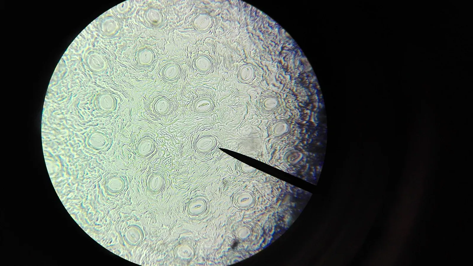
The nucleus is the membrane-bound organelle that stores a eukaryotic cell's genome and carries out transcription and RNA processing. The nucleolus is a non-membrane-bound structure inside the nucleus where ribosomal RNA is made and ribosomal subunits are assembled.
Those two sentences answer most exam questions on the topic. The rest of this guide explains how each structure actually works, why the division of labor between them matters, and where students most often mix them up.
Why the Nucleus and Nucleolus Matter
Every protein a cell makes depends on instructions copied from DNA. Those instructions leave the nucleus as RNA, and the machinery that reads them, the ribosome, is itself built inside the nucleus. That creates a dependency loop. A cell cannot translate RNA into protein without ribosomes, and it cannot build ribosomes without the nucleus and nucleolus working first.
The loop also explains why the nucleolus is one of the most sensitive structures in the cell. When ribosome assembly stalls, the nucleolus changes shape and material properties before almost anything else does [1]. Researchers use that sensitivity as a readout for cellular stress, growth rate, and biosynthetic demand.
The Nucleus: Structure and Core Functions

.svg)
The nucleus is partitioned from the cytosol by a nuclear envelope, a double membrane that encapsulates a highly structured internal space containing the organized genome and the nucleolus [2]. That compartmentalization is the physical basis for everything the nucleus does.
Genome Storage and Organization
The nucleus holds the cell's DNA. In mammals that genome is roughly 3 billion base pairs per haploid set, packed with histone proteins into chromatin and organized into chromosomes.
Storage is not passive. The nucleus maintains the genome in a state that permits selective access. Regions that need to be read stay relatively open. Regions that should stay silent are compacted. Nuclear type I myosins, motor proteins inside the nucleus, help maintain this three-dimensional genome organization. When they are depleted, the genome becomes disorganized and gene expression patterns shift [2].
Transcription
Transcription is the synthesis of RNA from a DNA template, and it happens in the nucleus. Three RNA polymerases divide the work in eukaryotes:
- RNA polymerase I transcribes the large ribosomal RNA precursor.
- RNA polymerase II transcribes messenger RNA and most small regulatory RNAs.
- RNA polymerase III transcribes small RNAs including 5S ribosomal RNA and transfer RNA.
The nucleolus is the specialized nuclear region where RNA polymerase I transcription occurs. Ribosome biogenesis begins with the rate-limiting step of Pol I transcription of the 47S ribosomal RNA genes [3]. That single transcript is later cut into the mature ribosomal RNAs.
Transcription factors, the proteins that control which genes are read, are themselves nuclear. A transcription factor such as the rice protein OsMYB73 is nucleus-localized and regulates downstream gene networks [4]. The same principle applies across eukaryotes: the regulatory decisions that shape a cell's behavior are made in the nucleus.
RNA Processing
Freshly transcribed RNA is not ready to use. The nucleus processes it before export.
Messenger RNA receives a protective cap at its 5' end, a poly-A tail at its 3' end, and has its non-coding introns removed by splicing. Ribosomal RNA follows a different but equally staged path. In the nucleolus, ribosomal RNA undergoes transcription, chemical modification, splicing, and folding in spatially and temporally segregated regions [5]. Each step changes the RNA's shape and its interactions with proteins, which is part of how the nucleolus maintains its layered internal architecture.
Processing defects have consequences that reach beyond the RNA itself. When cleavage of the 5' external transcribed spacer of pre-ribosomal RNA is impaired, nucleolar organization breaks down, nascent RNA diffuses through a disorganized nucleolus, and heterochromatin markers mislocalize [6]. In other words, RNA processing helps hold the nucleus together structurally, not just functionally.
Nuclear Envelope Transport Through Pores
The nuclear envelope would be an impermeable wall without gates. Those gates are nuclear pore complexes, large protein assemblies that control the movement of macromolecules into and out of the nucleus [7].
A nuclear pore is roughly 120 nanometers in diameter. That is wide enough in principle for many proteins to fit through, but the pore is filled with a meshwork of FG-nucleoporins, proteins containing phenylalanine-glycine repeat domains, that restricts what actually passes.
Assembly of these pores is a two-step process. During mitotic exit, hydrophobic interactions among FG-nucleoporins dilate the assembling pore and build the central transport channel. That dilation then permits nuclear-import-driven nuclear expansion and tight nuclear envelope spacing [8]. Pore formation is therefore coupled to the rebuilding of the nucleus itself after cell division.
Small molecules can diffuse through the pore. Larger cargo needs a shuttle, a transport receptor that binds the cargo and carries it through. Artificial pore systems built to mimic this behavior show that selective shuttle-cargo transport is possible when affinity is tuned correctly, with predicted rates above 1000 molecules per pore per second in one engineered system [7]. That number illustrates the throughput a real cell needs.
The Nucleolus: Structure and Core Functions
The nucleolus is the largest membraneless organelle in the nucleus and the site of ribosome biogenesis [5]. It has no surrounding lipid bilayer. Instead, it forms through phase separation, a process in which protein-protein and protein-RNA interactions create distinct liquid-like compartments.
Its diameter is typically 1 to 3 micrometers. Within that small volume, the nucleolus runs a multi-stage assembly line.
Ribosomal RNA Synthesis
The nucleolus is where ribosomal RNA is synthesized [9]. RNA polymerase I transcribes the ribosomal RNA genes, which cluster in the nucleolar organizer regions of specific chromosomes.
This is the rate-limiting step of the entire ribosome production pathway [3]. Because it is rate-limiting, ribosomal RNA synthesis is tightly regulated and responds to growth signals, nutrient status, and stress.
Ribosomal Subunit Assembly
Ribosomal RNA alone is not a ribosome. It must be modified, folded, cut, and packaged with dozens of ribosomal proteins.
The nucleolus handles most of this. The pre-40S and pre-60S ribosomal subunits are largely pre-assembled in the nucleolus before being exported to the cytoplasm for final maturation [10]. Some ribosomal proteins, including eL24 of the large subunit, are loaded only after export, which is why the nucleolus produces near-complete subunits rather than finished ribosomes.
The internal organization of the nucleolus makes this possible. Multicomponent phase separation creates multiple nucleolar sub-compartments that function from the inside out as a ribosome assembly line [5]. Key proteins including nucleolin, fibrillarin, and nucleophosmin mediate this compartmentalization through their structural features and multivalent interactions [5].
The granular component, one of the nucleolus's main regions, is itself subdivided. Super-resolution microscopy shows that nucleophosmin, SURF6, and ribosomal RNA are heterogeneously localized within granular component sub-phases. In reconstituted systems these molecules form multiphase condensates with a SURF6 and rRNA-rich core and a nucleophosmin-rich shell [11]. As subunits assemble, SURF6's association with rRNA weakens, allowing nucleophosmin to extract finished subunits from the condensate. The result is an assembly-line mechanism of subunit efflux [11].
Structural scaffolds reinforce the whole system. The protein NEPRO forms elongated fibers that contour the periphery of the dense fibrillar component. Depleting NEPRO disrupts nucleolar integrity and impairs 40S, 60S, and 80S ribosome assembly [12].
Stress Sensing and Cellular Signaling
The nucleolus is not only a factory. It is a sensor.
Nucleolar dynamics report on the state of ribosome assembly. When early pre-ribosomal intermediates accumulate, nucleophosmin dynamics slow and the condensate compacts. When abortive late precursors accumulate, dynamics accelerate and condensate integrity breaks down [1]. These opposing biophysical states correlate with the strength of interactions between nucleophosmin and pre-ribosomes [1].
That means the nucleolus physically registers whether assembly is proceeding correctly. Because its membraneless organization participates in diverse signaling pathways, it influences cell cycle regulation, proliferation, apoptosis, differentiation, and cellular stress responses [13].
Mechanical cues reach the nucleolus too. Extracellular matrix stiffness, cell migration, confinement, and external mechanical stress alter nuclear structure and thereby affect nucleolar organization and ribosome biogenesis [13]. The nucleolus is emerging as a mediator of nuclear mechano-adaptation, linking physical forces outside the cell to the biosynthetic program inside it.
Nucleus vs Nucleolus: A Direct Comparison
| Feature | Nucleus | Nucleolus |
|---|---|---|
| Location | The organelle itself, bounded by the nuclear envelope | A structure inside the nucleus |
| Membrane | Enclosed by a double membrane (nuclear envelope) | Not membrane-bound, forms by phase separation [5] |
| Typical size | Several micrometers across, depending on cell type | 1 to 3 micrometers in diameter |
| Main function | Genome storage, transcription, RNA processing, and gated transport through nuclear pores | Ribosomal RNA synthesis and ribosomal subunit assembly [9] |
| Key components | Nuclear envelope, nuclear pore complexes (about 120 nm diameter), chromatin, nucleoplasm | Ribosomal RNA genes, RNA polymerase I, nucleolin, fibrillarin, nucleophosmin, NEPRO, SURF6 [5][12] |
| Transport role | Controls all traffic between nucleus and cytosol through nuclear pore complexes [7] | No independent transport role, subunits exit to the cytoplasm after assembly in the nucleus |
| Stress role | Maintains genome organization and gene expression programs [2] | Senses ribosome assembly state and responds to mechanical and cellular stress [1][13] |
How the Two Structures Work Together
The relationship is sequential and directional.
- Ribosomal RNA genes are transcribed by RNA polymerase I inside the nucleolus [3].
- The long ribosomal RNA precursor is modified, folded, and cut within distinct nucleolar sub-compartments [5].
- Ribosomal proteins join the maturing RNA, and assembly factors guide the process. Most of the pre-40S and pre-60S subunits are built here [10].
- Assembled subunits are extracted from nucleolar condensates and released [11].
- Subunits travel through the nucleoplasm and exit through nuclear pore complexes.
- Final maturation steps, including loading of a few remaining ribosomal proteins such as eL24, occur in the cytoplasm [10].
Every step depends on the nucleus providing a sealed, organized compartment and on the nucleolus providing the assembly environment. Disrupt one and the other degrades. When nuclear type I myosin is depleted, nucleolar function declines first, including ribosomal RNA synthesis and processing, and genome disorganization follows [2]. Nucleolar failure precedes nuclear failure in that system.
How Researchers Observe Nucleus and Nucleolus Function
Several standard approaches show these structures at work.
Fluorescence microscopy with labeled nucleolar proteins reveals nucleolar number, size, and shape. Because the nucleolus is not membrane-bound, it appears as a dense region rather than an outlined compartment.
FRAP (fluorescence recovery after photobleaching) measures how quickly fluorescent molecules move back into a bleached area. A high-throughput version, HiT-FRAP, has been used to screen hundreds of genes for their effect on the dynamics of the nucleolar scaffold protein nucleophosmin [1]. Slower recovery indicates a more compact, less fluid condensate.
Super-resolution microscopy resolves sub-nucleolar compartments that conventional microscopy blurs together. It revealed that nucleophosmin, SURF6, and ribosomal RNA occupy distinct sub-phases within the granular component [11].
Electron tomography and MINFLUX super-resolution have been used together to watch nuclear pore assembly in dividing cells, showing how FG-nucleoporins dilate the pore and build the central transport channel [8].
Affinity purification with mass spectrometry identifies which proteins associate with pre-ribosomal complexes at specific assembly stages. This approach has been used to characterize small subunit assembly factors across organisms [14].
Reporter assays and transcriptomics track gene expression changes when nuclear or nucleolar proteins are depleted, connecting structural disruption to downstream effects on the genome [2].
Comparative and Applied Relevance
The core architecture is conserved across eukaryotes, but the details differ in ways that matter for research.
In trypanosomatids such as Trypanosoma brucei, ribosomal RNA expansions and additional ribosomal protein insertions suggest unique processing events. The region corresponding to the human 28S ribosomal RNA is fragmented into six molecules in these organisms [14]. Studying their assembly factors reveals both conserved machinery and lineage-specific adaptations.
In plants, ribosome biogenesis requires the OPENER complex, which localizes to both the nuclear envelope and mitochondria. Depletion of its components causes reproductive lethality and traps assembly factors on pre-60S ribosomes in the cytoplasm [15]. This points to subcellular complexity in plants that does not map neatly onto animal models.
In the alga Chlamydomonas, a nucleus-localized histone demethylase regulates carbon storage by modulating a transcription factor, linking nuclear gene regulation directly to starch and lipid accumulation [16]. Nuclear function extends into metabolic control, not just gene expression.
Ribosome biogenesis rates also track with cell growth state. The process is regulated by the PI3K, RAS, and MYC signaling network, and dysregulation of ribosome biogenesis and mRNA translation is a feature of many cancers [3]. That connection is why nucleolar size and activity are used as proxies for proliferative rate in cell biology research.
Common Mistakes and Limitations
Treating the nucleolus as an organelle with its own membrane. It has no lipid bilayer. It is a biomolecular condensate held together by phase separation and multivalent protein-protein and protein-RNA interactions [5]. This is the single most common error on exams.
Confusing where ribosomal RNA is made with where ribosomes finish. Ribosomal RNA synthesis and most subunit assembly happen in the nucleolus. Final maturation of the pre-40S and pre-60S particles happens in the cytoplasm [10].
Assuming the nucleolus only builds ribosomes. It also senses stress, responds to mechanical cues, and participates in cell cycle and apoptosis signaling [13]. Its role is broader than the textbook one-liner suggests.
Thinking nuclear pores are simple holes. They are selective channels. The FG-nucleoporin meshwork restricts passage, and most macromolecules need a shuttle to cross [7]. Pore assembly itself is an active, regulated process involving membrane fusion and pore dilation [8][17].
Assuming nucleolar structure is fixed. Nucleolar morphology changes with ribosome assembly state. Early intermediate accumulation compacts the condensate, while late precursor accumulation disrupts it [1].
Overgeneralizing from one organism. Ribosome biogenesis is conserved in outline but diverges in detail. Findings from yeast, trypanosomes, plants, and mammals do not always transfer directly [14][15].
Reading too much into a single measurement. Nucleolar size and number vary with cell type, growth conditions, and cell cycle stage. A single snapshot does not establish function.
Individual cells and experimental systems vary, and interpreting specific observations requires context that only a qualified researcher or clinician can provide.
Quick Review
- The nucleus stores the genome, performs transcription, processes RNA, and controls transport through nuclear pores.
- The nucleolus is a non-membrane-bound structure inside the nucleus that synthesizes ribosomal RNA and assembles ribosomal subunits.
- Nuclear pores are about 120 nanometers in diameter and use FG-nucleoporin meshworks plus shuttle proteins to control traffic [8][7].
- The nucleolus is typically 1 to 3 micrometers across and forms through phase separation, not a lipid bilayer [5].
- Ribosomal RNA synthesis by RNA polymerase I is the rate-limiting step of ribosome production [3].
- Most pre-40S and pre-60S subunit assembly occurs in the nucleolus, with final steps in the cytoplasm [10].
- Nucleolar dynamics report ribosome assembly state, making the nucleolus a stress sensor as well as a factory [1].
Frequently Asked Questions
What does the nucleus do?
The nucleus stores DNA, carries out transcription to make RNA, processes that RNA, and controls what enters and leaves through nuclear pore complexes. It also maintains three-dimensional genome organization, which shapes which genes are expressed [2].
What is the main function of the nucleolus?
The nucleolus synthesizes ribosomal RNA and assembles ribosomal subunits [9]. It also acts as a sensor that reports on ribosome assembly state and responds to cellular and mechanical stress [1][13].
Is the nucleolus membrane-bound?
No. The nucleolus is a membraneless organelle that forms through phase separation of proteins and RNA [5]. It is the largest membraneless structure in the nucleus.
How big are the nucleus, nucleolus, and nuclear pores?
Nuclear pores are about 120 nanometers in diameter. The nucleolus is typically 1 to 3 micrometers across. The nucleus itself is several micrometers wide and varies with cell type.
Where are ribosomes made?
Ribosomal subunits are largely pre-assembled in the nucleolus, then exported to the cytoplasm for final maturation [10]. Ribosomal RNA is transcribed inside the nucleolus by RNA polymerase I [3].
Why does the nucleolus change shape under stress?
Nucleolar morphology reflects the state of ribosome assembly. Accumulating early intermediates compact the condensate, while abortive late precursors disrupt it [1]. Those physical changes make the nucleolus a sensitive indicator of cellular condition.
Related Articles
- Anticodon Sequence: Definition, Function, and Role in Translation
- 5 Prime Cap: Definition, Function, and Role in mRNA
- DNA Polymerase 1 2 3: Functions and Roles in Replication
- Histone H1: Structure, Function, and Role in Chromatin
- Stop Codon: Definition, Function, and Role in Translation
- DNA Ligase Definition: Function, Mechanism, and Role in DNA Replication
- Greater Trochanter: Anatomy, Attachments, and Role
- Flavin Adenine Dinucleotide (FAD): Structure and Role
Sources
- Nucleolar dynamics are determined by the ordered assembly of the ribosome.
- Nuclear type I myosins actively support nucleolar and nuclear structure and function.
- Ribosome Biogenesis and Function in Cancer: From Mechanisms to Therapy.
- A novel transcription factor OsMYB73 affects grain size and chalkiness by regulating endosperm storage substances' accumulation-mediated auxin biosynthesis signalling pathway in rice.
- Phase separation via protein-protein and protein-RNA networks coordinates ribosome assembly in the nucleolus.
- Defining the impact of rRNA processing on nucleolar organization and function.
- Artificial Nuclear Pore Complexes with Exceptionally Selective Shuttle-Cargo Transport.
- Hydrophobic interactions of FG-nucleoporins are required for dilating nuclear membrane pores into selective transport channels after mitosis.
- Elucidating structure-function relationships in the mammalian nucleolus.
- The assembly factors Rei1 and Reh1 share a redundant function in loading ribosomal protein eL24.
- Granular component sub-phases direct ribosome biogenesis in the nucleolus.
- NEPRO forms fibrous scaffolds in the nucleolus to maintain nucleolar integrity and orchestrate ribosome biogenesis.
- Mechanobiology of the Nucleolus.
- Proteomic Identification of Small-Subunit Ribosome Assembly Factors in Trypanosoma brucei.
- Ribosome biogenesis in plants requires the nuclear envelope and mitochondria localized OPENER complex.
- A histone demethylase is involved in regulating the transcription factor PSR1 for carbon storage in Chlamydomonas.
- To fuse, or not to fuse: Closing in on the nuclear envelope fusion machinery.