Nictitating Membrane (Third Eyelid): Anatomy & Care
By Dr. Zubair Khalid, DVM, MS, PhD ·
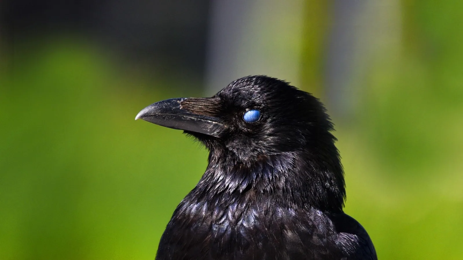
The nictitating membrane, also called the third eyelid or palpebra tertia, is a fold of conjunctiva supported by a T-shaped piece of cartilage that sweeps across the eye to spread tears and protect the cornea. In dogs and cats it usually hides in the inner corner of the eye, but when it slides across the globe and stays there, it is a visible signal that something is wrong.
This article is educational and is not a substitute for veterinary diagnosis or treatment.
What the Third Eyelid Is
The third eyelid is a conjunctival fold, not a true eyelid like the upper and lower lids. A thin sheet of conjunctiva, the mucous membrane that lines the eye and lids, doubles back on itself to form the membrane. Inside that fold sits a flexible T-shaped cartilage. The stem of the T anchors the membrane near the inner corner of the orbit, and the crossbar of the T curves along the leading edge, giving the membrane its shape and spring.
Because it is built from conjunctiva, the third eyelid has two surfaces. The bulbar surface faces the eyeball and the palpebral surface faces outward. Both are covered by epithelium that can respond to irritation with redness and swelling, which is why an inflamed third eyelid looks puffy and fleshy. The membrane also carries lymphoid tissue, and studies of canine third eyelid tissue have mapped populations of T cells and B cells within it, showing that this structure participates in local immune surveillance of the ocular surface [1].
The third eyelid is well supplied with blood. The artery that feeds it runs almost the entire length of the membrane to reach the free border before branching back inward toward a deeper segmental level, an arrangement that is unusual among ocular adnexa [1]. That vascular pattern helps explain why the membrane can swell and redden quickly when irritated.
The T-Shaped Cartilage
The cartilage is the structural core of the third eyelid. It is flexible, not rigid, so the membrane can glide across the cornea and then retract. When the cartilage scrolls or curls abnormally, the membrane cannot sit flat. A case report in a cat described exactly this problem: an 8-year-old British Blue cat was presumed to have a prolapsed gland, but surgery revealed an everted cartilage of the nictitating membrane instead. Removing the scrolled portion of cartilage through an incision on the bulbar side of the membrane resolved the irritation and the third eyelid returned to its normal position [2]. This is a useful reminder that not every red bump at the inner corner is a gland problem.
The Third Eyelid Gland
Tucked around the base of the cartilage is the third eyelid gland, also called the nictitans gland or the superficial gland of the third eyelid. In dogs, this gland and the lacrimal gland together produce the aqueous portion of the tear film, the watery layer that keeps the cornea moist and clear [3]. A gross anatomy study of dog heads measured the third eyelid gland at roughly 10.5 mm long, 11.0 mm wide and 3.3 mm thick, with a mean weight near 263 mg, making it a substantial tear-producing organ rather than a small accessory [3].
The gland is held in place by connective tissue. When that connective tissue fails, the gland pops out over the leading edge of the membrane. That is the condition commonly called cherry eye.
What the Third Eyelid Does
The nictitating membrane has two main jobs: spreading tears and protecting the eye.
Tear Spreading
Every blink does not just move the upper and lower lids. The third eyelid also sweeps across the corneal surface, helping to redistribute the tear film evenly. Because the third eyelid gland contributes a large share of the aqueous tears in dogs, the membrane is directly tied to tear production, not just tear movement [3]. This is why removing the gland rather than repositioning it can leave a dog with dry eye later in life.
Protection and Debris Removal
The membrane acts as a windshield wiper for the eye. It can flick across the cornea to clear dust, hair and small foreign material, and it can close over the globe when the eye is threatened. In species that peck, dig or root with their faces close to abrasive material, this protective sweep matters a great deal. A histologic study of six woodpecker species found that the nictitating membrane showed regional variation in its marginal plait, leading edge, epithelial surfaces and stromal pigmentation, features interpreted in the context of protecting the ocular surface during repeated mechanical loading and exposure to particulate material [4].
The membrane also contributes to the immune defense of the eye surface through the lymphoid tissue it contains [1].
How the Third Eyelid Moves: Active vs Passive
The way the third eyelid moves differs across species, and this difference matters clinically.
Passive Movement in Dogs and Cats
In dogs and cats, the third eyelid does not have its own dedicated muscle that pulls it across the eye in the way a bird does. Instead, its position depends on the pressure and volume relationships inside the orbit. When the globe is pushed forward, or when the fat and tissue behind the eye lose volume, the membrane passively slides across. The cat has smooth muscle strands extending into the third eyelid, and a histologic study of eight cats identified nine smooth muscle strands running into the membrane, with no striated muscle fibers found extending into or within it [5]. Separate work on cats, dogs and rabbits showed that retraction of the membrane can be driven by the extraocular muscles through their fascial connections, with the superior oblique and medial rectus producing the greatest movement in post mortem specimens [6]. In other words, the third eyelid in these species is moved indirectly by the muscles that move the eyeball.
This passive mechanism is the key to understanding protrusion. If the membrane only moves when orbital pressure changes, then a persistently protruded membrane tells you that orbital pressure or globe position has changed.
Active Protrusion in Birds and Some Reptiles
Birds can actively draw the nictitating membrane across the eye using a dedicated muscle, the pyramidalis muscle, which pulls on a tendon loop around the optic nerve. This lets a bird sweep the membrane across the cornea voluntarily and rapidly, often during flight, feeding or impact. Some reptiles have a comparable active mechanism. The result is that a bird can protect and clean its cornea on demand, while a dog or cat cannot.
Species Differences at a Glance
| Species | Third eyelid prominence | Movement | Notable features |
|---|---|---|---|
| Dog | Prominent, normally hidden at inner corner | Passive, driven by orbital pressure and extraocular muscle fascial connections | Large third eyelid gland contributing much of the aqueous tear film [3] |
| Cat | Prominent, normally hidden at inner corner | Passive, with smooth muscle strands in the membrane and no striated fibers within it [5] | Bilateral protrusion often points to systemic disease |
| Horse | Large and mobile | Passive but very free-moving | Easily seen during handling and head restraint |
| Bird | Highly mobile, actively controlled | Active via the pyramidalis muscle | Membrane sweeps the cornea on demand and protects against debris [4] |
| Ruminant | Prominent, especially when the eye is retracted | Passive | Large membrane that covers much of the globe when the eye is pulled back |
The prairie dog offers an interesting contrast. In captive black-tailed prairie dogs, the nictitating membrane is vestigial, meaning it is reduced and no longer fully functional, and it sits in the medial canthus area [7]. This shows that the third eyelid is not uniformly developed across all mammals.
Cherry Eye: Prolapse of the Third Eyelid Gland
Cherry eye is the common name for prolapse of the nictitans gland, the tear-producing gland that sits under the third eyelid in dogs [8]. The classic appearance is a red, fleshy protuberance at the inner corner of the eye that looks like a small cherry. The gland has been displaced from its normal position, and the underlying cause is thought to be a defect in the connective tissue that secures the gland in place [8].
Cherry eye is most often seen in dogs under two years of age, and certain breeds have a higher incidence, which points to a genetic component [8]. A large genome-wide association study using several thousand cases and controls across purebred and mixed-breed dogs found a single peak of genome-wide significance on canine chromosome 18, directly at the location of an FGF4 retrogene insertion already known to cause chondrodysplasia in several breeds [8]. That finding links cherry eye to a known genetic variant rather than to trauma or infection.
Treatment is usually surgical replacement of the gland. A survey of general practitioners and veterinarians with postgraduate ophthalmology training found that 96.2% recommended surgical replacement of cherry eye, with a pocketing technique the most frequently named procedure [9]. A retrospective study of 101 dogs and 126 eyes using a modified Morgan pocket technique with temporary ventral scleral gland fixation reported successful repositioning in 125 of 126 eyes after one surgery, with only one eye recurring and that recurrence managed by repeating the technique [10]. A separate technique anchoring the gland with a nonabsorbable suture around the insertion of the ventral rectus muscle reported no recurrences in 100 dogs and 122 eyes, and five dogs with preexisting keratoconjunctivitis sicca improved their Schirmer tear test results after correction and medical therapy [11].
The takeaway for owners is that the gland should be put back, not cut out. Removing the gland sacrifices a major source of tears and raises the risk of dry eye later.
What Protrusion of the Third Eyelid Means
A third eyelid that stays across the eye is a sign, not a disease by itself. The cause can be local to the eye, orbital, or systemic. The table below separates the common causes.
| Cause | Typical pattern | Key features |
|---|---|---|
| Cherry eye (gland prolapse) | Usually one eye, young dog | Red fleshy mass at inner corner, gland displaced [8] |
| Everted cartilage | Usually one eye | Membrane rolled or scrolled, mimics cherry eye [2] |
| Haw syndrome | Both eyes, often in dogs with gastrointestinal or other systemic illness | Bilateral protrusion with no primary eye disease |
| Tetanus | Both eyes | Protrusion with generalized muscle rigidity and other neurologic signs |
| Horner syndrome | One eye | Protrusion with miosis, ptosis and enophthalmos [12] |
| Retrobulbar mass | One eye | Protrusion with exophthalmos and resistance to retropulsion [13] |
| Orbital inflammatory disease | One or both eyes | Protrusion with exophthalmos and resistance to retropulsion [14] |
| Dehydration or weight loss | Both eyes | Sunken globes from loss of orbital fat, membrane passively slides across |
| Dysautonomia | Both eyes | Protrusion with mydriasis, reduced tear production and other autonomic signs [15] |
| Chemical or traumatic injury | Usually one eye | Protrusion with blepharospasm, corneal ulceration and conjunctival hyperemia [16] |
Cherry Eye vs Haw Syndrome
Cherry eye and Haw syndrome are easy to confuse because both produce a visible third eyelid, but they are different problems. Cherry eye is a prolapse of the gland, so you see a distinct red mass at the inner corner of one eye, most often in a young dog [8]. Haw syndrome is a bilateral protrusion of the third eyelid without a gland mass, and it is often associated with another illness, particularly gastrointestinal disease, weight loss or dehydration. In Haw syndrome the membrane is elevated because the globe has sunk back into the orbit, not because the gland has popped out.
Horner Syndrome
Horner syndrome results from loss of sympathetic nerve supply to the eye. The classic signs are miosis (a constricted pupil), ptosis (a drooping upper eyelid), enophthalmos (a sunken globe) and protrusion of the nictitating membrane [12]. In cats, there can also be peripheral vasodilation in the skin of the face on the same side [12]. Because the globe sinks back, the third eyelid passively slides across. Horner syndrome is a neurologic sign, and the underlying cause must be found.
Tetanus
Tetanus causes generalized muscle rigidity, and the third eyelids can protrude because of spasm of the muscles around the eye. Protrusion in tetanus is usually bilateral and comes with other signs such as a stiff gait, erect ears and difficulty opening the mouth.
Retrobulbar Mass and Orbital Disease
A mass behind the eye pushes the globe forward. The most common clinical signs of retrobulbar tumors in dogs and cats are exophthalmos in 84% of cases, conjunctival hyperemia in 40%, and protrusion of the nictitating membrane in 28% [13]. Orbital inflammatory disease and orbital pseudotumor produce the same trio of exophthalmos, third eyelid protrusion and resistance to retropulsion of the globe [14]. Any unilateral protrusion with a bulging eye needs imaging and a biopsy or aspirate.
Dehydration and Weight Loss
When an animal loses orbital fat through dehydration, chronic illness or weight loss, the globe sinks deeper into the orbit and the third eyelid passively slides across. This is a bilateral, symmetric protrusion with no redness or mass. Rehydration and treatment of the underlying condition usually resolve it.
Dysautonomia
Dysautonomia is a disease in which autonomic neurons degenerate. A retrospective review of 53 cats and dogs found reported clinical findings including gastrointestinal dysmotility and distension, urinary retention, reduced or absent tear production, third eyelid protrusion and inappropriate mydriasis [15]. Protrusion here is part of a widespread autonomic failure, not an eye problem alone.
Chemical and Traumatic Injury
Direct injury to the eye surface can cause the third eyelid to protrude. A study of dogs and cats injured by the Bronze Orange Bug found third eyelid protrusion in 39.4% of cases, alongside blepharospasm, corneal ulceration, conjunctival hyperemia and chemosis [16]. Any chemical exposure to the eye is an emergency and needs immediate flushing and veterinary care.
Why Bilateral Protrusion in Cats Deserves Special Attention
In cats, protrusion of the third eyelid is often bilateral, and bilateral protrusion frequently points to a systemic problem rather than a primary eye disease. Causes include Haw syndrome, dysautonomia, severe dehydration, weight loss and generalized illness. A cat with both third eyelids showing and no obvious eye redness should be examined for underlying disease, including a full physical examination, bloodwork and a neurologic assessment. Unilateral protrusion in a cat is more likely to be local, such as Horner syndrome, an orbital mass or an eye injury.
Caring for the Third Eyelid
When to See a Veterinarian
Any third eyelid that stays protruded for more than a short time needs evaluation. Seek care promptly if the protrusion is one-sided, if the eye is red, cloudy or painful, if there is discharge, if the pet is squinting or pawing at the eye, or if both third eyelids are showing. A red fleshy mass at the inner corner of a young dog's eye is likely cherry eye and should be assessed before the gland becomes chronically inflamed.
What You Can Do at Home
Keep the eye clean with a damp cotton ball and plain warm water if there is mild discharge. Do not try to push the third eyelid back into place, and do not apply human eye drops or ointments unless a veterinarian prescribes them. Prevent your pet from rubbing the eye, and use an Elizabethan collar if needed.
What Not to Do
Do not attempt to cut or remove the third eyelid or the gland at home. The third eyelid gland produces a large share of the aqueous tear film, and removing it can cause permanent dry eye [3]. Do not assume a red bump is cherry eye without confirmation, because an everted cartilage can look similar and needs a different surgery [2].
After Surgical Correction
After cherry eye surgery, most dogs are comfortable within a few days. A study of a modified pocket technique reported immediate postoperative complications in 7 of 126 eyes, including lacrimal cysts in 5 eyes and corneal ulceration in 2 eyes, all managed with local drainage or topical treatment [10]. A separate study of a suture anchor technique reported minor intraoperative conjunctival perforations in 21 of 122 eyes, repaired at the time of surgery [11]. Follow your veterinarian's instructions on rechecks and medications.
Clinical Relevance, Limitations and Common Mistakes
The third eyelid is a sensitive indicator of orbital and systemic health. Its position depends on the balance of pressure and volume inside the orbit, so a change in that balance shows up as protrusion. In dogs and cats the movement is passive, which is why the membrane slides across when the globe sinks or is pushed forward. In birds and some reptiles the movement is active, controlled by a dedicated muscle, so protrusion there has a different meaning.
Common mistakes include assuming every red mass at the inner corner is cherry eye, when an everted cartilage can look nearly identical [2]. Another mistake is treating bilateral protrusion in a cat as an eye problem when it often reflects systemic disease. A third mistake is removing the third eyelid gland instead of repositioning it, which sacrifices tear production and risks dry eye later [3].
Limitations: individual animals vary, and the same visible sign can have different causes. A veterinarian needs to examine the eye, test tear production, measure intraocular pressure and, when indicated, use imaging to reach a diagnosis. This article is educational and is not a substitute for veterinary diagnosis or treatment.
Frequently Asked Questions
What is the nictitating membrane?
The nictitating membrane is the third eyelid, a fold of conjunctiva supported by a T-shaped cartilage that sweeps across the eye to spread tears and protect the cornea.
Why is my dog's third eyelid showing?
Common causes include cherry eye (prolapse of the third eyelid gland), Haw syndrome, Horner syndrome, dehydration, weight loss, an orbital mass or eye injury. A veterinarian can narrow it down.
Is cherry eye the same as Haw syndrome?
No. Cherry eye is a prolapsed gland that appears as a red mass at the inner corner of one eye, usually in a young dog [8]. Haw syndrome is a bilateral protrusion of the membrane without a gland mass, often linked to another illness.
Can cherry eye go away on its own?
Mild cases may improve with anti-inflammatory medication, but surgical replacement of the gland is usually needed [8]. Waiting too long can lead to chronic inflammation.
Why is my cat's third eyelid showing in both eyes?
Bilateral protrusion in cats often points to a systemic problem such as Haw syndrome, dysautonomia, dehydration or weight loss. A full examination and bloodwork are usually recommended.
Does the third eyelid produce tears?
The third eyelid gland produces a large share of the aqueous tear film in dogs [3]. The membrane itself also helps spread tears across the cornea.
Can the third eyelid be removed?
It should not be removed unless there is a serious reason, because it protects the eye and its gland contributes to tear production. Removing the gland can cause dry eye later [3].
When should I take my pet to the vet for a third eyelid problem?
Take your pet promptly if the protrusion is one-sided, if the eye is red, cloudy or painful, if there is discharge or squinting, or if both third eyelids are showing.
Related Articles
- Cat Third Eyelid Showing Why
- Cat Third Eyelid Protrusion
- Cat Third Eyelid Showing Sick
- Cat Third Eyelid Showing Reddit
- Cat Third Eyelid Showing Sometimes
- Cat Third Eyelid Showing With Discharge
- Third Spacing Explained: Fluid Shifts in the Body
- Dog Eyelid Infection: Causes, Signs, and Treatment
Sources
- Immunophenotyping of lymphocyte subsets in the third eyelid tissue in dogs (Canis familiaris): morphological, microvascular, and secretory aspects of this ocular adnexa.
- Everted third eyelid cartilage in a cat: a case report and literature review.
- Gross anatomy and morphometric evaluation of the canine lacrimal and third eyelid glands.
- Histological Diversity of Eyelids and the Nictitating Membrane in Six Woodpecker Species (Picidae).
- Morphologic study of the musculature of the third eyelid in the cat (Felis catus).
- The roles of striated and smooth muscle in the movement of the cat's nictitating membrane.
- Clinical and histologic description of ocular anatomy in captive black-tailed prairie dogs (Cynomys ludovicianus).
- Association of FGF4L1 Retrogene Insertion with Prolapsed Gland of the Nictitans (Cherry Eye) in Dogs.
- Variation in the Reported Management of Canine Prolapsed Nictitans Gland and Feline Herpetic Keratitis.
- Surgical Correction of Prolapse of Nictitating Membrane Gland Using a Variant of the Pocket Technique: A Retrospective Study on 101 Dogs and 126 Eyes.
- Suture anchor placement technique around the insertion of the ventral rectus muscle for the replacement of the prolapsed gland of the third eyelid in dogs: 100 dogs.
- Horner's syndrome in the dog and cat as an aid to diagnosis.
- Retrobulbar tumors in dogs and cats: 25 cases.
- Orbital inflammatory disease and pseudotumor in dogs and cats.
- Dysautonomia in 53 cats and dogs: retrospective review of clinical data and outcome.
- Ocular findings in dogs and cats following chemical injury by the Bronze Orange Bug (Musgraveia sulciventris).