Molar Tooth Anatomy: Structure and Function
By Dr. Zubair Khalid, DVM, MS, PhD ·
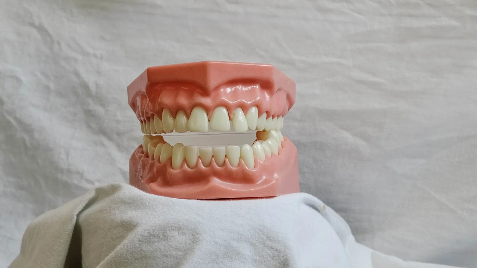
A molar tooth is a caudal (back) cheek tooth specialized for crushing, grinding, or shearing food, and it is built from four hard or soft tissues arranged into a crown, a neck, and one or more roots. Molar anatomy across domestic species reflects diet: carnivores carry relatively few, sharply cusped brachydont molars, while grazing herbivores such as horses and cattle carry many hypsodont molars with cementum-covered ridges that wear down as the animal ages.
Molar teeth matter in veterinary practice because they are the teeth most likely to develop periodontal disease, wear abnormalities, and malocclusion in companion animals, and the teeth most often examined to estimate age in horses and cattle. A clinician who can read crown height, cusp pattern, and root number can predict how a tooth will wear, where disease will start, and how extraction or floating will change the mouth. This article is educational and is not a substitute for veterinary diagnosis or treatment.
The Four Dental Tissues


Every mammalian tooth, including a molar tooth, is built from the same four tissues. Three are mineralized, one is soft.
Enamel is the hardest tissue in the body, formed almost entirely of hydroxyapatite crystals arranged in prisms. It covers the crown of brachydont teeth and forms ridges or folds in hypsodont teeth. Enamel is produced by ameloblasts before eruption and cannot be replaced after the tooth erupts. In a minipig distal molar model, enamel maturation continues for months before eruption, with crystallite thickness increasing by roughly 3.8 nm per month during the early maturation stage and the interprismatic matrix filling in during the final two months [1]. That long pre-eruption window explains why nutritional or systemic insults during development leave permanent enamel defects.
Dentin lies beneath enamel and cementum and forms the bulk of the tooth. It is less mineralized than enamel and contains microscopic tubules that radiate from the pulp outward. Dentin is produced by odontoblasts throughout life, so it can thicken as the tooth ages or responds to wear. Secondary and tertiary dentin are the tooth's slow repair response to attrition.
Cementum is a bone-like tissue covering the root surface. In brachydont teeth it stays below the gumline. In hypsodont teeth it also covers the crown and fills the spaces between enamel folds, which is why horse and cattle molars look like ridged grinding stones rather than smooth-crowned teeth.
Pulp is the soft tissue at the center of the tooth, containing blood vessels, nerves, and connective cells. It sits inside the pulp chamber in the crown and continues down each root as the root canal. Pulp is the reason a fractured tooth hurts, and it is the tissue that lays down dentin in response to injury.
Crown, Neck, and Root
The molar tooth is divided into three regions along its long axis.
Crown
The crown is the portion of the tooth covered by enamel (in brachydont teeth) or by enamel and cementum (in hypsodont teeth). It carries the cusps, which are the raised projections that contact the opposing tooth. Cusp number, height, and orientation determine whether a molar crushes, grinds, or shears.
Neck
The neck (cervix) is the narrow transition zone where the crown meets the root, usually at or just below the gumline. In brachydont teeth the neck is a clear landmark. In hypsodont teeth the neck is not a fixed anatomical line because the crown continues to erupt and wear, so the "neck" is best thought of as the current gum attachment level rather than a permanent structure.
Root
The root anchors the tooth in the alveolar bone of the jaw. Roots are covered by cementum and attached to bone by the periodontal ligament. Root number varies by species and by position in the dental arcade. Mandibular molar roots in particular show wide morphological variation, and a radiographic classification study of 512 molars found that inter-observer agreement on root morphology ranged from 75% to 91% among experts, with the lowest agreement on third molars because of atypical shapes [2]. That variability is clinically relevant: a veterinarian planning an extraction cannot assume a standard root count.
Brachydont Versus Hypsodont Molars
The single most useful distinction in comparative molar anatomy is crown height relative to the gumline.
Brachydont (Short-Crowned) Molars
Brachydont teeth have a distinct crown, a distinct neck, and roots that close at the apex. Once the crown is worn away, the tooth loses function. Dogs and cats have brachydont molars. These teeth are adapted to a diet that requires less prolonged grinding, and they rely on sharp cusps for shearing and crushing rather than on a continuously renewing grinding surface.
Hypsodont (High-Crowned) Molars
Hypsodont teeth have a tall crown that continues to erupt as the occlusal surface wears. The crown is covered by cementum, and enamel is folded into ridges or infundibula. Horses and cattle have hypsodont molars. The result is a self-sharpening grinding surface that can process abrasive plant material for years. The trade-off is that hypsodont teeth are more prone to uneven wear, cemental decay, and periodontal disease at the gingival margin.
A useful intermediate category is the radicular hypsodont tooth, which has a tall crown but also true roots. Horse cheek teeth are often described this way. Aradicular hypsodont teeth, such as rodent incisors, lack true roots and grow continuously. Molars in domestic herbivores are radicular hypsodont, not continuously growing.
Cusp Patterns and Diet
Cusp shape is a direct readout of diet. Four terms describe the main patterns.
- Bunodont: rounded, low cusps suited to crushing. Omnivores and pigs show this pattern.
- Selenodont: crescent-shaped cusps suited to grinding fibrous plant material. Ruminants such as cattle show this pattern.
- Lophodont: cusps connected by ridges (lophs) suited to shearing and grinding. Horses and other equids show this pattern.
- Secodont: sharp, blade-like cusps suited to shearing meat. Carnassial teeth in dogs and cats show this pattern.
The golden snub-nosed monkey, a leaf-eating primate, has larger molars with blunt cusp crests, tiny incisors, and strong mandibles with well-developed masticatory muscles, features interpreted as adaptations to a fibrous diet [3]. The same logic applies to domestic species: the more fibrous and abrasive the diet, the flatter and more ridged the molar crown.
Species Comparison Table
| Feature | Dog | Cat | Horse | Cow |
|---|---|---|---|---|
| Molar type | Brachydont | Brachydont | Hypsodont (radicular) | Hypsodont |
| Crown coverage | Enamel only | Enamel only | Enamel plus cementum | Enamel plus cementum |
| Cusp pattern | Bunodont to secodont | Secodont (carnassial) | Lophodont | Selenodont |
| Typical molar count per quadrant | 2 upper, 3 lower | 1 upper, 2 lower | 3 upper, 3 lower | 3 upper, 3 lower |
| Root form | Distinct, closed apex | Distinct, closed apex | Long, open for years | Long, open for years |
| Main function | Crushing and shearing | Shearing | Grinding | Grinding |
| Wear pattern | Crown loss is permanent | Crown loss is permanent | Continuous eruption compensates | Continuous eruption compensates |
Counts vary with breed and with the presence of wolf teeth or missing teeth, so the table gives typical values rather than fixed rules.
Eruption Timing by Species
Eruption timing is one of the most practical pieces of molar anatomy because it lets a clinician estimate age and anticipate when a tooth will become functional.
Dog
Deciduous teeth begin erupting at about 3 weeks of age and are complete by about 6 weeks. Permanent incisors, canines, and premolars erupt between 3 and 5 months. Permanent molars erupt slightly later, with the first molar typically appearing around 4 months and the third molar by about 6 to 7 months. By 7 months, most dogs have a full permanent dentition.
Cat
Deciduous teeth erupt from about 2 to 4 weeks and are complete by 6 to 8 weeks. Permanent teeth begin erupting at about 3.5 months, and the permanent molars are typically in place by 5 to 6 months. Cats finish the permanent dentition slightly earlier than dogs.
Horse
Deciduous premolars erupt shortly after birth and are replaced by permanent cheek teeth between about 2 and 4 years. The permanent molars erupt in sequence: the first molar around 9 to 12 months, the second molar around 2 years, and the third molar around 3.5 to 4 years. Because hypsodont cheek teeth continue to erupt and wear, eruption timing and wear pattern together are used to estimate age in horses.
Cow
Deciduous teeth erupt before or shortly after birth, and permanent incisors and cheek teeth replace them over the first few years. The first permanent molar typically erupts around 5 to 6 months, the second around 12 to 15 months, and the third around 24 months. As in horses, wear on the occlusal surface is used alongside eruption to age cattle.
These timings are approximate. Nutrition, breed, and individual variation shift them by weeks to months, so eruption should be combined with other age markers rather than used alone.
How Molar Anatomy Is Examined in Practice
A complete molar examination combines visual inspection, probing, and imaging.
Visual inspection looks at crown height, cusp wear, cementum coverage, and the gingival margin. In horses, the occlusal surface is examined for sharp enamel points, hooks, and ramps that form when the upper and lower arcades do not align perfectly.
Periodontal probing measures the depth of the gingival sulcus and detects pockets around the roots. This is the standard method for staging periodontal disease in dogs and cats.
Dental radiography shows roots, pulp chambers, alveolar bone, and retained deciduous teeth. Root morphology is variable enough that radiographs are essential before extraction. The atlas-based classification study noted that inter-observer agreement on molar root morphology was substantial to almost perfect (Gwet's AC1 0.62 to 0.88) when a structured framework was used, which supports using a systematic approach rather than ad hoc description [2].
Computed tomography is used in research and in complex cases to map root canals and enamel thickness. A study of human teeth over ten centuries found a strong linear correlation between endodontic system volume and enamel thickness at the support cusp (r = 0.723), showing that hard tissue thickness and pulp volume are linked [4]. The same principle applies in animals: a tooth with a large pulp chamber has thinner surrounding dentin and is more vulnerable to fracture.
Dental wear analysis uses the pattern of pits and scratches on the occlusal surface to infer diet. In dinosaurs, scratch-dominated microwear indicated high-fiber herbivory, while pit-and-scratch mixtures suggested mixed feeding [5]. The method is used in paleontology and comparative anatomy more than in routine practice, but it illustrates how directly wear reflects function.
Development and the Jaw
Molar shape is not determined by the tooth alone. A study comparing mouse and vole molars showed that cusp offset, the alternating left-right displacement of cusps, depends on mechanical constraint from the growing jaw. Vole molars cultured without jaw support lost their cusp offset, and mouse molars cultured with braces developed one [6]. This means that some aspects of molar crown geometry are regulated by the surrounding bone and soft tissue, not just by the tooth germ. For veterinary anatomy, the practical point is that jaw conformation and molar crown shape are developmentally linked. A breed with a shortened jaw may have molars that are normal in shape but crowded or maloccluded.
Vascularization of the developing tooth germ is another developmental factor. In fetal mice, c-Mpl mRNA is expressed in the dental papilla, enamel organ, and dental lamina in a spatiotemporally controlled pattern that precedes endothelial invasion [7]. Blood supply to the pulp is established early and is essential for dentin formation.
Clinical Relevance, Limitations and Common Mistakes
Molar disease is common and often underdiagnosed because the caudal mouth is hard to see without sedation or anesthesia.
Periodontal disease starts at the gingival margin and progresses to the periodontal ligament and alveolar bone. It is the most common dental disease in dogs and cats and the leading reason for tooth loss in small animal practice.
Wear abnormalities are the main molar problem in horses. Uneven wear produces sharp enamel points that lacerate the cheek and tongue. Routine floating smooths these points, but over-floating can remove too much crown and reduce the grinding surface.
Fractured molars expose dentin and pulp, leading to infection and pain. In brachydont teeth, a crown fracture that reaches the pulp requires treatment or extraction. In hypsodont teeth, fractures may be tolerated longer because the tooth continues to erupt, but they still disrupt occlusion.
Retained deciduous teeth can cause the permanent molar to erupt in an abnormal position. This is most common in small breed dogs.
Misconceptions students often hold:
- That all molars are grinding teeth. In carnivores, the carnassial molar is a shearing tooth.
- That hypsodont means continuously growing. Horse and cattle molars erupt for years but are not aradicular like rodent incisors.
- That root number is constant. Third molars in particular show atypical morphology and lower observer agreement [2].
- That enamel can regrow. Enamel is formed before eruption and is not replaced.
- That eruption timing is exact. It varies with breed, nutrition, and individual development.
- That a normal-looking crown means a healthy root. Root and periodontal disease can be advanced with minimal crown changes.
Limitations: eruption tables give population averages, not individual predictions. A single examination cannot establish age or dental health with certainty. Individual cases need a veterinarian.
Quick Review
- A molar tooth has four tissues: enamel, dentin, cementum, and pulp.
- The crown, neck, and root divide the tooth along its long axis.
- Brachydont molars (dogs, cats) have a fixed crown and closed roots. Hypsodont molars (horses, cattle) have tall cementum-covered crowns that erupt as they wear.
- Cusp pattern reflects diet: bunodont for crushing, selenodont and lophodont for grinding, secodont for shearing.
- Eruption timing is species-specific and is used with wear pattern to estimate age.
- Root morphology is variable, especially in third molars, so radiographs are needed before extraction.
- Molar shape and jaw shape develop together, so jaw conformation affects occlusion.
Frequently Asked Questions
What is the difference between a molar and a premolar?
Molars are the caudal cheek teeth that are not preceded by deciduous teeth in most species, while premolars have deciduous predecessors. Molars typically have a larger grinding or shearing surface and sit further back in the arcade.
Do dogs have hypsodont molars?
No. Dogs have brachydont molars with a distinct crown, neck, and closed roots. Their molars are adapted for crushing and shearing rather than prolonged grinding.
Why do horse molars have cementum on the crown?
Cementum fills the spaces between the enamel folds and covers the crown surface, creating a durable grinding surface that wears evenly as the tooth erupts. This is an adaptation to a highly abrasive grass diet.
How many molars does a cat have?
Cats typically have one upper molar and two lower molars per side, for a total of six molars. The upper molar and lower first molar form the carnassial pair used for shearing.
Can a veterinarian tell a horse's age from its molars?
Eruption timing and occlusal wear pattern together give a reasonable age estimate, but nutrition, breed, and individual variation shift the timeline. Molars are one of several age indicators, not a precise clock.
What happens if a molar is fractured?
A fractured molar exposes dentin and possibly pulp, which can become infected and painful. Treatment depends on the species, the tooth, and how much crown remains, and may include extraction or endodontic care.
Related Articles
- Equine Hoof Anatomy: Structures and Function
- Equine Larynx Anatomy and Function in Respiration
- Canine Lymphatic System: Anatomy and Function
- Canine Liver Anatomy and Lobular Structure
- Fish Gills: Anatomy, Function, and Common Health Issues
- Snake Anatomy: Do Snakes Have Teeth, Spines, and Other Structures?
- Condyloid Joint: Structure and Movement
- Papillary Dermis: Structure and Function Explained
- Anatomical Heart: Structure and Chambers
Sources
- Mammalian enamel maturation: Crystallographic changes prior to tooth eruption.
- Atlas-based radiographic classification of mandibular molar root morphology for forensic dental identification.
- Current Knowledge on Craniodental Anatomy and Dental Pathology in Golden Snub-Nosed Monkeys (Rhinopithecus roxellana): A Comparative Review.
- [[Changes in the morphological characteristics of the endodontic system and parameters of human dental hard tissues in the North-Western region of Siberia during the X-XX centuries].](https://pubmed.ncbi.nlm.nih.gov/39704991/)
- The functional and palaeoecological implications of tooth morphology and wear for the megaherbivorous dinosaurs from the Dinosaur Park Formation (upper Campanian) of Alberta, Canada.
- Mechanical constraint from growing jaw facilitates mammalian dental diversity.
- Spatiotemporal expression profiles of c-Mpl mRNA in the tooth germ: Comparative expression dynamics of vascularization-related genes.