Meissner Cells: Tactile Corpuscle Structure and Function
By Dr. Zubair Khalid, DVM, MS, PhD ·
A Meissner corpuscle (also called a tactile corpuscle) is an encapsulated, rapidly adapting, low-threshold mechanoreceptor that sits in the dermal papillae of glabrous skin and signals low-frequency flutter and slip. Its core is a stack of lamellar Schwann cells that wrap a single myelinated afferent terminal, and its transduction channel is Piezo2.
This structure matters because it is the receptor that lets a fingertip, a paw pad, or a hoof wall read fine texture and detect the moment an object begins to slide. Students meet it in histology practicals, in comparative anatomy of the digital pads, and in neurology cases where a patient cannot feel a light moving touch. Getting the layered architecture right, and keeping Meissner cells separate from Pacinian corpuscles, is the difference between a correct answer and a common exam error.
What a Meissner Corpuscle Is and Where It Sits

Meissner corpuscles occupy the dermal papillae, the finger-like projections of superficial dermis that interdigitate with the overlying epidermis. Each papilla is a narrow connective tissue peg capped by epidermis, and a tactile corpuscle fills much of that peg. Because the corpuscle sits so close to the epidermal surface, mechanical events at the skin surface reach it with very little attenuation.
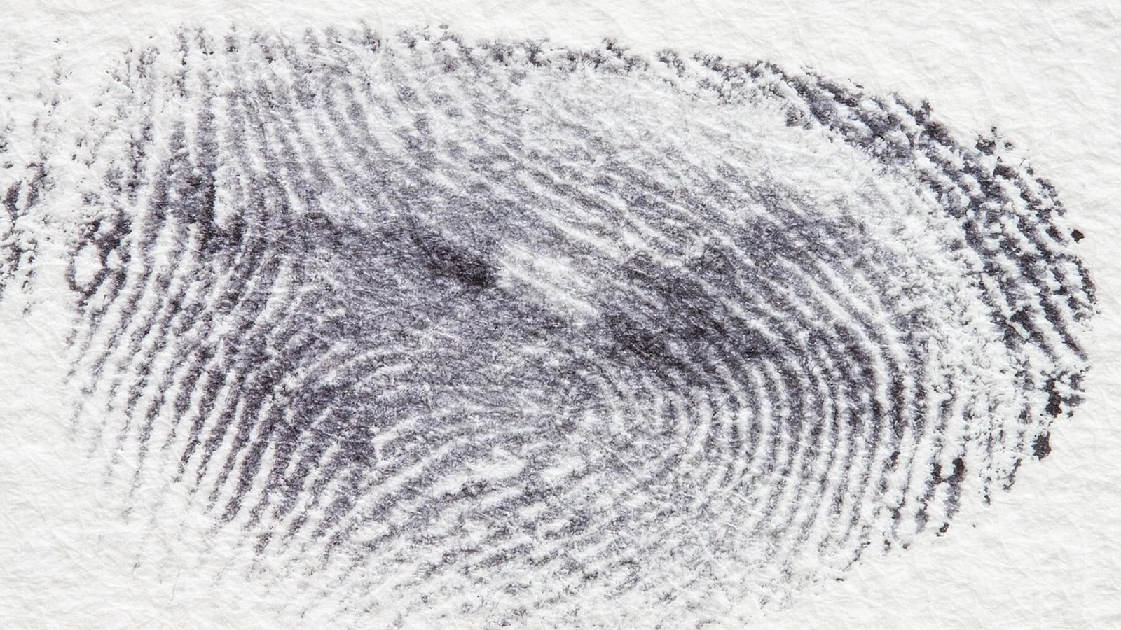
The epidermis above the papilla is thin and, in glabrous skin, carries a thick stratum corneum. A ridge of epidermis, the intermediate ridge, arches over each corpuscle. This anatomy is why glabrous skin has its characteristic fingerprint pattern: the dermal papillae and their corpuscles sit in rows beneath the epidermal ridges, and the ridges are the visible surface expression of that underlying order.
A tactile corpuscle is roughly oval to ellipsoid, oriented with its long axis perpendicular to the skin surface. Reported dimensions cluster around 30 to 140 micrometres in length, with the long axis often two to three times the short axis. That range reflects real biological variation between sites and between species, and it overlaps with the size of other end organs, so size alone does not identify a corpuscle in a micrograph.
Meissner corpuscles are a feature of glabrous (hairless) skin. They are densely packed in the digital pads of primates and in the equivalent friction surfaces of other mammals, and they are absent from hairy skin. Where hairy skin needs a rapidly adapting receptor, it uses a different end organ, typically a hair follicle afferent or a field receptor. This is a comparative point worth memorizing: the presence of Meissner corpuscles is a marker of glabrous skin, and their absence does not mean a hairy skin region lacks fine touch.
Density is highest where tactile acuity is highest. On human fingertips, Meissner corpuscle density is roughly 100 to 150 per square centimeter. That figure is a useful anchor for exam answers and for understanding why the fingertip is the reference standard for two-point discrimination. Density falls as you move proximally along the digit and onto the palm, and it falls further on the forearm.
Receptive fields are small. A single Meissner afferent typically responds to stimulation over a few millimetres of skin. Small receptive fields plus high receptor density produce fine spatial resolution. The afferents are A-beta fibers, which are large, myelinated, and fast conducting.
Layered Architecture of the Tactile Corpuscle
The corpuscle is a layered structure, and each layer has a mechanical or trophic role. Work through it from the outside in.
Epidermis and the Dermal Papilla
The epidermis is not part of the corpuscle, but it is the mechanical interface. The stratum corneum and the viable epidermis transmit and filter the deformation that reaches the papilla. The papilla itself is loose connective tissue carrying capillaries, and the corpuscle is anchored within it. Because the papilla is narrow, the corpuscle is mechanically coupled to a small patch of skin surface, which is one reason receptive fields stay small.
The Capsule and Lamellar Schwann Cells
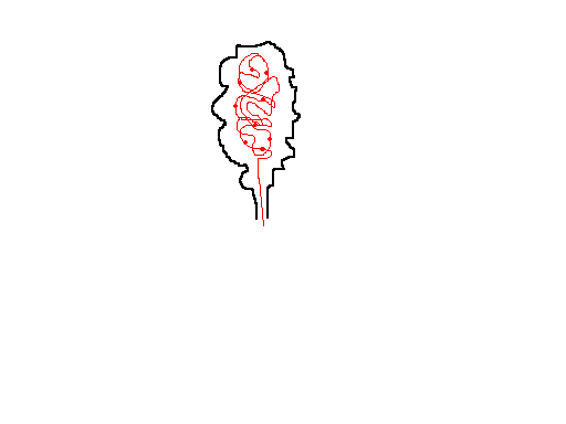
The outer boundary of the corpuscle is a thin connective tissue capsule. Inside it, lamellar Schwann cells form stacked, flattened layers that resemble a stack of coins or a rolled-up sleeve. These Schwann cells are the "Meissner cells" of the older literature. They are not neurons. They are terminal glial cells that ensheath the afferent ending.
The lamellae are arranged transversely across the long axis of the corpuscle, so the stack looks like a column of discs. Between and around the lamellae is a small amount of extracellular matrix. The number of lamellae varies between corpuscles and between species.
Terminal glial cells of cutaneous end organs were long treated as purely structural and trophic elements. Current work treats them as potentially active participants in mechanosensory processing. An immunohistochemical mapping study of human Meissner and Pacinian corpuscles examined mechanosensitivity-associated ion channel proteins in these terminal glial cells and in the mechanoreceptor axons separately [1]. In Meissner corpuscles, terminal glial cell immunoreactivity for ASIC2 and TRPV4 appeared in subsets of evaluable corpuscles, and TRPA1 immunoreactivity appeared in selected specimens. PIEZO1 and PIEZO2 immunoreactivity appeared in subsets of corpuscles from the single available labia minora specimen. The authors describe these findings as hypothesis-generating and note that they do not demonstrate protein function or mechanotransduction [1]. Treat the glial channel data as an open question, not as established mechanism.
Separately, Piezo2 has been identified in Schwann cells, and a study of Schwann cell mechanosensitivity and neurotrophic release regulation examined undifferentiated and differentiated Schwann cell subtypes, including expression of Piezo1, Piezo2, and TRP channels, with functional testing under hypotonic conditions [2]. That work places Piezo2 in Schwann cells as well as in the sensory ending, which is relevant to how the corpuscle environment might contribute to signaling.
The Axon Terminal
At the center of the lamellar stack is the afferent terminal. It is the ending of a single myelinated A-beta fiber that has lost its myelin sheath as it enters the corpuscle. The terminal is not a simple point. It branches and runs between the Schwann cell lamellae, so the ending is in intimate contact with many lamellar cells along the length of the corpuscle.
This geometry matters. The terminal is long relative to the corpuscle and is coupled to the lamellar stack along its whole course. Mechanical deformation of the stack deforms the terminal membrane, and the terminal membrane carries the mechanically gated ion channels that convert force into current.
Putting the Layers Together
Read the corpuscle as a mechanical chain:
- Skin surface deforms under a moving stimulus.
- The epidermis and dermal papilla transmit that deformation to the corpuscle.
- The capsule and lamellar Schwann cell stack distribute the deformation along the terminal.
- The afferent terminal membrane is stretched or indented, opening mechanically gated channels.
- Cation current depolarizes the terminal, and if threshold is reached, action potentials travel centrally along the A-beta fiber.
Each layer can in principle filter or shape the mechanical signal. The traditional view of encapsulated end organs assigned most filtering to the outer layers. Recent work on Pacinian corpuscles has revised that view, and the revision is instructive for how we think about Meissner corpuscles as well.
Mechanotransduction: From Force to Action Potential
Mechanotransduction is the conversion of mechanical force into an electrical signal. In Meissner corpuscles, the channel that carries this conversion is Piezo2, a mechanically gated cation channel.
The steps are worth learning as a sequence:
- A moving stimulus, such as a fingertip sliding across a textured surface, produces a time-varying deformation of the skin.
- The deformation reaches the corpuscle through the epidermis and papilla.
- The lamellar stack and capsule couple the deformation to the afferent terminal membrane.
- Membrane tension or curvature changes open Piezo2 channels.
- Sodium and calcium enter the terminal, producing a generator (receptor) potential.
- The generator potential spreads electronically along the terminal.
- If the generator potential reaches threshold at the spike initiation site, action potentials are generated.
- Action potentials travel along the A-beta fiber to the dorsal column nuclei and onward to somatosensory cortex.
Two properties of this sequence define the Meissner receptor's behavior.
First, it is rapidly adapting. The receptor potential rises at the onset of a mechanical step and falls again during a maintained step. A static indentation produces a brief burst of spikes and then silence. A moving or vibrating stimulus produces maintained firing because the stimulus keeps changing. Rapid adaptation is the physiological basis for detecting change rather than steady state.
Second, it is low threshold. The force needed to activate a Meissner afferent is small, which is why a light brush is enough. Low threshold plus rapid adaptation makes the receptor a detector of flutter and slip rather than of sustained pressure.
The frequency range is the point where students most often overreach. Meissner afferents respond best to low-frequency skin deformation, in the flutter range, and they do not carry the high-frequency vibration sense. High-frequency vibration detection is the job of Pacinian corpuscles. A recent study of Pacinian corpuscles showed that high-frequency tuning results from the afferent terminal's sensitivity to stimulus velocity rather than to stimulus cycle rate, and that these properties are largely independent of the corpuscular environment, reflecting the biophysics of the mechanically gated channels in the terminal, such as Piezo2 [3]. That is a Pacinian result. Do not transfer it to Meissner corpuscles. The practical rule is that Meissner cells signal flutter and slip, and vibration above roughly 50 Hz belongs to Pacinian afferents.
A frequency-band framework used in a recent tactile-acuity study decomposed friction-induced vibrations into bands attributed to Merkel, Meissner, Ruffini, and Pacinian mechanoreceptors [4]. That study used the framework to build a tactile acuity index and to examine how texture and age change mechanosensory activity. The strongest correlations in that work were for Ruffini and Pacinian receptors, reflecting sensitivity to macroscopic strain and high-frequency vibration respectively [4]. The takeaway for this article is that Meissner afferents occupy a defined low-frequency band within a multi-receptor system, not the whole spectrum.
Comparison With Other Cutaneous Mechanoreceptors
Four low-threshold mechanoreceptor classes dominate the glabrous skin literature. The table below compares them by adaptation, adequate stimulus, and location. Use it as a single summary reference.
| Receptor | Afferent type | Adaptation | Adequate stimulus | Location |
|---|---|---|---|---|
| Meissner corpuscle | A-beta, rapidly adapting | Rapid | Low-frequency flutter, slip, light moving touch | Dermal papillae of glabrous skin |
| Merkel cell-neurite complex | A-beta, slowly adapting | Slow | Sustained pressure, edges, fine form and texture | Basal epidermis of glabrous and hairy skin |
| Pacinian corpuscle | A-beta, rapidly adapting | Rapid | High-frequency vibration | Deep dermis and subcutaneous tissue, widespread |
| Ruffini ending | A-beta, slowly adapting | Slow | Skin stretch, sustained pressure | Dermis, especially in hairy skin and joints |
Three points about this table deserve emphasis.
Meissner and Pacinian corpuscles are both rapidly adapting, so adaptation alone does not separate them. The separating features are stimulus frequency band and depth. Meissner corpuscles are superficial and low frequency. Pacinian corpuscles are deep and high frequency.
Merkel cells and Ruffini endings are both slowly adapting, so adaptation alone does not separate them either. The separating features are location and stimulus quality. Merkel cells sit at the epidermal base and signal edges and sustained pressure. Ruffini endings sit in the dermis and signal stretch.
Meissner corpuscles are the only one of the four that is essentially restricted to glabrous skin. Merkel cells, Pacinian corpuscles, and Ruffini endings all occur in hairy skin as well.
How Meissner Corpuscles Are Studied and Observed
Histology
The classic demonstration is a thick skin section, such as fingertip or paw pad, stained to show connective tissue and cells. Meissner corpuscles appear as oval bodies in the dermal papillae, aligned in rows beneath the epidermal ridges. The lamellar stack shows as transverse bands within the corpuscle, and the capsule shows as a thin boundary. A histology reference slide of thick skin is a standard teaching resource for this appearance [5].
When you look at a micrograph, work through a checklist:
- Is the skin glabrous? Look for a thick stratum corneum and epidermal ridges.
- Are the corpuscles in the dermal papillae, immediately beneath the epidermis?
- Is the long axis perpendicular to the surface?
- Do you see transverse lamellae within the body?
- Is there a single central terminal region rather than a central cavity?
If the answer to the first two is no, you are probably looking at Pacinian corpuscles, which sit deeper and have a more obviously layered, onion-like outer core.
Immunohistochemistry
Antibodies against mechanosensitive channel proteins and Schwann cell markers can be used to map the corpuscle. The human mapping study cited above assessed immunoreactivity separately in mechanoreceptor axons and in terminal glial cells, which is the right experimental design if you want to know which cell type carries which channel [1]. Remember that immunoreactivity shows protein presence, not function.
Physiology
Single-unit recording from A-beta afferents, or microneurography in human subjects, characterizes adaptation, threshold, and receptive field size. Vibrotactile psychophysics maps the frequency bands that different receptor classes serve. The tactile acuity index approach uses friction-induced vibrations recorded during controlled touch and decomposes them into receptor-specific bands [4]. That is an indirect, non-invasive way to estimate mechanoreceptor recruitment.
Pathology and Comparative Material
Tactile corpuscle-like bodies are structures that resemble Meissner corpuscles on histology and can appear in unexpected sites. They are a recognized finding in pathology practice and are a reminder that morphology alone can mislead [6]. In veterinary material, glabrous friction surfaces such as digital pads and the equine hoof are the places to look for Meissner-type corpuscles, and species differences in density and distribution are substantial.
Comparative and Clinical Relevance
Comparative Anatomy
Glabrous friction surfaces across species carry encapsulated mechanoreceptors, but the mix and density vary. Primates have the highest Meissner corpuscle densities on fingertips, which tracks with fine manipulative ability. Carnivore digital pads and the equine hoof wall are other glabrous surfaces where encapsulated endings are found. In hairy skin, the rapidly adapting channel is served mainly by hair follicle afferents, which is why a light brush on a furry flank is detected by a different mechanism than a light brush on a fingertip.
For veterinary students, the practical comparative lesson is that you cannot assume a receptor map from one species applies to another. The functional categories (rapidly adapting low threshold, slowly adapting low threshold) are conserved. The anatomical packaging is not.
Clinical Relevance
Loss of Meissner corpuscle function contributes to loss of fine tactile discrimination. Because the corpuscle depends on an intact A-beta afferent, a peripheral neuropathy that affects large myelinated fibers will degrade flutter and slip detection before it degrades pain and temperature sensation. This is why large-fiber neuropathy testing uses vibration and light touch tasks.
Age changes mechanosensory performance. The tactile acuity study that decomposed friction-induced vibrations found a pronounced reduction in mechanosensory activity across all receptor types with age, with the largest declines in deep mechanotransductive pathways [4]. That is a whole-system finding, not a Meissner-specific one, but it supports the general principle that tactile acuity declines with age.
Tactile corpuscle-like bodies in biopsy material are a diagnostic consideration in pathology and should not be mistaken for a neoplastic process without context [6].
Clinical Relevance, Limitations and Common Mistakes
The most common mistake is confusing Meissner corpuscles with Pacinian corpuscles. Both are rapidly adapting A-beta receptors, so students who classify by adaptation alone cannot tell them apart. Use location and frequency band instead. Meissner corpuscles are superficial, in dermal papillae, and serve low-frequency flutter. Pacinian corpuscles are deep and serve high-frequency vibration.
The second mistake is assigning high-frequency vibration detection to Meissner cells. Meissner afferents do not carry vibration above roughly 50 Hz. That range belongs to Pacinian afferents, whose high-frequency tuning has been shown to depend on the velocity sensitivity of mechanotransduction in the afferent terminal rather than on the outer core acting as a filter [3].
The third mistake is treating the lamellar Schwann cells as passive packing. They are terminal glial cells, and there is active investigation into whether they contribute to mechanosensory processing. The human immunohistochemical mapping study found channel protein immunoreactivity in terminal glial cells of Meissner and Pacinian corpuscles, but the authors explicitly describe the findings as hypothesis-generating and note that they do not demonstrate function [1]. Do not present glial mechanotransduction as established.
The fourth mistake is assuming Piezo2 is exclusive to one receptor. Piezo2 is the principal transduction channel in Meissner corpuscles and in other low-threshold mechanoreceptors, and it has also been identified in Schwann cells, where it has been studied in the context of volume regulation and neurotrophic release [2]. Channel presence in a cell type is not the same as a defined sensory role.
The fifth mistake is reading size as identity. Corpuscle dimensions overlap between classes, and 30 to 140 micrometres does not by itself tell you which corpuscle you are looking at. Use position, lamellar pattern, and skin type.
This article is educational and is not a substitute for veterinary diagnosis or treatment. Individual animals need a veterinarian for any clinical problem.
Quick Review
- A Meissner corpuscle is a rapidly adapting, low-threshold mechanoreceptor in the dermal papillae of glabrous skin.
- Its core is a stack of lamellar Schwann cells (the Meissner cells) wrapped around a single A-beta afferent terminal.
- Piezo2 is the mechanically gated transduction channel.
- It signals low-frequency flutter and slip, not high-frequency vibration.
- Density reaches roughly 100 to 150 per square centimeter on human fingertips, and receptive fields are a few millimetres across.
- Corpuscles are about 30 to 140 micrometres long and sit perpendicular to the skin surface.
- They are absent from hairy skin, where other end organs and hair follicle afferents take over rapidly adapting touch.
Frequently Asked Questions
Are Meissner cells the same as Meissner corpuscles?
No. Meissner cells are the lamellar Schwann cells inside the corpuscle, and the Meissner corpuscle is the whole end organ including those cells, the capsule, and the afferent terminal.
Do Meissner corpuscles detect vibration?
They detect low-frequency flutter and slip. High-frequency vibration detection belongs to Pacinian corpuscles.
Where are Meissner corpuscles found?
They sit in the dermal papillae of glabrous skin, such as fingertips and digital pads, and they are absent from hairy skin.
What ion channel drives Meissner mechanotransduction?
Piezo2, a mechanically gated cation channel in the afferent terminal membrane.
How big is a Meissner corpuscle?
Roughly 30 to 140 micrometres in length, with the long axis oriented perpendicular to the skin surface.
Why do fingertips have so many Meissner corpuscles?
High density plus small receptive fields gives fine spatial resolution, which supports texture discrimination and slip detection during manipulation.
Related Articles
- Chromatin in a Cell: Structure, Function, and Dynamics
- Ribosome in Cell: Structure, Function, and Synthesis
- Diagram of Yeast Cell: Structure, Function, and Labeling Guide
- p53 Protein: Structure, Function, and Regulation in Cell Signaling
- Cell Membrane Proteins: Structure, Function, and Experimental Analysis
- Prokaryotic DNA: Location, Structure, and Function
- Cell Image Guide: Labeled Diagrams of Cell Structures
- Pacinian Corpuscle: Structure, Function and Location
- Muscle Cell: Structure, Types, and Function
Sources
- Exploratory Immunohistochemical Mapping of Mechanosensitivity-Associated Ion Channel Proteins in Terminal Glial Cells of Human Meissner and Pacinian Corpuscles.
- Role of Piezo2 in Schwann Cell Volume Regulation and Its Impact on Neurotrophic Release Regulation.
- Velocity sensitivity of mechanotransduction in the afferent terminal underlies vibration detection in the Pacinian corpuscle.
- How Touch Triggers Mechanotransduction in Cutaneous Mechanoreceptors.
- Meissner Corpuscles - Nervous Tissue
- Pathology Outlines - Tactile corpuscle-like bodies