Loop of Henle: Function and Structure
By Dr. Zubair Khalid, DVM, MS, PhD ·
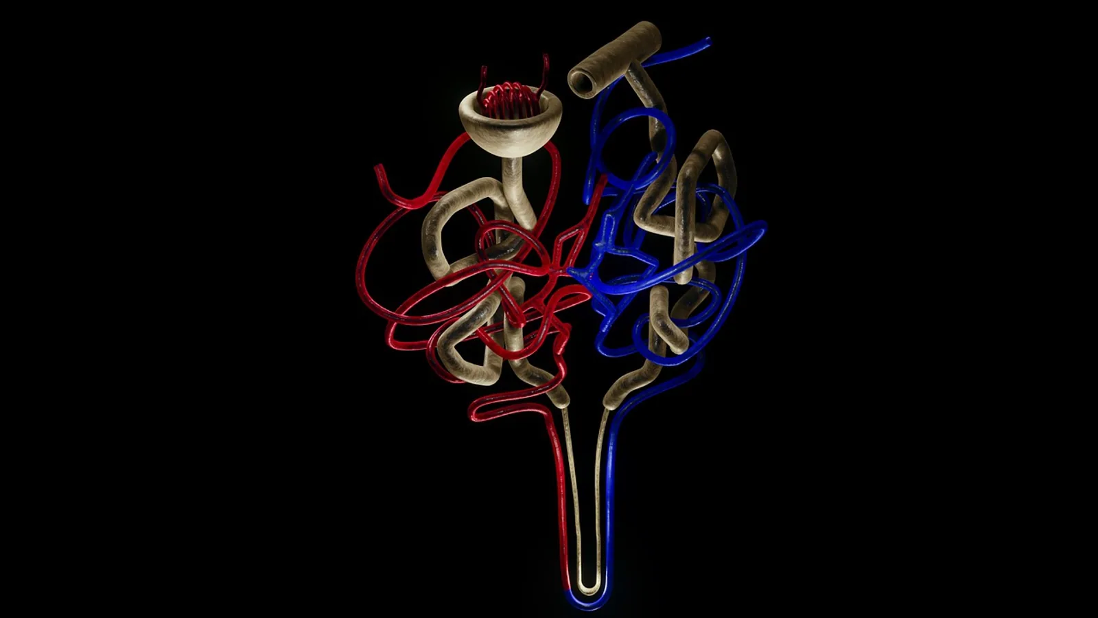
The loop of Henle is the U-shaped segment of the nephron that runs from the pars recta of the proximal tubule down into the renal medulla and back up to the distal convoluted tubule. Its function is to create and maintain a steep osmotic gradient in the medullary interstitium, so that water can be reclaimed from the collecting duct and a concentrated urine can be produced.
No other part of the nephron does this job. The proximal tubule reclaims the bulk of filtered salt and water, and the collecting duct makes the final decision about how much water leaves the body, but neither can generate the gradient on its own. The loop supplies the energy and the geometry. When loops are short, as in the cat, or damaged, as in chronic interstitial nephritis, urine concentrating ability falls even though glomerular filtration may look acceptable on a chemistry panel. That is why the loop matters clinically as much as it matters physiologically.
What the Loop of Henle Is and Where It Sits
The nephron loop (also written Henle loop, Henle's loop, or loop of nephron) begins at the end of the proximal straight tubule and ends where the distal straight tubule meets the macula densa. It has four named parts in mammals:
- Descending thick limb (pars recta of the proximal tubule), present only in nephrons with long loops.
- Descending thin limb.
- Ascending thin limb.
- Ascending thick limb, which ends at the macula densa.
The hairpin bend sits at the deepest point of the loop, and the depth determines how far into the medulla the tubule reaches. Juxtamedullary nephrons, whose glomeruli sit near the corticomedullary junction, have long loops that descend almost to the papilla. Superficial cortical nephrons have short loops that barely enter the outer medulla. The mix of long and short loops in a kidney sets the upper limit of that species' urine concentrating ability.
Jacob Henle described the U-shaped tubule in 1862, but the structure sat largely unexplained for eighty years. Werner Kuhn, a physical chemist, proposed in 1942 that the loop behaves as a hairpin countercurrent multiplier, and Kuhn, Hargitay, and Wirz showed experimentally in 1951 that the loop is the central element of the mammalian urine concentrating mechanism. Carl Gottschalk and Margaret Mylle confirmed the theory in 1958, and Gottschalk summarized the accumulated evidence in 1962, three centuries after the first description of renal tubules [1].
The Countercurrent Multiplier in Plain Terms
Countercurrent means that fluid flows in opposite directions in the two limbs that lie side by side. Multiplication means that a small local effect, repeated along the length of the tubule, becomes a large axial gradient.
Three properties are required.
- The descending limb must lose water and retain solute, so the fluid inside it becomes progressively more concentrated as it descends.
- The ascending limb must remove solute without allowing water to follow, so the fluid inside it becomes progressively more dilute as it ascends.
- The two limbs must be close enough that the interstitial solute left behind by the ascending limb can pull water out of the descending limb.
Each pass of fluid through the hairpin adds a little more solute to the interstitium. Over time, the medullary interstitium reaches a stable gradient that rises from roughly isotonic at the corticomedullary junction to several times plasma osmolality at the papilla. In the dog, the medullary gradient runs from approximately 300 mOsm/kg in the cortex to about 1200 mOsm/kg at the tip of the papilla. Desert rodents with much longer loops can exceed this considerably, and mathematical models of the inner medulla suggest that urine osmolalities on the order of 5000 mOsm/L can be generated when the passive thin limb properties are favorable [2].
The Descending Limb
The descending limb is permeable to water and nearly impermeable to solutes. Water leaves by osmosis because the interstitium around it is hypertonic. Solute stays behind. The result is that tubular fluid becomes more concentrated as it approaches the bend, and the fluid arriving at the hairpin is the most concentrated fluid in the nephron.
The thin descending limb contains no significant active transport machinery. Its water permeability depends on aquaporin-1 in the apical and basolateral membranes. Solute permeability is not zero, and measurements show that NaCl and urea permeability vary along the length of the descending thin limb rather than staying constant, which is one reason simple models of the inner medulla underperform compared with real kidneys [3].
The Thin Ascending Limb
The thin ascending limb is impermeable to water. It is highly permeable to sodium and chloride, moderately permeable to urea, and it dilutes tubular fluid without any movement of water across the epithelium [4]. The classic question is whether this segment pumps salt actively. The experimental answer is no. Active sodium reabsorption in the thin ascending limb amounts to only a few percent of net sodium reabsorption there, and the segment maintains a low intracellular sodium concentration through a ouabain-sensitive Na+/K+-ATPase in the basolateral membrane while the luminal membrane has a furosemide-insensitive sodium permeability [4].
Chloride permeability in this segment is extremely high, with saturation kinetics, and it is inhibited by halogens and by anion transport inhibitors including DIDS, phloretin, and NPPB. The chloride conductance is pH sensitive and activated by calcium, and it operates in series across the apical and basolateral membranes [5]. This arrangement lets salt diffuse passively out of the thin ascending limb into the interstitium, which is exactly what a passive inner medullary multiplier needs.
The Thick Ascending Limb
The thick ascending limb is the engine of the whole system. It is water-impermeable and it reabsorbs NaCl actively, in excess of water, which dilutes the tubular fluid and delivers solute to the medullary interstitium [6]. It has at least three roles: it dilutes urine, it produces the concentration gradients that drive countercurrent multiplication, and it reabsorbs large amounts of potassium, calcium, and magnesium in an energy-efficient way [7].
The apical step is the Na+-K+-2Cl- cotransporter, usually abbreviated NKCC2 and encoded by SLC12A1. The transporter is electroneutral and moves one sodium, one potassium, and two chloride ions into the cell. It is inhibited by loop diuretics such as furosemide and bumetanide. Potassium that enters the cell recycles back into the lumen through the renal outer medullary potassium channel, ROMK, which keeps the luminal potassium concentration high enough for NKCC2 to keep working [8]. Chloride exits across the basolateral membrane through a conductive pathway, and sodium exits through the basolateral Na+/K+-ATPase [6].
Two consequences follow from this arrangement. First, the thick ascending limb generates a lumen-positive transepithelial voltage, and the epithelium has high paracellular conductance. That positive voltage drives roughly half of net sodium absorption through the paracellular route, which reduces the metabolic cost of salt reabsorption compared with a purely transcellular mechanism [6]. Second, the same positive voltage drives paracellular reabsorption of calcium and magnesium [7].
The thick ascending limb is also the site where ammonium is reclaimed. Apical Na+-NH4+-2Cl- cotransport absorbs ammonium, and that absorption provides a single effect for countercurrent multiplication of ammonium in the medulla, which in turn drives ammonium secretion into medullary collecting ducts [9].
The Vasa Recta as a Countercurrent Exchanger
The loop does not work alone. The vasa recta are the hairpin capillary loops that run alongside the nephron loops in the medulla. They behave as a countercurrent exchanger. Blood descending into the medulla loses water to the hypertonic interstitium and gains solute. Blood ascending out of the medulla does the reverse. The net effect is that the vasa recta carry away the water that was reabsorbed without washing out the solute gradient they pass through.
Without this exchanger, medullary blood flow would carry the accumulated solute straight out of the kidney and the gradient would collapse. The hairpin geometry of the vasa recta, not any active transport, is what preserves the gradient.
Summary Table: The Three Thin and Thick Segments Compared
| Feature | Descending limb | Thin ascending limb | Thick ascending limb |
|---|---|---|---|
| Water permeability | High (aquaporin-1) | Very low | Essentially zero |
| Solute permeability | Low for NaCl, variable along length | Very high for NaCl, moderate for urea | Low paracellular for water, high paracellular for Ca2+ and Mg2+ |
| Main transporter | None (passive) | Passive Na+ and Cl- conductance, furosemide-insensitive | Apical NKCC2 (Na+-K+-2Cl-), apical ROMK, basolateral Na+/K+-ATPase and Cl- channel |
| Active transport | No | Minimal, a few percent of net Na+ reabsorption | Yes, primary engine of the multiplier |
| Effect on tubular fluid | Becomes concentrated | Becomes dilute | Becomes more dilute |
| Hormonal response | Regulated mainly by medullary blood flow and interstitial osmolality | Not a major hormonal target | Vasopressin (V2) increases ROMK abundance. Cyclooxygenase inhibitors increase NKCC2 abundance. cGMP promotes NKCC2 ubiquitination and reduces surface NKCC2 |
| Diuretic sensitivity | Not a diuretic target | Furosemide-insensitive | Furosemide and bumetanide sensitive |
How the Gradient Is Built, Step by Step
- Fluid enters the descending limb at roughly 300 mOsm/kg, isotonic with plasma.
- As the fluid descends, the surrounding interstitium is hypertonic. Water leaves through aquaporin-1. Solute stays. The fluid becomes progressively more concentrated.
- At the hairpin, the fluid is at its most concentrated.
- Fluid enters the thin ascending limb. Water cannot follow. Sodium and chloride diffuse out through the highly permeable passive pathway.
- Fluid enters the thick ascending limb. NKCC2 actively moves NaCl into the cell. The lumen becomes dilute, potentially as low as 100 mOsm/kg by the time the fluid reaches the distal tubule.
- The NaCl removed in step 5 enters the interstitium, making it more concentrated. This is the single effect that the multiplier amplifies.
- The concentrated interstitium pulls more water out of the descending limb in the next pass, which concentrates the fluid arriving at the bend further.
- The vasa recta remove reabsorbed water without carrying away the solute, so the gradient is preserved.
- When vasopressin is present, the collecting duct inserts aquaporin-2 and water leaves the duct down the osmotic gradient into the hypertonic medulla, producing concentrated urine.
The whole system reaches steady state when the solute delivered to the medulla by the thick ascending limb equals the solute removed by the vasa recta and by urine leaving the papilla. Anything that increases medullary blood flow, damages the thick ascending limb, or shortens the loops reduces the maximum achievable gradient.
How the Loop Is Studied and Observed
Isolated perfused tubule preparations remain the gold standard for measuring transport properties. Investigators dissect a single tubule segment, perfuse it at a controlled rate, and measure the flux of sodium, chloride, or water across the epithelium. This technique established the lumen-to-bath chloride flux coefficients in the thick ascending limb and showed that increasing the perfusion flow rate increases sodium and chloride flux, and that ouabain reduces sodium flux [10].
Immunoblotting and immunocytochemistry quantify transporter abundance. Antibodies raised against ROMK recognize a protein of the predicted 45 kD size in rat kidney and in transfected COS cells, and infusion of the vasopressin V2 receptor agonist dDAVP for seven days into Brattleboro rats dramatically increases apical ROMK labeling in the thick ascending limb, with more than a threefold increase in immunoreactive ROMK in the outer medulla [8]. Restricting water intake to raise endogenous vasopressin produces the same effect [8].
Semiquantitative immunoblotting has also been used to show that cyclooxygenase inhibitors such as indomethacin and diclofenac, given over 48 hours, strongly increase expression of the apical Na-K-2Cl cotransporter in both outer medulla and cortex. The EP3-selective PGE2 analog misoprostol reverses that effect, which indicates a tonic EP3-receptor-mediated inhibition of NKCC2 expression that cyclooxygenase inhibitors relieve [11].
More recent work has combined single-nucleus RNA sequencing with immunolocalization and electrophysiology to identify at least three major thick ascending limb cell types in rats, mice, and humans, distinguished by potassium channel expression and by which claudins border them. One type has apical potassium channels, low basolateral potassium conductance, and is bordered by a monovalent cation-permeable claudin. A second lacks apical potassium channels, has high basolateral potassium conductance, and is bordered by calcium- and magnesium-permeable claudins. A third also lacks apical potassium channels and has high basolateral potassium conductance but is ringed by monovalent cation-permeable claudins [12]. This heterogeneity helps explain how the segment can modulate monovalent and divalent cation transport independently.
Comparative Species Notes
Loop length tracks urine concentrating ability across species. This is one of the clearest structure-function relationships in comparative renal physiology.
Desert rodents have extremely long loops that extend deep into a large papilla, and they produce the most concentrated urine of any mammal. Mathematical modeling of the inner medulla suggests that forces on the order of 20 mOsm/L can generate urine osmolalities near 5000 mOsm/L when the passive thin limb properties are favorable, and desert rodents are thought to use a variant of this basic mechanism [2].
Dogs have intermediate loop lengths and a well-developed medullary gradient, running from about 300 mOsm/kg in the cortex to roughly 1200 mOsm/kg at the papilla. This supports the dog's substantial concentrating ability.
Cats have relatively short loops compared with dogs, and correspondingly lower maximal urine osmolality. The practical consequence is that a cat with a concentrating defect may still produce urine that looks unremarkable by canine standards, and a urine specific gravity that would be flagged in a dog may be closer to normal for a cat. Clinicians who interpret urine concentration without adjusting for species will misjudge feline renal function.
Birds take a different route. Avian kidneys contain both loopless and looped nephrons, lack a thin ascending limb entirely, and build a corticomedullary gradient that consists primarily of NaCl without urea. The countercurrent multiplier operates between the descending and ascending limbs by recycling a single solute, NaCl, with no water accompaniment [13]. The Japanese quail thick ascending limb has a higher lumen-to-bath chloride flux than that of mammals, and salt loading increases sodium and chloride flux coefficients, with ouabain reducing sodium flux [10]. Avian aquaporin-2 expression increases in response to arginine vasotocin, the avian antidiuretic hormone, but the vasotocin-induced increase in cAMP production and water permeability is less marked than in mammals [13].
Clinical Relevance, Limitations and Common Mistakes
The thick ascending limb is the target of loop diuretics. Furosemide and bumetanide inhibit NKCC2, which shuts down the apical entry step for sodium and chloride. The downstream effects are predictable from the physiology. Salt reabsorption falls, the medullary gradient dissipates because the single effect is gone, and the ability to concentrate urine falls with it. The lumen-positive voltage that normally drives paracellular calcium and magnesium reabsorption also falls, which is why loop diuretics increase urinary calcium and magnesium loss.
Loop diuretics are used in veterinary medicine for congestive heart failure, pulmonary edema, and other conditions where rapid diuresis is needed. Because they act on the segment that builds the concentrating gradient, they produce a dilute, high-volume urine rather than a concentrated one. This is a pharmacological consequence of the transport mechanism, not a side effect.
Loop function is also central to several disease states. Loss-of-function mutations in SLC12A1, the gene encoding NKCC2, cause a salt-wasting tubulopathy with hypokalemic metabolic alkalosis and hypercalciuria, the same physiological picture that loop diuretics produce pharmacologically. In chronic kidney disease, interstitial fibrosis and loss of medullary architecture shorten or destroy functional loops, which is why concentrating ability falls early in the disease course, often before azotemia becomes marked.
The calcium-sensing receptor in the thick ascending limb adjusts paracellular calcium transport in response to blood calcium. Hypercalcemia activates the receptor, which upregulates claudin-14, a tight junction protein that selectively blocks paracellular divalent cation reabsorption. Loss of the renal calcium-sensing receptor impairs the ability to excrete calcium during hypercalcemia [14]. This is a feedback loop that protects against hypercalcemia but, when chronically activated, promotes hypercalciuria and stone formation.
Common mistakes students make:
- Treating the descending limb as an active segment. It is not. Water leaves passively and solute stays behind.
- Assuming the thin ascending limb pumps salt. It does not. The passive chloride conductance does the work, and active sodium reabsorption there is a few percent of the total [4].
- Confusing the loop of Henle with the vasa recta. The loop builds the gradient, the vasa recta preserve it. Both are needed.
- Forgetting that the thick ascending limb reabsorbs calcium and magnesium paracellularly. This is why loop diuretics cause hypercalciuria.
- Assuming all species concentrate urine equally well. Loop length is the main determinant, and cats sit well below dogs.
This article is educational and is not a substitute for veterinary diagnosis or treatment.
Quick Review
- The descending limb is permeable to water and nearly impermeable to solutes, so tubular fluid concentrates as it descends.
- The thin ascending limb is impermeable to water and highly permeable to NaCl, and it dilutes fluid passively without significant active transport.
- The thick ascending limb actively reabsorbs NaCl through apical NKCC2, with potassium recycling through ROMK and chloride exiting basolaterally.
- The lumen-positive voltage in the thick ascending limb drives paracellular reabsorption of calcium and magnesium and accounts for about half of net sodium absorption.
- The hairpin geometry plus the vasa recta countercurrent exchanger builds and preserves the medullary gradient, roughly 300 to 1200 mOsm/kg in dogs.
- Loop diuretics such as furosemide and bumetanide inhibit NKCC2 directly.
- Loop length tracks urine concentrating ability. Desert rodents have very long loops, dogs are intermediate, and cats have relatively short loops.
Frequently Asked Questions
What is the main function of the loop of Henle?
The loop of Henle creates and maintains the osmotic gradient in the renal medulla that allows the kidney to produce concentrated urine. It does this by reabsorbing NaCl without water in the ascending limb while allowing water to leave the descending limb.
Why is the descending limb permeable to water but the ascending limb is not?
The descending limb expresses aquaporin-1 water channels, so water follows the osmotic gradient into the hypertonic interstitium. The ascending limb lacks these channels, so salt can be removed without water following, which is what makes the fluid dilute and what deposits solute in the medulla.
What transporter does the thick ascending limb use to reabsorb salt?
The apical Na+-K+-2Cl- cotransporter, known as NKCC2 and encoded by SLC12A1. It moves one sodium, one potassium, and two chloride ions into the cell and is the target of loop diuretics.
What is countercurrent multiplication?
Countercurrent multiplication is the process by which fluid flowing in opposite directions in the two limbs of the loop amplifies a small local osmotic effect into a large axial gradient. Each pass through the hairpin deposits more solute in the interstitium, and the gradient builds until solute delivery equals solute removal.
How does the vasa recta help the loop of Henle?
The vasa recta act as a countercurrent exchanger. Their hairpin shape lets them deliver blood to the medulla and remove reabsorbed water without washing out the solute gradient that the loop has built.
Do all animals have the same loop of Henle structure?
No. Loop length varies widely and tracks urine concentrating ability. Desert rodents have very long loops and produce highly concentrated urine, dogs have intermediate loops with a medullary gradient of roughly 300 to 1200 mOsm/kg, and cats have relatively short loops. Birds lack a thin ascending limb entirely and build their gradient mainly from NaCl.
Related Articles
- G-Quadruplex Telomere Structure and Function
- Equine Hoof Anatomy: Structures and Function
- Plasmid in Bacteria: Structure, Types, and Functions
- Histone Variants: Structure, Function, and Implications
- Chromatin in a Cell: Structure, Function, and Dynamics
- Nucleosome Protein: Structure, Function, and Assembly
- Condyloid Joint: Structure and Movement
- Papillary Dermis: Structure and Function Explained
- Anatomical Heart: Structure and Chambers
Sources
- The loop of Henle as the milestone of mammalian kindey concentrating ability: a historical review.
- Externally driven countercurrent multiplication in a mathematical model of the urinary concentrating mechanism of the renal inner medulla.
- Effect of varying salt and urea permeabilities along descending limbs of Henle in a model of the renal medullary urine concentrating mechanism.
- Functions of ascending thin limb of Henle's loop with special emphasis on mechanism of NaCl transport.
- Regulation of Cl- conductance in the thin ascending limb of Henle's loop.
- Control of NaCl transport in the thick ascending limb.
- Thick ascending limb of Henle's loop.
- Regulation of potassium channel Kir 1.1 (ROMK) abundance in the thick ascending limb of Henle's loop.
- Ammonium transport by the loop of Henle.
- Control of sodium and chloride transport in the thick ascending limb in the avian nephron.
- Cyclooxygenase inhibitors increase Na-K-2Cl cotransporter abundance in thick ascending limb of Henle's loop.
- Distinct cell types along thick ascending limb express pathways for monovalent and divalent cation transport.
- Urine concentration and avian aquaporin water channels.
- Role of the calcium-sensing receptor in regulating calcium transport in the thick ascending limb.