Jugular Notch: Suprasternal Landmark Anatomy
By Dr. Zubair Khalid, DVM, MS, PhD ·
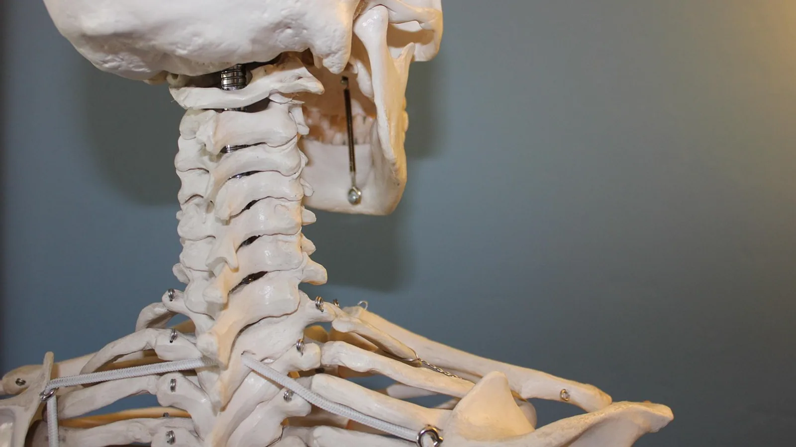
The jugular notch, also called the suprasternal notch or sternum notch, is the palpable midline depression on the superior border of the manubrium of the sternum, sitting between the medial ends of the two clavicles. It marks the junction where the manubrium meets the cervical soft tissues and serves as the surface gateway to the superior mediastinum.
This small depression punches far above its size in clinical importance. It is the entry point for suprasternal ultrasound views of the aortic arch, a reference plane for front-of-neck airway procedures, a surface marker for tracheal position, and a measuring anchor for airway prediction indices used before intubation. In veterinary anatomy, the same region shows striking species differences because the clavicle is reduced or absent in most domestic mammals, which reshapes the entire cranial border of the thorax.
This article is educational and is not a substitute for veterinary diagnosis or treatment.
What the Jugular Notch Is and Where It Sits
The jugular notch is a shallow, roughly U-shaped or V-shaped concavity on the cranial (superior) border of the manubrium sterni. In humans, the manubrium is the uppermost of the three bony sternal segments, and its cranial border is thickened and concave centrally. That concavity is the notch. The medial (sternal) ends of the clavicles articulate with the manubrium on either side of it at the sternoclavicular joints.
The notch is not a foramen, canal, or joint. It is a surface feature, a depression in bone that you can feel through skin and subcutaneous tissue. Its floor is the dorsal (posterior) surface of the manubrium, and immediately behind that thin plate of bone lies the superior mediastinum.
The Suprasternal Space Behind the Notch
Deep to the notch is a small compartment sometimes called the interclavicular suprasternal space. A 2026 computed tomography angiography study of 100 adults classified the great vessels relative to a horizontal plane drawn through the suprasternal notch [1]. The brachiocephalic trunk was the most variable structure, sitting at or above the notch in 64% of cases and fully cervical (high-riding) in 49%. The aortic arch was high-riding in only 3% and the left brachiocephalic vein in 5%. High-riding brachiocephalic trunks were more common in women (63.5% versus 33.3%) [1].
That variability matters. A landmark that feels like a safe, superficial dimple can sit directly in front of a major artery in a large fraction of individuals. This is why suprasternal procedures demand imaging or careful technique rather than blind needle passage.
Levels and Adjacent Structures
The notch corresponds roughly to the level of the second to third thoracic vertebra (T2 to T3) in humans. Structures that lie at or near this level include the aortic arch, the brachiocephalic trunk, the left brachiocephalic vein, the trachea, the esophagus (dorsal to the trachea), and the inferior thyroid veins. The trachea is palpable just cranial to and slightly deep to the notch, which is why the notch is used as a starting point for tracheal palpation.
| Landmark | Approximate level | Adjacent structures | Clinical use |
|---|---|---|---|
| Jugular notch (suprasternal notch) | T2 to T3 | Manubrium, sternoclavicular joints, brachiocephalic trunk, left brachiocephalic vein, aortic arch, trachea | Tracheal palpation, central venous access reference, mediastinoscopy entry, suprasternal ultrasound window |
| Sternal angle (angle of Louis) | T4 to T5 | Manubriosternal joint, aortic arch, tracheal bifurcation | Rib counting, auscultation reference |
| Thyroid notch | Cervical, above sternum | Thyroid cartilage, vocal folds | Airway assessment, endotracheal tube depth estimation |
| Xiphoid process | T9 to T10 | Diaphragm, liver, pericardium | Caudal boundary of the heart, CPR hand placement |
How the Notch Forms and What It Tells You
The sternum develops from paired sternal bars of mesenchyme that migrate ventromedially and fuse in the midline. In humans and many mammals, the sternum ossifies from a series of segmental centers called sternebrae. The manubrium is the most cranial sternebra. The jugular notch forms at the cranial end of the manubrium where the two sternal bars fail to completely close the cranial border, leaving a concave edge.
Because the notch is a developmental remnant of midline fusion, its shape varies. A deep notch can reflect altered mechanical forces on the anterior chest wall. A 2025 study of idiopathic pleuroparenchymal fibroelastosis (iPPFE), a chronic interstitial lung disease with upper-lobe elastofibrosis, found that notch depth correlated anatomically with pre-tracheal soft tissue thickness and with posterior or rightward deviation of the trachea [2]. The authors reported that notch depth discriminated iPPFE from idiopathic pulmonary fibrosis with 75% sensitivity and 86.4% specificity at a cutoff of 9.5 mm, and that patients with a deep notch had significantly shorter survival [2]. This is a human disease finding, but it demonstrates a general principle: surface landmarks of the sternum can encode information about deeper thoracic pathology.
Step-by-Step Palpation
- Position the subject supine with the neck slightly extended.
- Identify the thyroid cartilage (Adam's apple) and slide your finger caudally along the midline.
- The first bony resistance you meet is the cranial border of the manubrium.
- The central depression in that border is the jugular notch.
- Move laterally to confirm the sternoclavicular joints on each side.
- Just cranial to the notch, palpate the trachea as a cartilaginous, slightly mobile tube.
In animals without clavicles, step 5 changes. You will not find a sternoclavicular joint because there is no clavicle to articulate. You will instead feel the cranial end of the manubrium blending into the pectoral musculature and the first pair of sternebrae or costal cartilages.
Comparative Sternum Anatomy Across Species
The jugular notch as a distinct, palpable depression is essentially a human feature because it depends on a well-developed clavicle and a broad, flat manubrium. Domestic mammals diverge in several ways.
Dogs and Cats
Dogs and cats have a sternum built from a series of sternebrae, typically eight in the dog, arranged in a segmented chain from the manubrium cranially to the xiphoid process caudally. The manubrium in the dog is more elongated and projects further cranially than in humans, forming a prominent bony prow at the front of the chest. The clavicle is vestigial or absent. In the cat, a small, free-floating clavicular remnant may exist in the musculature, but it does not articulate with the sternum or form a sternoclavicular joint.
Because there is no clavicle and the manubrium is elongated, the cranial border of the sternum does not present a clean transverse notch between two clavicles. Instead, the manubrium tapers to a point or a rounded edge. The "notch" region is replaced by the manubrial apex and the adjacent costal cartilages of the first rib.
Horses
The equine sternum is compressed laterally, meaning it is narrow from side to side and deeper dorsoventrally than the broad, flat human sternum. The manubrium is elongated and projects cranially, sometimes called the sternal spur or manubrial cartilage. The clavicle is absent. The cranial border of the sternum in the horse is not a wide transverse plate with a central depression. It is a narrow, keel-like structure. Palpation of a jugular notch in the horse is not a standard landmark because the geometry does not produce one.
Ruminants
Goats and other ruminants show breed and age variation in sternal segmentation. A radiographic study of West African dwarf and Danish Landrace goats found that young Danish Landrace kids had six or seven sternebrae, with the fifth or penultimate sternebra doubled in some animals due to bilateral ossification centers [3]. West African dwarf goats had five, six, or seven sternal elements at birth, and those with fewer segments showed bilateral indentations interpreted as prenatal cranial coalescence of the last segments [3]. Both breeds had eight sternal and five asternal ribs [3]. This variability shows that even within a species, the number and shape of sternal segments is not fixed.
Species Without Clavicles
The jugular notch is absent or modified in species without clavicles. The clavicle provides the lateral bony pillars between which the notch is defined. Remove the clavicles and the manubrium becomes the sole cranial boundary of the thoracic inlet. The depression may persist as a subtle concavity on the manubrial border, but it loses the paired articular landmarks that make it clinically useful in humans.
| Species | Clavicle | Manubrium shape | Jugular notch |
|---|---|---|---|
| Human | Complete, articulates with manubrium | Broad, flat | Distinct, palpable between clavicles |
| Dog | Vestigial or absent | Elongated, projects cranially | Absent or modified |
| Cat | Small free remnant, non-articulating | Elongated | Absent or modified |
| Horse | Absent | Laterally compressed, keel-like | Absent |
| Goat | Absent | Segmented sternebrae, variable count | Absent |
Clinical and Procedural Relevance
The suprasternal notch is a working landmark in several clinical contexts. Most published evidence comes from human medicine, but the anatomy translates directly to veterinary patients with appropriate species adjustments.
Airway Confirmation
After endotracheal intubation, confirming tube placement is critical. A 2023 diagnostic study compared color Doppler epigastric ultrasound and linear probe suprasternal notch ultrasound against standard capnography in 104 patients [4]. Suprasternal notch ultrasound had 98.98% sensitivity and 66.67% specificity, while epigastric ultrasound had 97.96% sensitivity and 100% specificity. The combination of both methods achieved 96.94% sensitivity and 100% specificity [4]. Mean time to confirmation was 5.08 seconds for suprasternal notch ultrasound, 10.38 seconds for epigastric ultrasound, and 17.95 seconds for capnography [4]. The suprasternal view is fast because the trachea lies immediately deep to the notch, and an air-filled tube produces a characteristic artifact.
Difficult Airway Prediction
The acromio-axillo-suprasternal notch index (AASI) uses the notch as one geometric anchor. The index is calculated by drawing a vertical line from the acromion to the superior border of the axilla (line A), then a perpendicular line from the suprasternal notch to line A (line B). The portion of line A above the intersection is line C. AASI equals C divided by A [5].
Multiple studies have evaluated AASI for predicting difficult laryngoscopy. A 2020 study of 108 emergency patients reported sensitivity of 84.6%, specificity of 77.7%, and overall accuracy of 81.13% at a cutoff of 0.515 [6]. A 2020 study of 240 patients found AASI sensitivity of 93.94%, specificity of 97.58%, and diagnostic accuracy of 97.08% [7]. A 2020 comparison of 150 patients reported AASI specificity of 93.2%, positive predictive value of 55%, and accuracy of 89.3% [8]. A 2020 observational study of 200 patients found AASI 92% sensitive and 97.71% specific, with an area under the curve of 0.97 [9]. A 2026 study comparing AASI with combined Mallampati score and thyromental distance in 142 patients found AASI sensitivity of 84%, specificity of 88.9%, and accuracy of 88% [10]. A pediatric case series of 20 patients aged 1 to 6 years found AASI positively correlated with Cormack-Lehane grade [11].
The consistent finding is that AASI performs well as a screening test, particularly in patients who cannot cooperate with conventional airway exams. Its weakness is that it requires a tape measure and a few seconds of patient positioning, which is why some studies note longer administration time compared with simpler tests [8].
Central Venous Access and Front-of-Neck Procedures
The notch is a conventional surface landmark for front-of-neck airway access and anterior cervical surgery [1]. In central venous catheterization, the notch helps estimate the level of the brachiocephalic vein and superior vena cava. Because the brachiocephalic trunk can ride at or above the notch in a majority of adults [1], any needle directed deeply at this level risks arterial puncture. Ultrasound guidance is standard for this reason.
Suprasternal Notch Ultrasound Views
The suprasternal notch view (SSNV) is an additional echocardiographic window that visualizes the aortic arch and ascending aorta. A 2016 pilot study of 79 patients found the SSNV was obtainable in 97% of cases, rated easy in 64.5%, and very difficult in only 7.6% [12]. Mean differences between ultrasound and CT angiography measurements were 1.2 mm at the sinus of Valsalva, 1.0 mm at the sinotubular junction, and 0.8 mm at the ascending aorta [12].
The SSNV has diagnostic value in aortic dissection. A 2020 case report described a patient with ascending aortic dissection detected via emergency echocardiography using the suprasternal notch view after a standard transthoracic exam was normal [13]. A 2019 case report described type B aortic dissection diagnosed on the same view [14]. The view allows visualization of structures that standard parasternal windows cannot reach.
Mediastinal Evaluation
Suprasternal notch sonography can assess mediastinal lymphadenopathy. A 2014 pediatric radiology report described point-of-care sonography through the notch for tuberculous mediastinal lymphadenopathy in children [15]. The notch provides a narrow acoustic window into the superior mediastinum without ionizing radiation.
Sternal Pathology
The notch can be a site of rare pathology. A 2025 case report described primary tubercular osteomyelitis of the sternum presenting as a chronic ulcer over the suprasternal notch [16]. Sternal tuberculosis is rare, and the presentation was indolent with localized swelling and pain. Magnetic resonance imaging delineated the extent of bone and soft tissue involvement, and diagnosis was confirmed by cytopathological and microbiological examination [16]. A second branchial cleft cyst has also been reported in the suprasternal notch region, an unusual location for a congenital anomaly that typically appears along the anterior border of the sternocleidomastoid muscle [17].
Wearable Sensing
A 2019 study in Nature Biomedical Engineering described a soft wireless device placed conformally at the suprasternal notch that captures mechano-acoustic signals from the underlying trachea and great vessels [18]. The device measured accelerations from about 10⁻³ m/s² to about 10 m/s² at frequencies up to 800 Hz. Using frequency-domain analysis and machine learning, it extracted heart rate, respiration rate, energy intensity, talking time, swallow counts, and other biomarkers from human subjects during daily activities [18]. The notch is an ideal site because it sits directly over the trachea and major vessels, making it a natural acoustic window.
Clinical Relevance, Limitations and Common Mistakes
The jugular notch is a reliable surface landmark in humans and a useful conceptual landmark in veterinary anatomy, but it has limits.
First, the notch is not a fixed point. Its depth and shape vary with body condition, age, and disease. The iPPFE study showed that notch depth can change with underlying lung pathology [2]. In veterinary patients, body fat, pectoral muscle mass, and sternal conformation all affect palpability.
Second, the great vessels behind the notch are variable. The brachiocephalic trunk reaches or crosses the notch in 64% of adults [1]. Blind needle procedures at this level are unsafe.
Third, species differences are not cosmetic. A dog or cat does not have a jugular notch in the human sense because the clavicle is absent and the manubrium is elongated. A horse has a laterally compressed sternum with a keel-like manubrium. Applying human surface anatomy directly to these species leads to errors in landmark identification.
Fourth, the AASI and similar indices are screening tools, not diagnostic tests. They estimate risk and guide preparation. They do not replace direct laryngoscopy findings or clinical judgment.
Fifth, the suprasternal notch view in ultrasound is operator-dependent. The 2016 study found it was very difficult in 7.6% of cases [12]. Training and practice matter.
Common mistakes students make include confusing the jugular notch with the sternal angle (angle of Louis), which is lower at T4 to T5 and marks the manubriosternal joint. Another mistake is assuming the notch is the same as the thoracic inlet. The thoracic inlet is the bony ring formed by the first ribs, the manubrium, and the first thoracic vertebra. The notch is a surface feature on the manubrium, not the entire inlet. A third mistake is palpating too deeply and mistaking the tracheal rings for a mass or the brachiocephalic artery for a thyroid nodule.
Individual patients vary, and clinical decisions require a veterinarian who can examine the whole animal.
Quick Review
- The jugular notch is the central depression on the superior border of the manubrium between the clavicles.
- It sits at approximately T2 to T3 and overlies the brachiocephalic trunk, left brachiocephalic vein, aortic arch, and trachea.
- The brachiocephalic trunk is at or above the notch in 64% of adults, so deep blind needle placement is risky [1].
- Dogs and cats have sternebrae and an elongated manubrium with no functional clavicle, so the notch is absent or modified.
- Horses have a laterally compressed sternum and no clavicle.
- The notch is a key landmark for tracheal palpation, central venous access, mediastinoscopy entry, and suprasternal ultrasound views.
- AASI uses the notch as a geometric anchor and performs well as a difficult airway screening test, but it is not a diagnosis.
Frequently Asked Questions
What is the jugular notch?
The jugular notch is the palpable depression on the superior border of the manubrium of the sternum, located between the medial ends of the clavicles. It is also called the suprasternal notch or sternum notch.
Can you feel the jugular notch in dogs and cats?
No, dogs and cats do not have a jugular notch in the human sense. Their manubrium is elongated and their clavicle is vestigial or absent, so the cranial border of the sternum does not form a depression between two clavicles.
Why is the jugular notch important in emergency medicine?
It is a fast surface landmark for confirming endotracheal tube placement with ultrasound, for estimating central venous anatomy, and for obtaining suprasternal echocardiographic views of the aortic arch. Suprasternal notch ultrasound confirmed tube placement in a mean of 5.08 seconds in one study [4].
What is the acromio-axillo-suprasternal notch index?
AASI is a screening measurement that uses the suprasternal notch as one geometric point to predict difficult laryngoscopy. It is calculated from the distance between the acromion and axilla relative to a perpendicular line from the notch [5].
Is the jugular notch the same as the sternal angle?
No. The jugular notch is the cranial depression on the manubrium at about T2 to T3. The sternal angle is the manubriosternal joint at about T4 to T5, where the second rib attaches.
What structures lie behind the jugular notch?
The brachiocephalic trunk, left brachiocephalic vein, aortic arch, trachea, and esophagus lie deep to the notch. The brachiocephalic trunk is the most variable and can sit above the notch in a majority of adults [1].
Related Articles
- Bovine Heart Anatomy and Auscultation Landmarks
- Notch Signaling in Reproduction: Mechanisms and Roles
- Notch Signaling in Neurogenesis: Mechanisms and Roles
- VEGF Notch Signaling: Mechanisms and Roles in Angiogenesis
- Notch Signaling in Cancer: Mechanisms and Therapeutic Implications
- Notch Delta Signaling: Mechanisms, Methods, and Misconceptions
- Sternum Bones: Anatomy, Parts and Comparative Notes
- Jugular Foramen: Anatomy, Contents, and Clinical Notes
Sources
- Major Vessels of the Interclavicular Suprasternal Space: A Suprasternal Notch-Referenced Topographic Classification with Potential Procedural Relevance.
- Diagnostic and prognostic implications of a deep suprasternal notch in idiopathic pleuroparenchymal fibroelastosis.
- Comparative structure and morphology of the goat sternum in West African dwarf and Danish Landrace breeds.
- Diagnostic Value of Epigastric Ultrasound and Suprasternal Notch Ultrasound in Comparison with Standard Capnography in Confirmation of Endotracheal Tube Placement after Intubation
- Acromio-Axillo-Suprasternal Notch Index: A New Screening Test to Predict Difficult Laryngoscopy in General Population
- How to Predict Difficult Tracheal Intubation: The Application of Acromio-axillo-suprasternal Notch Index
- Comparison of acromio-axillo-suprasternal notch index with upper lip bite test and ratio of height to thyromental distance for prediction of difficult intubation: a prospective study
- A comparative study of acromio-axillo-suprasternal notch index with upper lip bite test and modified Mallampati score to predict difficult laryngoscopy
- Is Acromio-Axillo-Suprasternal Notch Index a Reliable Test to Predict Difficult Tracheal Intubation in Adults in Supine Position - an Observational Study
- Comparison of Acromio-Axillo-Suprasternal Notch Index with Combination of Modified Mallampati Score and Thyromental Distance in Predicting Difficult Visualization of Larynx
- Acromioaxillo suprasternal notch index as new screening test to predict difficult laryngoscopy in pediatric population: A case series
- EVALUATION OF THE AORTIC ARCH FROM THE SUPRASTERNAL NOTCH VIEW USING FOCUSED CARDIAC ULTRASOUND
- Ascending Thoracic Aortic Dissection: A Case Report of Rapid Detection Via Emergency Echocardiography with Suprasternal Notch Views
- Point-of-care Ultrasound Diagnosis of Type B Aortic Dissection on the Suprasternal Notch View
- Through the eye of the suprasternal notch: point-of-care sonography for tuberculous mediastinal lymphadenopathy in children
- Primary Tubercular Osteomyelitis of Sternum Presenting as a Chronic Ulcer over Suprasternal Notch: A Rare Case Report
- Unusual location of a second branchial cleft cyst presenting in the suprasternal notch
- Mechanoacoustic sensing of physiological processes and body motions via a soft wireless device placed at the suprasternal notch