Sternum Bones: Anatomy, Parts and Comparative Notes
By Dr. Zubair Khalid, DVM, MS, PhD ·
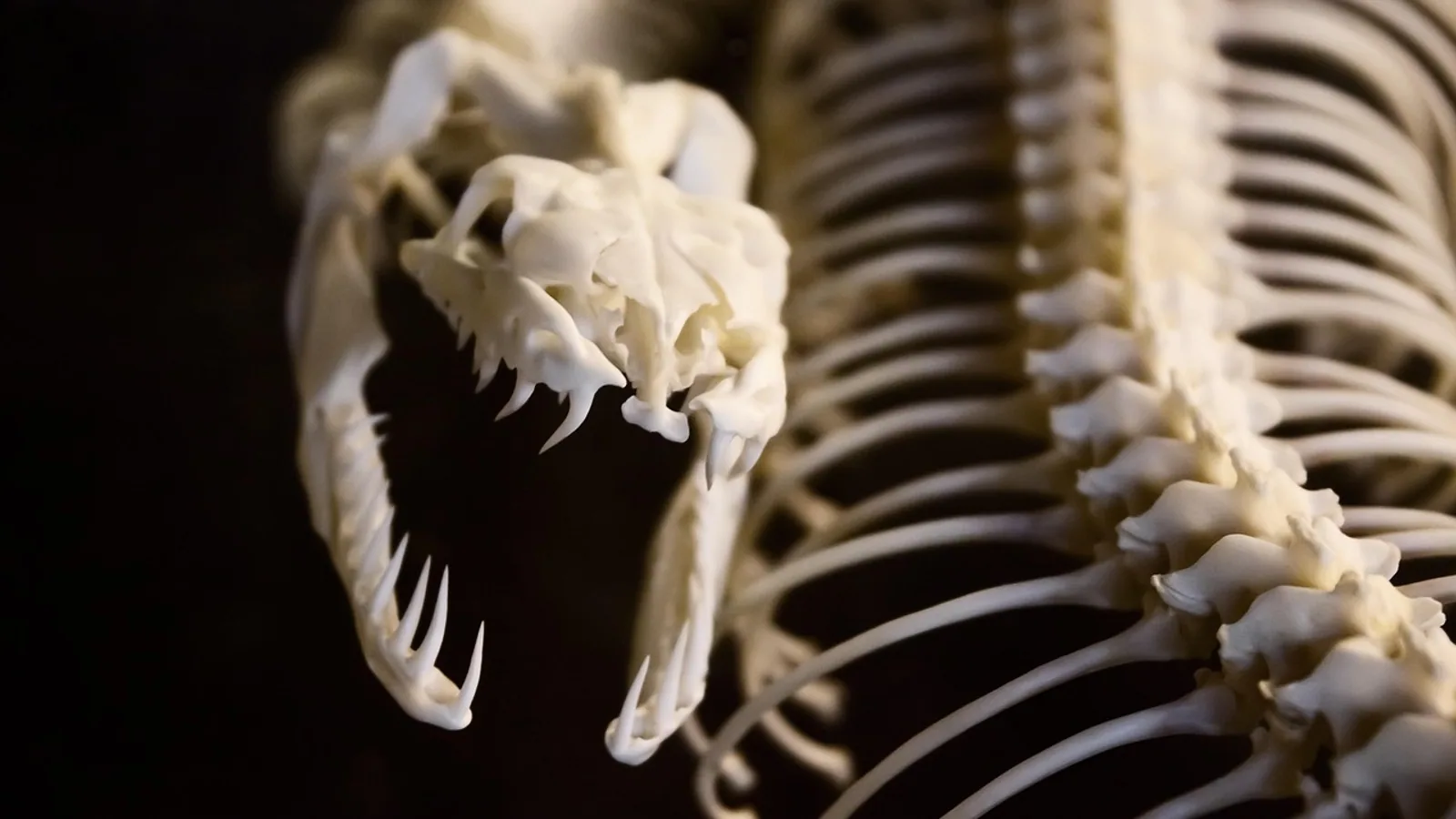
The sternum bones form the unpaired, ventral midline floor of the thorax, built from three regions: the manubrium cranially, the body (mesosternum) in the middle, and the xiphoid process caudally. In domestic mammals the body is assembled from a series of segmental sternebrae that ossify from separate centers and fuse with age, while the sternum as a whole anchors the costal cartilages of the true ribs and gives the heart and lungs a bony shield that still flexes with every breath.
What the Sternum Is and Where It Sits
The sternum, or breast bone, is a flat, segmental bone lying in the ventral thoracic wall between the paired costal cartilages. It closes the thoracic inlet ventrally, meets the clavicle in species that have one, and completes the bony rib cage together with the thoracic vertebrae and ribs. The ribs, sternum, and costal margin form a rigid but flexible chest wall that protects the cardiothoracic organs while allowing respiratory excursion [1].
That flexibility matters. The accessory muscles of inspiration use the downward slope and outward curve of each rib. When they contract, the ribs swing upward and outward, which pushes the sternum cranially and ventrally and increases thoracic volume [1]. The sternum is therefore not a static plate. It is the moving anterior hinge of the chest pump, and its position changes measurably between inspiration and expiration.
The anatomy of the sternum follows a simple rule across mammals: a cranial manubrium, a segmented body, and a caudal xiphoid process. The relative size, shape, and degree of fusion of these three parts differ enormously between species, and those differences carry direct clinical weight for restraint, imaging, cardiopulmonary resuscitation, and bone marrow sampling.
The Three Regions of the Sternum
Manubrium
The manubrium is the most cranial segment. It is typically wider and thicker than the sternebrae behind it, and it carries the costal notch or facet for the first rib. In humans the manubrium is the widest part of the sternum, and sternal measurements taken from it are used in forensic sex estimation, with manubrium width contributing to discriminant functions that reach about 90 percent accuracy when combined with body length and body width [2]. The same principle of a robust cranial segment holds across domestic mammals, though the shape varies. Ruminants have a notably prominent manubrium, and in the horse the manubrium is compressed and elongated, forming the cranial end of a sternum that is shaped like a boat keel.
The manubrium also articulates with the first costal cartilage and, where present, with the clavicle. Comparative dissection of the capuchin monkey showed that the first rib's primordium continues into the sternum without a visible border, and the clavicle connects to the sternal anlage without a clear interzone before the sternoclavicular articulation and disc form [3]. That detail explains why the first sternocostal junction is the least mobile and the least likely to be injured by shear.
Body (Mesosternum)
The body, or mesosternum, is the longest region. It is built from a stack of sternebrae, the segmental units of the sternum. Each sternebra ossifies from its own center, and the number of centers, the order in which they appear, and the age at which they fuse are all used in developmental and forensic work [4]. In humans, sternal development begins in intrauterine life and continues until about age 25, and the sternal body shows the most pronounced age-related ossification of any region [4]. The manubrium and body reach their optimal ossification cut-off ages at 4 and 15 years in females and 5 and 15 years in males [4].
The body carries costal notches along each side for the true ribs. Between the sternebrae, the intersternebral cartilage persists for years and can appear as a lucent band on radiographs. Fusion between adjacent segments is age-dependent, and the S1 to S2 junction shows the strongest age signal of any sternal fusion in humans [4]. In animals the same segmental logic applies, and the number of sternebrae is a standard comparative landmark.
Xiphoid Process
The xiphoid process is the most caudal part. It commonly presents as a small, solid bone shaped like an inverted triangle, but the clinical literature reports wide variation in size, shape, and presentation, largely because it develops from cartilage into fully ossified bone over a long period [5]. The xiphoid may remain partly cartilaginous into adulthood, may be bifid, duplicated, or trifurcated, and may contain foramina [6].
The xiphoid cartilage is the cartilaginous extension of the xiphoid process. It is the most caudal and most mobile part of the sternum, and it is the landmark used for external chest compressions in small animals. Because it is flexible and often incompletely ossified, it is also the sternal region most likely to be misread as abnormal on imaging. Variant xiphoid morphology such as bifid, duplicated, or trifurcated processes may be mistaken for fractures during imaging [6].
Sternebrae: How the Sternum Forms
The sternum does not begin as a single bone. It develops by fusion of paired sternal bars, and those bars arise from the anterior bent parts of the ribs, lying one over another like tiles [3]. The newly formed sternum has different proportions from its definitive state. In human fetuses the manubrium is wider and bigger, the xiphoid process is longer, and the xiphoid is doubled or at least bifurcated in almost all cases [3]. The sternocostal junctions are provided with interzones, with the exception of the first rib, whose primordium continues into the sternum without any border [3].
The medial part of the sternal anlage contains a cartilaginous model that ossifies very early, becoming the first bone of the body [3]. From there, ossification proceeds in a predictable sequence through the remaining sternebrae, and the number of ossification centers in the manubrium and body can be counted on computed tomography [4]. Postnatal maturation continues for years. Multidetector CT studies of 250 patients found significant variation between individuals in ossification centers, manubrial and body shape, xiphoid direction and calcification, and manubriosternal and sternoxiphoidal fusion [7]. Manubrial thickness and sagittal dimensions, body sagittal dimension, and total sternal sagittal dimension all differed significantly between age groups [7].
Two practical consequences follow. First, an incompletely fused sternum in a young animal is normal, not a fracture. Second, the sternum retains developmental variants into adulthood. Sternal foramina, which are developmental defects of the sternum, are usually radiologic or postmortem accidental findings, and they can be misinterpreted as osteolytic lesions on cross-sectional imaging [8]. In a Kenyan dissection series of 80 adult sterna, foramina were present in 11 specimens, or 13.8 percent, with the highest frequency in the sternal body at the fifth intercostal segment, and xiphoid foramina in 2 specimens, or 2.5 percent [6].
Rib Articulations at the Costal Cartilages
The sternum articulates with ribs indirectly, through costal cartilages. Ribs are classified as true, false, or floating based on their anterior attachment. Ribs 1 through 7 are true ribs because their costal cartilage directly articulates with the manubrium and sternum. Ribs 8 through 10 are false ribs because their costal cartilage connects to the cartilage of the immediately superior rib instead of the sternum itself. Ribs 11 and 12 are floating ribs because they have rudimentary cartilage caps and no sternal attachment [1].
That complex cartilaginous structure is the costal margin, which provides attachments for the diaphragm and various abdominal muscles [1]. The costal margin was historically thought to be a consistent structure, but cadaveric studies have defined its variable nature [1]. For the veterinary clinician this means the palpable edge of the chest is not a fixed line. It shifts with body condition, age, and species, and it is a poor substitute for imaging when sternal pathology is suspected.
The sternocostal junctions are also a source of normal imaging artifact. On CT, normal sternal variants include cortical unsharpness along the posterior aspect of the manubrium, along the lateral surfaces of the body, and at the sternal fibrocartilaginous articulations, plus soft tissue prominence at the junction of the sternum and costochondral cartilage and bony sclerosis at the manubrium-to-body and body-to-xiphoid transitions [9]. In a series of 35 patients, part of the posterior cortical margin of the manubrium was unsharp and irregular in 34, and part of the anterior cortical margin was indistinct in 20 [10]. Angling the CT gantry more nearly perpendicular to the manubrium improved cortical definition [10]. These are normal appearances, and recognizing them prevents false calls of sternal disease.
Comparative Anatomy of the Sternum Across Species
The table below summarizes the sternal features that veterinary students are expected to recognize across the major domestic species.
| Species | Manubrium | Body and sternebrae | Xiphoid and cartilage | Ventral profile | Key comparative note |
|---|---|---|---|---|---|
| Dog | Short, with a rounded cranial end | Relatively short sternum with eight sternebrae | Xiphoid cartilage prominent, used as CPR landmark | Slight ventral convexity, no keel | Sternal lymph nodes most often at the level of the second sternebra [11] |
| Cat | Short, similar to dog | Relatively short sternum with eight sternebrae | Xiphoid cartilage thin and flexible | Flat to slightly convex | Same segmental plan as dog, smaller scale |
| Horse | Compressed and elongated cranially | Body long, sternebrae fused early | Xiphoid flattened | Keel-like ventral crest | The ventral crest is the equine equivalent of a low keel and gives the sternum a boat-like profile |
| Ruminant (cattle, sheep, goat) | Prominent and broad | Body long, sternebrae well defined | Xiphoid broad, often with a thick cartilage | Flattened to slightly keeled | The prominent manubrium is a reliable identification feature on the disarticulated skeleton |
| Pig | Short and stout | Body with several sternebrae | Xiphoid robust | Slight keel | Sternum is relatively short relative to the long thorax |
| Bird | Reduced or fused into the body | Body fused into a single carina-bearing plate | Xiphoid reduced | Large carina (keel) | The carina provides the attachment surface for the flight muscles |
The dog and cat pattern is the one most students encounter first. Both have a relatively short sternum with eight sternebrae, which means the sternum spans fewer segments than the thoracic vertebral column. The sternum is a site of hematopoietically active trabecular bone marrow in dogs, alongside the proximal humerus, femur, pelvis, and rib [12]. Bone marrow samples from rib and sternum show higher cellularity than samples from humerus and ilium in dogs, and most differences between sites are attributable to individual animal variation rather than to the site itself [12]. Proliferative marrow activity is distributed across the skeleton in adult dogs, and the sternum is one of the axial sites that contributes to that distribution [13].
The horse pattern is defined by the keel-like ventral crest. The equine sternum is compressed laterally and carries a prominent ventral ridge, which gives it a shape often described as boat-like or keel-like. This crest is not a flight adaptation. It is a surface for muscle attachment and a structural response to the loads carried by the equine thorax. The equine manubrium is elongated and lies at the cranial end of this keel.
The ruminant pattern is defined by a prominent manubrium. In cattle, sheep, and goats the manubrium is broad and robust, and it is one of the easiest sternal features to identify on a disarticulated skeleton. The body is long and the sternebrae are well defined, and the xiphoid carries a broad cartilage.
The bird pattern is the most divergent. Birds have a large carina, the keel of the sternum, which provides the attachment surface for the flight muscles. The avian sternum is a single fused plate in most species rather than a chain of separate sternebrae, and the carina projects ventrally to increase the lever arm and attachment area for the pectoral muscles. This is the same anatomical principle as the equine ventral crest, scaled up for powered flight.
Comparative work in non-domestic species reinforces the segmental rule. The greater cane rat has a sternum consisting of a manubrium, a xiphoid process, and four sternebrae, with 13 thoracic vertebrae and corresponding rib pairs [14]. The black-striped capuchin has a sternum composed of five or six sternebrae, with the ninth pair of ribs as the last sternal pair and the last two pairs buoyant [15]. The number of sternebrae is therefore a species-specific character, and it is worth memorizing for the species you handle most.
Development, Ossification and Imaging
Sternal ossification is a slow, staged process, and its timing is used in both clinical and forensic settings. In humans, development begins in intrauterine life and continues until about age 25, and the sternal body shows the most significant age-related ossification of the three regions [4]. Complete and partial synostosis of the manubrium and body, synostosis of body segments, and synostosis of the body and xiphoid process all occur in a sequence that correlates with age, and the strongest single age correlates include overgrowths on the articular edges of the body and calcification foci in the sternal edges of ribs two through seven [16].
For veterinary imaging, the practical points are these. First, the sternum of a young animal will show multiple separate ossification centers, and these should not be called fractures. Second, the manubriosternal and sternoxiphoidal junctions can show irregular mottled calcifications and indistinct margins that simulate bony lesions [10]. Third, sternal foramina can be misinterpreted as osteolytic lesions on cross-sectional imaging [8]. Fourth, CT is the imaging study of choice when sternal abnormality is suspected, because it resolves the cortical contour and the surrounding soft tissue better than plain radiography [9].
Sternal lymph nodes are a normal finding on canine thoracic CT. In a study of 27 dogs with no abnormality on blood work, urinalysis, or CT, sternal lymph nodes were identified in all but one dog, with a mean of 2.1 nodes per dog and the most frequent location at the level of the second sternebra in 23 dogs, or 85 percent [11]. Node dimensions correlated positively with body weight, and the dorsoventral dimension correlated negatively with age [11]. These reference values matter when staging thoracic disease, because an enlarged sternal node can be the first sign of spread from the thorax or abdomen.
Clinical Relevance, Limitations and Common Mistakes
The sternum is a landmark bone as much as a structural one. In small animal practice, the xiphoid cartilage is the caudal landmark for external chest compressions during cardiopulmonary resuscitation. Compressions are delivered over the heart, which in dogs and cats lies between the third and sixth intercostal spaces, and the xiphoid marks the caudal boundary of that window. Placing hands too far caudal over the xiphoid itself wastes effort on the flexible cartilage and the cranial abdomen rather than the heart.
Sternal recumbency is the other daily use of this anatomy. When a dog or cat is placed in sternal recumbency, the weight of the thorax rests on the sternum and the costal cartilages. The xiphoid cartilage and the manubrium are the two points most likely to contact a hard table surface. Prolonged sternal recumbency on a rigid surface can cause pressure injury over these thin soft tissue areas, which is why padded surfaces are used for anesthetized patients and for animals with limited mobility.
Bone marrow sampling is a third clinical application. The sternum is an axial site of hematopoietically active trabecular bone marrow in dogs, and samples from rib and sternum show higher cellularity than samples from humerus and ilium [12]. Sternal bone marrow aspirates are therefore a reasonable choice when a cellular sample is needed, provided the operator is familiar with the local anatomy and the risk of inadvertent cardiac or great vessel injury from sternal foramina [6].
Common mistakes in sternal anatomy are predictable. Students confuse the manubrium with the xiphoid because both are wider than the intervening sternebrae. Students count the sternal segments on a radiograph and mistake unfused sternebrae for fractures. Students assume the costal margin is a fixed line when it is a variable structure [1]. Students treat the sternum as immobile when it moves cranially and ventrally with every inspiration [1]. Students also assume that a sternal foramen is a lesion rather than a developmental variant [8].
Limitations deserve a direct statement. Sternal anatomy varies between individuals and between breeds, and normal variants overlap with pathologic appearances on imaging [7][9]. The comparative notes here describe typical patterns, not fixed rules for every animal. Any animal with thoracic pain, a palpable sternal swelling, or an abnormal thoracic image needs assessment by a veterinarian. This article is educational and is not a substitute for veterinary diagnosis or treatment.
Frequently Asked Questions
What are the three parts of the sternum?
The three parts are the manubrium cranially, the body or mesosternum in the middle, and the xiphoid process caudally. The body is built from segmental sternebrae that ossify separately and fuse with age.
How many sternebrae do dogs and cats have?
Dogs and cats have a relatively short sternum with eight sternebrae. The number is a standard comparative landmark and is used to identify the species on a disarticulated skeleton.
What is the keel of the sternum in a horse?
The keel is the prominent ventral crest of the equine sternum. It is a ridge on the ventral surface that gives the bone a boat-like profile and serves as a muscle attachment surface.
Why do birds have a large carina?
The carina is the keel of the avian sternum, and it provides the attachment surface for the flight muscles. Its size reflects the mechanical demand of powered flight.
What is the xiphoid cartilage?
The xiphoid cartilage is the cartilaginous extension of the xiphoid process at the caudal end of the sternum. It is flexible, often incompletely ossified, and serves as a CPR landmark in small animals.
How do ribs attach to the sternum?
Ribs attach indirectly through costal cartilages. True ribs have cartilage that articulates directly with the manubrium and sternum, false ribs connect to the cartilage of the rib above, and floating ribs have no sternal attachment.
Can a sternal foramen be mistaken for disease?
Yes. Sternal foramina are developmental defects that can be misinterpreted as osteolytic lesions on cross-sectional imaging. They are usually incidental findings and are not lesions.
Why does the sternum matter in CPR?
The xiphoid cartilage marks the caudal boundary of the cardiac compression window in dogs and cats. Compressions placed too far caudal land on the flexible xiphoid and the cranial abdomen instead of the heart.
Related Articles
- Comparative Anatomy of the Mammalian Kidney
- Comparative Anatomy of the Mammalian Heart
- Ruminant Digestive Anatomy and Physiology: A Clinical Reference
- Equine Hindlimb Anatomy: Bones, Joints, and Ligaments
- Do Frogs Have Bones? Skeletal Anatomy and Pet Care
- Veterinary Anatomy Book Selection: A Comparative Guide for NAVLE
- Testis Anatomy: Structure, Function, and Comparative Notes
Sources
- Anatomy of the Ribs, Sternum, and Costal Margin.
- Sex estimation of the sternum by automatic image processing of multi-slice computed tomography images in a Croatian population sample: a retrospective study.
- Contribution to the study of the development and ossification of human sternum.
- Evaluating sternum development and ossification through computed tomography images: a comparative analysis across age and sex.
- Discovery of an Anteriorly Deviated, Partially Ossified Xiphoid Process With a Large, Teardrop-Shaped Foramen in a Male Cadaver.
- Sternal foramina and variant xiphoid morphology in a Kenyan population.
- Evaluation of the postnatal development of the sternum and sternal variations using multidetector CT.
- Double sternal foramina in a dried sternum: a rare normal variant and its radiologic assessment.
- Computed tomography of the sternum and its articulations.
- Computed tomography of the normal sternum.
- Computed tomography measurements of presumptively normal canine sternal lymph nodes.
- Comparison of the bone marrow composition in samples from rib, ilium, sternum, and humerus of dogs.
- [Relative skeletal distribution of proliferating marrow in the adult dog determined using 3'-deoxy-3'-[(18) F]fluorothymidine.](https://pubmed.ncbi.nlm.nih.gov/30353574/)
- Thoracic and Abdominal Radiologic Anatomy of the Greater Cane Rat (Thryonomys swinderianus).
- Comparative morphofunctional analysis of axial skeleton excluding the skull of primates based on the anatomical, radiographic, and tomographic description of the black-striped capuchin (Sapajus libidinosus Spix, 1823).
- [[Forensic age diagnosis by assessment of sternum ossification processes].](https://pubmed.ncbi.nlm.nih.gov/39189495/)