Ischial Tuberosity: Anatomy and Clinical Notes
By Dr. Zubair Khalid, DVM, MS, PhD ·
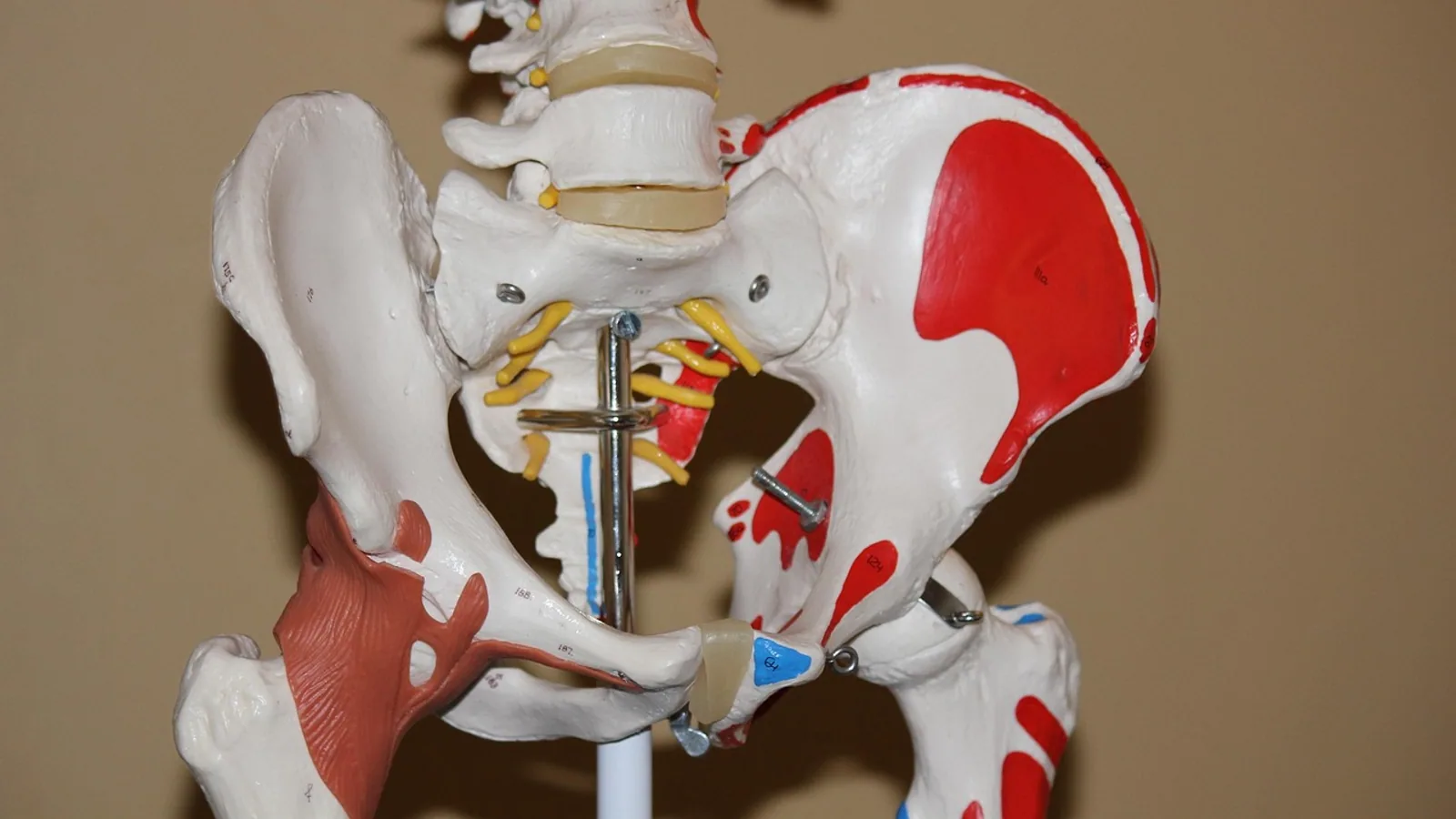
The ischial tuberosity is the thickened, caudally projecting mass of bone on the ischium that serves as the proximal attachment for the hamstring muscle group and the sacrotuberous ligament. In dogs and cats it forms a prominent, easily palpated landmark on the caudolateral pelvis, while in horses it fuses into the pelvic girdle and becomes less distinct, and in ruminants it broadens into a wide, flat surface.
This article covers the osteology, muscle attachments, comparative species anatomy, and clinical significance of the ischial tuberosity for veterinary students and practitioners.
Osteology: The Tuberosity of the Ischium
The pelvis of domestic mammals is built from three paired bones that meet at the acetabulum: the ilium cranially, the pubis ventromedially, and the ischium caudally. The ischium contributes the caudal third of the pelvic floor and the caudal margin of the obturator foramen. Its most conspicuous feature is the ischial tuberosity, a roughened, thickened eminence at the caudolateral corner of the bone.
The tuberosity of the ischium marks the junction between the body of the ischium and its caudal ramus. In the dog, the tuberosity is a distinct, dorsoventrally flattened ridge that projects caudally and slightly laterally. It sits dorsal to the ischial arch, the curved ventral border of the ischium that forms the caudal limit of the pelvic outlet. The two ischial tuberosities flank the ischial arch and, together with the sacrum, define the bony boundaries of the perineum.
The tuberosity is not a single smooth surface. It carries a series of ridges and impressions where tendons and ligaments attach. In humans, the ischial tuberosity is the bony point you sit on, which is why it is sometimes called the "sit bone." In quadrupeds, the weight-bearing mechanics differ, but the tuberosity remains a key palpable landmark for pelvic examination.
Blood Supply and Ossification
The ischial tuberosity receives blood from branches of the internal pudendal artery and the caudal gluteal artery. In young animals, the tuberosity develops as a separate apophysis (a secondary ossification center) that fuses with the main body of the ischium during skeletal maturation. In humans, this apophysis begins to ossify around age 13 to 15 years, and before fusion it is the weakest link in the hamstring attachment chain [1]. The same principle applies in veterinary species: in immature dogs and cats, the apophyseal growth plate at the ischial tuberosity is mechanically weaker than the hamstring tendons, so forceful contraction can avulse the apophysis rather than tear the tendon.
Muscle Attachments at the Ischial Tuberosity
The ischial tuberosity is best known as the proximal origin of the hamstring muscle group. In dogs, cats, and humans, the hamstrings comprise three muscles: biceps femoris, semitendinosus, and semimembranosus. All three arise from or near the ischial tuberosity.
Biceps Femoris
The biceps femoris is a large, broad muscle on the lateral aspect of the thigh. Its long head originates from the ischial tuberosity via a strong tendon. The muscle extends distally to insert on the patella, the tibial crest, and the calcaneus via the crural fascia. In dogs, the biceps femoris is a major extensor of the hip and a flexor of the stifle (knee). The tendon of origin of the long head of the biceps femoris can subluxate across the ischial tuberosity, a rare condition described as "snapping bottom" in humans, which causes pain and swelling in the buttock region [2].
Semitendinosus
The semitendinosus lies caudal to the biceps femoris and is also a hip extensor and stifle flexor. Its proximal origin is consistently divided into two parts: a common part shared with the long head of the biceps femoris and a solitary part that originates directly from the ischial tuberosity [3]. This dual origin is present in both adult and fetal specimens, and the proportions between the two parts are similar across age groups [3]. The semitendinosus inserts on the medial aspect of the tibia, and its myotendinous junctions are common sites of strain injury [3].
Semimembranosus
The semimembranosus is the most medial of the hamstrings. It originates from a superolateral impression on the ischial tuberosity [4]. The muscle is present in 100% of specimens in anatomical studies, though its distal tendon morphology varies. A 2025 classification system identified three main morphological types of the semimembranosus tendon: Type I (66%), a single main tendon with three subtypes based on attachment shape and location; Type II (24%), a double main tendon with two subtypes; and Type III (10%), other configurations [4]. This variability matters for surgical planning and for understanding why some semimembranosus injuries behave differently from others.
Adductor Magnus (Ischiocondylar Portion)
In humans, the ischiocondylar portion of the adductor magnus also arises from the ischial tuberosity. This deep muscle can be a source of ischial pain that mimics hamstring injury. A 2026 case report described a 63-year-old woman with persistent proximal posterior thigh pain after sprinting. Ultrasound revealed a partial tear of the ischiocondylar portion of the adductor magnus at its tendon insertion, with surrounding inflammation and ischial bursal fluid [5]. The lesion was identified by starting with a short-axis view of the ischial tuberosity and then using an oblique long-axis view of the ischiocondylar tendon [5]. This condition is uncommon but should be considered in the differential diagnosis for ischial pain.
Ischiocavernosus Muscle
The ischiocavernosus muscle is a small, paired muscle of the perineum. In rats, it begins at the ischial tuberosity and ends at the crus of the penis, covering the surface of the crus [6]. Unilateral or bilateral amputation of this muscle in male rats affects erectile function and conception, confirming its role in penile erection [6]. While this specific study is in rats, the ischiocavernosus muscle has similar attachments across mammalian species, including dogs and cats.
Sacrotuberous Ligament
The sacrotuberous ligament is a strong band of connective tissue that extends from the sacrum to the ischial tuberosity. In dogs and cats, it helps stabilize the sacroiliac joint and forms part of the boundary of the lesser sciatic foramen. The ligament also provides a broad surface for gluteal muscle attachment. In horses and ruminants, the sacrotuberous ligament is more extensive and blends with the broad sacrosciatic ligament, which converts the lesser sciatic notch into a foramen.
Comparative Anatomy Across Species
The ischial tuberosity varies in prominence and shape across domestic species. These differences reflect each species' posture, locomotion, and pelvic biomechanics.
Dogs
In dogs, the ischial tuberosity is a prominent, palpable ridge on the caudolateral pelvis. It is easily felt through the skin and is a useful landmark for pelvic examination, perineal surgery, and nerve blocks. The tuberosity is separated from the sacrum by the sacrotuberous ligament, and the sciatic nerve passes just cranial and dorsal to it as it exits the pelvis through the greater sciatic foramen. The distance between the ischial tuberosity and the greater trochanter of the femur is a standard morphometric measurement used in anatomical studies [7].
Cats
Cats have a similar pelvic anatomy to dogs, with a prominent and palpable ischial tuberosity. The tuberosity is slightly more pointed in cats and serves as the origin for the same hamstring muscle group. The sacrotuberous ligament is well developed and helps maintain the integrity of the pelvic outlet during parturition and defecation.
Horses
In horses, the ischial tuberosity is fused into the pelvic girdle and is less distinct than in dogs or cats. The equine pelvis is elongated and the ischium is largely incorporated into the broad pelvic floor. The tuberosity is not a separate, palpable projection but rather a smooth continuation of the ischial ramus. The sacrotuberous ligament in horses is extensive and merges with the sacrosciatic ligament, forming a broad sheet that separates the greater and lesser sciatic foramina. This arrangement provides a large surface for the attachment of the gluteal muscles and the biceps femoris.
Ruminants (Cattle, Sheep, Goats)
In ruminants, the ischial tuberosity is broad and flattened. It forms a wide, transverse ridge that is less projecting than in carnivores but more substantial than in horses. The broad tuberosity provides a large attachment area for the hamstring muscles and the sacrotuberous ligament. In cattle, the tuberosity is palpable per rectum and is used as a landmark during obstetrical examinations and epidural anesthesia.
Pigs
Pigs have a relatively short ischium with a modest ischial tuberosity. The pelvic anatomy is adapted for a squatting posture, and the tuberosity is less prominent than in dogs. The sacrotuberous ligament is present but not as extensive as in horses.
Small Mammals (Rats, Mice)
In the southern giant pouched rat, a large rodent used in landmine detection and kept as a pet, the ischial tuberosities are less prominent than in dogs [8]. The pelvis has a relatively short pelvic symphysis, and the obturator foramina and cranial opening of the pelvis are pear-shaped [8]. This reduced prominence reflects the different biomechanical demands of a rodent pelvis.
Table: Species Comparison of the Ischial Tuberosity
| Species | Prominence | Palpable Landmark | Key Muscle Attachments | Sacrotuberous Ligament |
|---|---|---|---|---|
| Dog | Prominent, ridge-like | Yes, caudolateral pelvis | Biceps femoris, semitendinosus, semimembranosus | Well developed |
| Cat | Prominent, pointed | Yes, caudolateral pelvis | Biceps femoris, semitendinosus, semimembranosus | Well developed |
| Horse | Fused, less distinct | Difficult to palpate separately | Biceps femoris, semitendinosus, semimembranosus | Extensive, merged with sacrosciatic ligament |
| Ruminant | Broad, flattened | Yes, per rectum | Biceps femoris, semitendinosus, semimembranosus | Broad, extensive |
| Pig | Modest | Yes, but less prominent | Biceps femoris, semitendinosus, semimembranosus | Present |
| Rat (giant pouched) | Less prominent | Difficult | Hamstring group | Reduced |
Clinical Relevance, Limitations and Common Mistakes
The ischial tuberosity is a clinically important landmark for several reasons. It is the origin of the hamstring muscles, which are among the most commonly injured muscle groups in active animals and humans [9]. Hamstring injuries typically occur during sudden, explosive movements such as sprinting, kicking, jumping, or lunging. The onset of pain is usually acute in the proximal posterior aspect of the upper thigh. Injured individuals may describe a "pop" or "snap" or a sensation as if someone kicked them in the back of the thigh [9].
In veterinary patients, hamstring strain is most common in racing greyhounds, working dogs, and athletic cats. Diagnostic musculoskeletal ultrasound (MSKUS) is a valuable, non-invasive imaging modality for evaluating proximal hamstring injuries, including tendinopathy, muscle strains (partial tears), and ruptures [9]. MSKUS is excellent at detecting changes in tendon and muscle composition and continuity [9]. MRI and ultrasound are key complementary modalities for the diagnosis, treatment, and prognosis of hamstring injuries, as injury length, connective tissue involvement, and tear location determine evolution, recovery strategies, and return to play [10].
The origin tendon structure of the hamstrings is characteristic. The semitendinosus and the long head of the biceps femoris share a conjoint tendon (CT), while the semimembranosus attaches to the ischial tuberosity independently [11]. This anatomical arrangement means that injuries to the conjoint tendon affect both the semitendinosus and the biceps femoris, while semimembranosus injuries are isolated. A classification system focusing on the origin tendon has been developed to standardize the diagnosis of proximal hamstring injuries [11].
Avulsion Fractures
In immature animals, the ischial tuberosity apophysis is the weakest part of the hamstring attachment complex. An avulsion injury of the hamstring muscle group in an adolescent results in a fracture of the apophysis rather than a tendon tear [1]. Ischial tuberosity fractures have a higher rate of non-union than other pelvic apophyseal injuries [1]. In humans, surgical fixation is considered for fractures displaced more than 20 mm or that remain symptomatic despite at least 3 months of conservative management [1]. In veterinary patients, similar principles apply, though the decision to operate depends on the species, the degree of displacement, and the functional demands on the animal.
Avulsion stress fractures can also occur after repetitive training. A 2016 case report described a 19-year-old conscripted policeman who developed an ischial tuberosity avulsion fracture after 5 days of 6-hour running with hip hyperflexion [12]. This case highlights that relatively short periods of intense, repetitive activity can cause stress fractures at the ischial tuberosity, even without a specific traumatic event [12].
Ischiogluteal Bursitis and "Snapping Bottom"
The ischial tuberosity is covered by a bursa, the ischiogluteal bursa, which reduces friction between the bone and the overlying gluteal muscles. Inflammation of this bursa (ischiogluteal bursitis) causes pain in the buttock region, sometimes called "weaver's bottom" in humans. Subluxation of the tendon of origin of the long head of the biceps femoris across the ischial tuberosity can cause a snapping sensation and may be a cause of ischiogluteal bursitis [2]. This condition, called "snapping bottom," occurs in patients who wear out the tendinous attachment through a lifetime of hyperflexion hip movements, such as bending to reach the floor [2]. If conservative measures fail, simple tenotomy is the treatment of choice in older nonathletes [2].
Sciatic Nerve Relationship
The sciatic nerve exits the pelvis through the greater sciatic foramen, passing just cranial and dorsal to the ischial tuberosity. The relationship between the sciatic nerve and the piriformis muscle is variable. In a study of 80 specimens, anatomical variations of the sciatic nerve (types B, C, and D according to Beaton and Anson's classification) were observed in 19.7% of specimens, with no significant sex-related difference in prevalence [7]. The distance between the posterior superior iliac spine and the ischial tuberosity is statistically greater in male specimens [7]. These anatomical relationships are relevant for surgical approaches to the caudal pelvis and for understanding piriformis muscle syndrome.
Common Mistakes
A common mistake is to assume that all ischial pain is hamstring-related. The ischiocondylar portion of the adductor magnus can also be injured, producing similar symptoms [5]. Another mistake is to overlook the ischial tuberosity as a source of lameness in dogs. Because the hamstrings are hip extensors and stifle flexors, animals with ischial tuberosity pain may show subtle gait abnormalities rather than obvious lameness. Palpation of the tuberosity should be part of every orthopedic examination of the hindlimb.
Pressure ulcers can develop over the ischial tuberosity in recumbent animals, particularly those with spinal cord injuries or prolonged anesthesia. These ulcers can become infected and lead to serious complications such as necrotizing fasciitis [13]. Prevention requires regular repositioning and padding of recumbent patients.
This article is educational and is not a substitute for veterinary diagnosis or treatment.
Frequently Asked Questions
What is the ischial tuberosity?
The ischial tuberosity is the caudally projecting, thickened eminence on the ischium bone of the pelvis. It serves as the origin for the hamstring muscles and the sacrotuberous ligament.
Which muscles attach to the ischial tuberosity?
The hamstring muscles attach to the ischial tuberosity: the biceps femoris, semitendinosus, and semimembranosus. In humans, the ischiocondylar portion of the adductor magnus also attaches there.
Is the ischial tuberosity palpable in dogs and cats?
Yes. In dogs and cats, the ischial tuberosity is a prominent, easily palpable landmark on the caudolateral pelvis. It can be felt through the skin during a physical examination.
Why is the ischial tuberosity less distinct in horses?
In horses, the ischial tuberosity is fused into the pelvic girdle and is not a separate, projecting structure. The equine pelvis is elongated, and the ischium is largely incorporated into the broad pelvic floor.
How does the ischial tuberosity differ in ruminants?
In ruminants such as cattle, sheep, and goats, the ischial tuberosity is broad and flattened. It forms a wide transverse ridge that provides a large attachment area for the hamstring muscles and the sacrotuberous ligament.
What injuries are associated with the ischial tuberosity?
Avulsion fractures, hamstring strains, ischiogluteal bursitis, and snapping bottom syndrome are associated with the ischial tuberosity. Avulsion fractures are more common in immature animals because the apophyseal growth plate is the weakest link.
Can the ischial tuberosity be a source of lameness in dogs?
Yes. Because the hamstrings are hip extensors and stifle flexors, pain at the ischial tuberosity can cause subtle gait abnormalities. Palpation of the tuberosity should be part of every hindlimb orthopedic examination.
What is snapping bottom?
Snapping bottom is subluxation of the tendon of origin of the long head of the biceps femoris across the ischial tuberosity. It causes pain and swelling in the buttock region and may lead to ischiogluteal bursitis.
Related Articles
- Clinical Research Notes Templates
- Veterinary Anatomy Notes: How to Create and Use Them for NAVLE
- The Best Note-Taking Tools for Clinical Research
- Project-Based Note Organization for Clinical Research
- Bovine Hoof Anatomy and Lameness Examination: A Clinical Approach
- Bovine Respiratory System: Anatomy and Clinical Examination
- Ischial Spines: Anatomy, Landmarks, and Clinical Use
- Epiphyseal Plate: Growth, Anatomy and Clinical Notes
Sources
- Endoscopic Repair of Ischial Tuberosity Avulsion Fracture.
- "Snapping bottom": subluxation of the tendon of the long head of the biceps femoris muscle.
- The complex architecture of the semitendinosus muscle belly: insights from adult and fetal anatomy with clinical implications.
- The semimembranosus muscle: A novel classification system of morphology and attachments with implications.
- Ultrasonographic Diagnosis of an Ischiocondylar Adductor Magnus Partial Tear: An Uncommon Cause of Ischial Pain.
- Effects of unilateral/bilateral amputation of the ischiocavernosus muscle in male rats on erectile function and conception.
- Anatomical variations in the relationship between the sciatic nerve and the piriformis muscle: clinical implications.
- Radiological Anatomy of the Pelvis and Hind Limb of the Southern Giant Pouched Rat (Cricetomys ansorgei).
- Diagnostic Musculoskeletal Ultrasound in the Evaluation of the Proximal Hamstrings at the Ischial Tuberosity.
- MRI and US in Hamstring Sports Injury Assessment: Anatomy, Imaging Findings, and Mechanisms of Injury.
- Ultrasound Diagnosis of Hamstring Muscle Complex Injuries Focus on Originate Tendon Structure-Male University Rugby Players.
- Ischial Tuberosity Avulsion Stress Fracture after Short Period of Repetitive Training.
- A Rare Case of Ischial Tubercle Pressure Sore with Secondary Periperineal Necrotizing Fasciitis.