Ischial Spines: Anatomy, Landmarks, and Clinical Use
By Dr. Zubair Khalid, DVM, MS, PhD ·
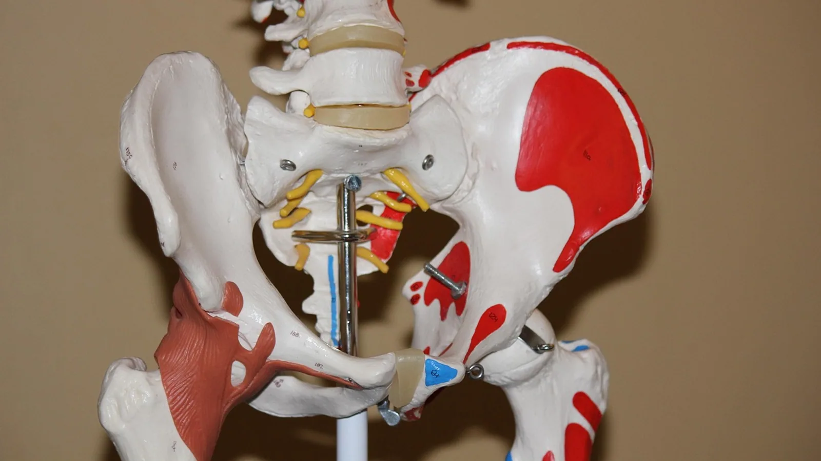
The ischial spine (spine of the ischium) is a small, pointed bony projection on the dorsal and lateral surface of the ischium, positioned between the greater sciatic notch and the lesser sciatic notch. It serves as the attachment site for the sacrotuberous and sacrospinous ligaments and acts as a fixed landmark that divides the two sciatic notches and helps define the greater and lesser sciatic foramina.
This article is educational and is not a substitute for veterinary diagnosis or treatment.
Why the Ischial Spine Matters
The ischial spine is one of the most useful bony landmarks in the pelvis because it sits at a crossroads of soft tissue attachments, nerve pathways, and surgical approaches. In human medicine, it is a standard reference point for pelvic imaging, orthopedic surgery, and obstetric assessment. In veterinary medicine, the same structure exists but with important species differences in prominence and clinical utility.
For veterinary students and practitioners, understanding the ischial spine means understanding:
- How the sacrotuberous and sacrospinous ligaments stabilize the pelvis.
- Where the sciatic nerve exits the pelvis relative to bony landmarks.
- How pelvic shape varies across species and why that variation affects clinical decision-making.
- Why obstetric relevance in dogs, cats, and horses differs fundamentally from human obstetrics.
Anatomy and Osteology
Location on the Ischium
The ischium is one of the three bones that form the os coxae (hip bone), along with the ilium and the pubis. The ischium contributes to the caudal portion of the acetabulum and forms the caudal body of the pelvis. Its major features include:
- Ischial body: the main plate of bone.
- Ischial tuberosity: the caudal projection that supports the animal in sitting.
- Ischial arch: the curved caudal border between the two ischial tuberosities.
- Ischial spine: a dorsal projection on the lateral surface, cranial to the ischial tuberosity.
The spine of the ischium sits on the dorsal ischial margin. It separates the greater sciatic notch (cranial) from the lesser sciatic notch (caudal). When the sacrotuberous and sacrospinous ligaments bridge these notches, they convert them into the greater sciatic foramen and the lesser sciatic foramen, respectively.
Ligament Attachments
Two major ligaments attach to or pass over the ischial spine:
- Sacrotuberous ligament: extends from the sacrum and dorsal ilium to the ischial tuberosity. It forms the caudal boundary of the greater sciatic foramen and the dorsal boundary of the lesser sciatic foramen.
- Sacrospinous ligament: extends from the sacrum to the ischial spine. It forms the ventral boundary of the greater sciatic foramen and separates the greater from the lesser sciatic foramen.
These ligaments are not just passive tethers. They resist rotation of the sacrum under load and help maintain pelvic stability during weight-bearing and locomotion. In humans, the sacrospinous ligament is a key structure in pelvic organ prolapse surgery. Giraudet et al. (2023) emphasized that surgeons should stay approximately 2 cm away from the ischial spine during sacrospinous fixation to avoid nerve and vascular injury, because the coccygeus and levator ani nerves can course along the medial portion of the ligament [1].
Foramina and Neurovascular Relationships
The greater sciatic foramen transmits:
- The sciatic nerve.
- The cranial and caudal gluteal nerves and vessels.
- The internal pudendal vessels and pudendal nerve (which re-enter the pelvis through the lesser sciatic foramen).
- The piriformis muscle.
The lesser sciatic foramen transmits:
- The tendon of the obturator internus muscle.
- The internal pudendal vessels and pudendal nerve.
The ischial spine sits between these two foramina. Its position makes it a reliable intraoperative landmark for identifying the sciatic nerve and avoiding iatrogenic injury.
Comparative Pelvic Anatomy
Humans
In humans, the ischial spine is a well-developed, palpable landmark. It projects medially into the pelvic cavity and is easily identified on computed tomography (CT) and magnetic resonance imaging (MRI). The human pelvis is short and wide, adapted for bipedal locomotion and childbirth. The ischial spines are prominent and are used as reference points for measuring the interischial spine distance, which correlates with pelvic capacity. Ichikawa et al. (2022) found that a shorter interischial spine distance (mean 93.3 mm in their surgical cohort) correlated with longer operative times during laparoscopic rectal resection, demonstrating how this measurement reflects pelvic narrowness [2].
In human fetal development, sexual dimorphism of the pelvis is already apparent at the onset of primary ossification. Kanahashi et al. (2024) showed that female fetuses have a larger distance between the ischial spines relative to the transverse diameter of the greater pelvis compared with males, suggesting that pelvic adaptations for childbirth begin before birth [3].
Dogs
The canine pelvis is more elongated and narrower than the human pelvis. The ischial spine in dogs is less prominent and is not palpable per rectum as a distinct obstetric landmark. The overall pelvic canal is relatively straight and cylindrical, which is why canine dystocia is more often caused by fetal size or fetal malposition than by pelvic inlet narrowing.
The sacrotuberous ligament in dogs is well developed and helps stabilize the sacroiliac joint. The sciatic nerve exits the greater sciatic foramen caudal to the ischial spine, and its relationship to the spine is relevant in cases of deep gluteal pain or sciatic nerve compression. However, the ischial spine is not a standard landmark for canine obstetric assessment.
Cats
The feline pelvis is similar to the dog but even more slender. The ischial spine is small and often difficult to identify on plain radiographs. The pelvic canal is narrow, and cats are at risk for pelvic canal stenosis after pelvic fractures. In these cases, the ischial spine may be displaced or obscured by callus formation, making it an unreliable landmark.
Horses
The equine pelvis is large and heavily muscled. The ischial spine is present but is not a prominent feature on the dorsal ischium. The sacrotuberous ligament in horses is broad and strong, and the ischial spine contributes to its attachment. The equine pelvic canal is long and curved, and obstetric assessment relies on external landmarks (such as the tuber coxae and tuber ischii) rather than on the ischial spine. Dystocia in mares is typically managed by manual correction or cesarean section, and the ischial spine does not serve as a useful obstetric reference point.
Comparative Summary Table
| Feature | Human | Dog | Cat | Horse |
|---|---|---|---|---|
| Prominence of ischial spine | Prominent, palpable | Moderate, not palpable per rectum | Small, often indistinct | Present but not prominent |
| Sacrotuberous ligament attachment | Yes | Yes | Yes | Yes |
| Sacrospinous ligament attachment | Yes | Yes (variably developed) | Yes (variably developed) | Yes |
| Separates greater and lesser sciatic notches | Yes | Yes | Yes | Yes |
| Obstetric landmark | Yes (interischial distance, station) | No | No | No |
| Clinical relevance | Pelvic imaging, surgery, obstetrics | Sciatic nerve localization, pelvic fracture assessment | Pelvic fracture assessment | Pelvic fracture assessment, ligament anatomy |
Development and Ossification
The ischium develops from endochondral ossification. In dogs and cats, the ossification center for the ischium appears late in gestation and fuses with the ilium and pubis at the acetabulum. In horses, ossification follows a similar pattern but on a larger scale. The ischial spine forms as a secondary projection from the dorsal ischial surface and is typically fully formed by the time of skeletal maturity.
In humans, the ischial spine is cartilaginous at birth and ossifies during childhood. By adolescence, it is a distinct bony projection. The timing of ossification is relevant in pediatric radiology, where an unfused ischial spine may be mistaken for a fracture.
Clinical Use and Landmarks
Radiographic Landmarks
In human medicine, the ischial spine sign is a radiographic finding on anteroposterior pelvis radiographs. It refers to the medial projection of the ischial spine into the pelvic inlet. Jackson et al. (2016) noted that pelvic tilt affects the presence of the ischial spine sign and the crossover sign, which are used in the evaluation of pincer femoroacetabular impingement [4]. Brockmeyer et al. (2019) evaluated positioning devices for pelvis radiographs and found that the ischial spine sign was one of the categorical measurements used to assess acetabular orientation, though positioning accuracy varied [5].
In veterinary radiology, the ischial spine is less commonly used as a standalone landmark. However, it can be identified on ventrodorsal and lateral pelvic radiographs and may help orient the clinician when assessing pelvic fractures or hip dysplasia.
Surgical Landmarks
The ischial spine is a key intraoperative landmark in several human surgical procedures:
- Periacetabular osteotomy (PAO): The ischial osteotomy line is defined by the inferior border of the posterior obturator tubercle and the highest point of the ischial spine. Bilgili et al. (2013) described this relationship in cadaveric dissections, noting that the ischial osteotomy must be performed with awareness of the pudendal neurovascular structures [6].
- Ischial screw placement in total hip arthroplasty: Bellas et al. (2024) established a safe zone for ischial screw placement with reference to the anterior superior iliac spine and acetabular landmarks, though the ischial spine itself was not the primary reference [7].
- Sacrospinous ligament fixation: Giraudet et al. (2023) recommended staying approximately 2 cm from the ischial spine during suture placement to avoid nerve and vascular injury [1].
- Radical hysterectomy: Kanao et al. (2026) demonstrated the ischial spine as one of the pivotal pelvic anatomical structures exposed during laparoscopic radical hysterectomy, along with the sacrospinous ligament and pelvic nerves [8].
In veterinary surgery, the ischial spine is less commonly used as a named landmark, but it remains relevant in:
- Pelvic fracture repair: Identifying the ischial spine helps orient the surgeon to the dorsal pelvic canal.
- Sciatic nerve exploration: The ischial spine marks the caudal boundary of the greater sciatic foramen, where the sciatic nerve exits.
- Perineal surgery: The ischial spine is near the origin of the internal obturator muscle, which is relevant in perineal hernia repair.
Neuroanatomical Relationships
The relationship between the sciatic nerve and the ischial spine is clinically significant. Hanna et al. (2024) analyzed MRI scans and found that patients with deep gluteal syndrome who underwent piriformis release had sciatic nerves that were significantly closer to the ischial spine than controls. The sciatic nerve was classified as zone A (medial to the spine), zone B (on the spine), or zone C (lateral to the spine). Patients in the surgical group were more likely to have the nerve located in zone B, suggesting that bone-neural conflict at the ischial spine may contribute to deep gluteal pain [9].
Tempski et al. (2025) performed a meta-analysis of the inferior gluteal nerve and found that in the mid-gluteal region, the nerve was on average 22.6 mm from the ischial spine. This distance is relevant for surgeons performing hip surgeries or gluteal procedures, as iatrogenic injury to the inferior gluteal nerve can cause postoperative pain or weakness [10].
In veterinary medicine, similar relationships exist. The sciatic nerve in dogs exits the pelvis through the greater sciatic foramen, caudal to the ischial spine. The inferior gluteal nerve and cranial gluteal nerve also exit near this region. Although deep gluteal syndrome is not a recognized diagnosis in dogs, sciatic nerve entrapment or compression can occur after pelvic trauma or surgery, and the ischial spine is a useful reference point for locating the nerve.
Obstetric Relevance
In human obstetrics, the ischial spines are used to assess fetal station (the level of the fetal head relative to the spines) and to estimate pelvic diameter. Arthuis et al. (2016) measured the symphysis-left ischial spine angle and distance on CT scans, finding a median angle of 106 degrees and a median distance of 26.1 mm. These measurements help standardize the interpretation of intrapartum translabial ultrasound for assessing fetal head descent [11].
In dogs, cats, and horses, the ischial spine is not used as an obstetric landmark. The pelvic canal in these species is more elongated, and the spine is less prominent. Canine dystocia is assessed by digital examination of the pelvic canal, fetal size, and fetal position. Feline dystocia is similarly assessed. Equine dystocia is assessed by manual examination and external landmarks. The ischial spine does not serve as a reliable reference point in any of these species for obstetric purposes.
How the Ischial Spine Is Examined in Practice
Palpation
In humans, the ischial spine can be palpated during a pelvic examination. In dogs and cats, it is not palpable per rectum because of the pelvic anatomy and the amount of surrounding soft tissue. In horses, it is not palpable externally.
Imaging
- Radiography: The ischial spine can be identified on ventrodorsal and lateral pelvic radiographs. In humans, it is used in the ischial spine sign for acetabular retroversion.
- CT: CT provides the most accurate assessment of ischial spine morphology and its relationship to the sciatic nerve and pelvic organs. Ichikawa et al. (2022) used axial CT slices to measure the interischial spine distance [2].
- MRI: MRI is used to assess the relationship between the sciatic nerve and the ischial spine in deep gluteal syndrome [9].
- Ultrasound: Translabial ultrasound in humans uses the ischial spine as a landmark for assessing fetal head descent [11].
Surgical Exposure
The ischial spine can be exposed during several surgical approaches:
- Medial approach to the ischium: Bilgili et al. (2013) described a medial approach between the adductor longus and pectineus muscles for ischial and pubic osteotomies, using the highest point of the ischial spine as a landmark [6].
- Laparoscopic approach: Kanao et al. (2026) demonstrated the ischial spine during laparoscopic radical hysterectomy, exposing it along with the sacrospinous ligament and pelvic nerves [8].
- Vaginal approach: Zoorob et al. (2026) measured the distance from a vNOTES retractor to the ischial spine, finding a mean sagittal distance of 1.0 cm and a transverse distance of 3.6 cm. These measurements help surgeons orient themselves during apical suspension procedures [12].
Common Misconceptions
Misconception 1: The ischial spine is the same as the ischial tuberosity
The ischial tuberosity is the caudal, weight-bearing projection of the ischium. The ischial spine is a separate, more cranial projection on the dorsal surface. They are distinct landmarks with different ligament attachments and clinical significance.
Misconception 2: The ischial spine is prominent in all species
The ischial spine is prominent in humans but less so in dogs, cats, and horses. In these species, the pelvis is more elongated and the spine is reduced. This affects its clinical utility, particularly in obstetrics.
Misconception 3: The ischial spine is only relevant in human medicine
The ischial spine is a conserved anatomical feature across mammals. It is relevant in veterinary medicine for surgical landmarking, radiographic interpretation, and understanding neurovascular relationships, even though its obstetric role is limited.
Misconception 4: The ischial spine is always easy to identify on radiographs
In small animals, the ischial spine can be obscured by overlying structures or by pelvic pathology. In horses, it is not a prominent feature. In humans, pelvic tilt can affect its appearance on radiographs [4].
Misconception 5: The ischial spine has no clinical relevance in dogs and cats
The ischial spine is a useful landmark for locating the sciatic nerve and for orienting pelvic fracture repair. It also helps define the greater sciatic foramen, which is relevant in cases of nerve entrapment or pelvic trauma.
Clinical Relevance, Limitations and Common Mistakes
The ischial spine is a reliable anatomical landmark when used appropriately. However, there are limitations and common mistakes to avoid:
- Species differences: Do not assume that the ischial spine is as prominent or as clinically useful in dogs, cats, and horses as it is in humans.
- Radiographic interpretation: Pelvic tilt and rotation can alter the appearance of the ischial spine on radiographs. Always assess pelvic positioning before making judgments about bony landmarks.
- Surgical safety: When operating near the ischial spine, be aware of the sciatic nerve, pudendal nerve, and internal pudendal vessels. In humans, staying approximately 2 cm from the ischial spine during sacrospinous fixation reduces the risk of nerve injury [1].
- Obstetric assessment: In veterinary obstetrics, rely on digital examination, fetal assessment, and species-specific landmarks rather than on the ischial spine.
- Imaging modality: CT and MRI provide more accurate assessment of the ischial spine and its relationships than plain radiography.
Individual cases require veterinary assessment. This article provides anatomical and clinical context but does not replace professional diagnosis or treatment.
Quick Review
- The ischial spine is a bony projection on the dorsal ischium between the greater and lesser sciatic notches.
- It is the attachment site for the sacrotuberous and sacrospinous ligaments.
- It separates the greater sciatic foramen from the lesser sciatic foramen.
- In humans, it is a prominent obstetric and surgical landmark. In dogs, cats, and horses, it is less prominent and has limited obstetric relevance.
- The sciatic nerve exits the greater sciatic foramen near the ischial spine. Its proximity to the spine is associated with deep gluteal syndrome in humans [9].
- The interischial spine distance is a measure of pelvic width in humans and correlates with surgical difficulty [2].
- Surgical approaches near the ischial spine must respect the pudendal nerve, sciatic nerve, and internal pudendal vessels.
Frequently Asked Questions
What is the ischial spine?
The ischial spine is a pointed bony projection on the dorsal surface of the ischium, located between the greater sciatic notch and the lesser sciatic notch. It serves as an attachment site for the sacrotuberous and sacrospinous ligaments.
Do dogs have ischial spines?
Yes, dogs have ischial spines, but they are less prominent than in humans. They are not used as obstetric landmarks in dogs because the canine pelvis is more elongated and the spine is not palpable per rectum.
What ligaments attach to the ischial spine?
The sacrotuberous ligament and the sacrospinous ligament attach to or pass over the ischial spine. These ligaments help stabilize the pelvis and define the greater and lesser sciatic foramina.
Why is the ischial spine important in human medicine?
In humans, the ischial spine is used as a landmark for pelvic imaging, orthopedic surgery, and obstetric assessment. It helps define pelvic dimensions and is used to assess fetal station during labor.
Is the ischial spine relevant in horses?
The ischial spine is present in horses but is not a prominent landmark. It contributes to the attachment of the sacrotuberous ligament but is not used for obstetric or surgical landmarking in routine equine practice.
Can the ischial spine be seen on radiographs?
Yes, the ischial spine can be seen on radiographs, but its appearance depends on species, pelvic positioning, and imaging technique. In humans, pelvic tilt can affect its radiographic appearance. In small animals, it may be obscured by overlying structures.
Related Articles
- Bovine Heart Anatomy and Auscultation Landmarks
- Snake Anatomy: Do Snakes Have Teeth, Spines, and Other Structures?
- Veterinary Anatomy Notes: How to Create and Use Them for NAVLE
- Bovine Hoof Anatomy and Lameness Examination: A Clinical Approach
- Bovine Respiratory System: Anatomy and Clinical Examination
- Feline Renal Anatomy and Physiology: A Clinical Correlation
- Ischial Tuberosity: Anatomy and Clinical Notes
- Dorsal Recumbency: Definition and Clinical Use
Sources
- The anatomy of the sacrospinous ligament: how to avoid complications related to the sacrospinous fixation procedure for treatment of pelvic organ prolapse.
- Interischial Spine Distance Is a Simple Index of the Narrow Pelvis That Can Predict Difficulty During Laparoscopic Low Anterior Resection.
- Sexual dimorphism of the human fetal pelvis exists at the onset of primary ossification.
- Supine and Standing AP Pelvis Radiographs in the Evaluation of Pincer Femoroacetabular Impingement.
- "Simple mechanical devices did not improve pelvis positioning in AP pelvis radiographs for reliable assessment of the acetabular orientation".
- Ischial and pubic osteotomies performed by medial approach during periacetabular osteotomies: an anatomical study.
- Determination of a Safe Zone for Ischial Screw Placement in Total Hip Arthroplasty.
- Relationship between the lateral dissection lines of various radical hysterectomies and anatomical structures in the female pelvis: an educational video from the Sapporo Cadaver Surgical Workshop.
- Anatomical Proximity Between Sciatic Nerve and Ischial Spine and its Relationship to the Development of Deep Gluteal Pain Syndrome.
- Clinical anatomy of the inferior gluteal nerve - systematic review and meta-analysis.
- Computed tomographic study of anatomical relationship between pubic symphysis and ischial spines to improve interpretation of intrapartum translabial ultrasound.
- Spatial Mapping of Pelvic Anatomy Focused on Vaginal Natural Orifice Transluminal Endoscopic Surgery (vNOTES) Apical Suspension Prolapse Procedures.