Greater Trochanter: Anatomy, Attachments, and Role
By Dr. Zubair Khalid, DVM, MS, PhD ·
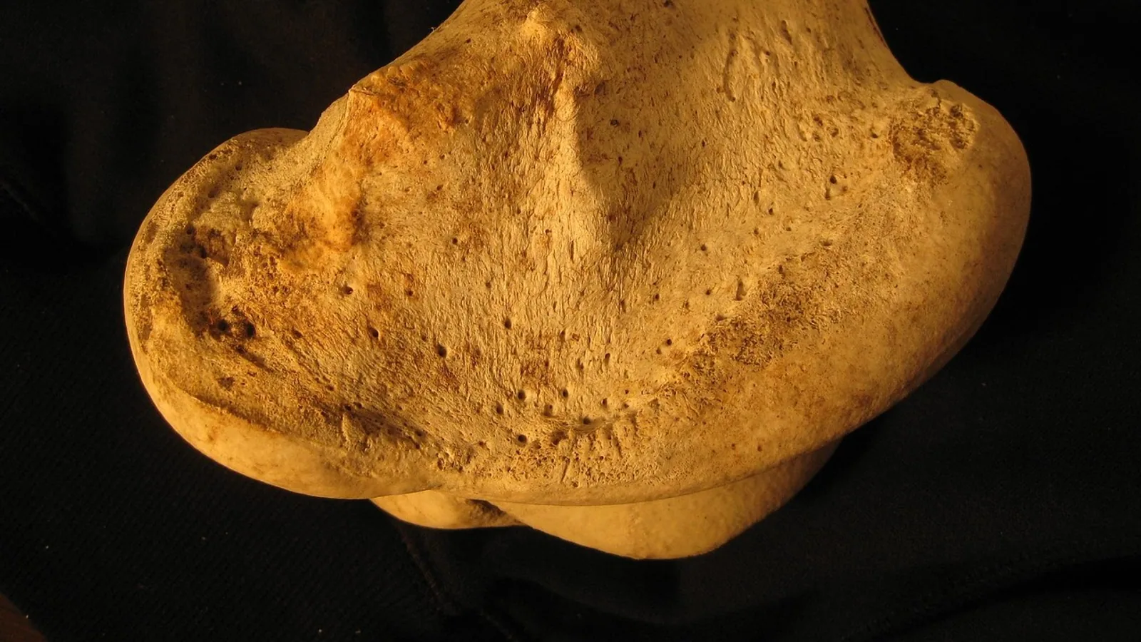
The greater trochanter is the large, blunt, craniolateral prominence of the proximal femur that serves as the principal insertion site for the hip abductor and external rotator muscles. It functions as a lever arm that converts gluteal muscle contraction into abduction, extension, and stabilization of the hip joint during weight bearing.
Every time a dog rises from a lying position, a horse pushes off during the gallop, or a cow shifts weight to escape a slippery floor, the greater trochanter is doing mechanical work. It is the bony handle that the gluteal muscles pull on to swing the limb outward and drive the body forward. In small animal practice it is a routine palpation landmark for hip radiography and for assessing muscle atrophy in orthopedic and neurologic disease. In large animal practice the trochanteric region is a site of clinically significant bursitis and fracture. Understanding its anatomy means understanding why hip disease produces the gait abnormalities that veterinarians look for on examination.
Orientation and Basic Anatomy
The greater trochanter sits at the proximal end of the femur, lateral to the femoral head and neck. It projects cranially and laterally, which places it outside the line of the femoral shaft. The femoral head faces medially and articulates with the acetabulum of the pelvis. The greater trochanter is therefore the most lateral palpable bony point of the hip region in most domestic species.
Several other landmarks share the proximal femur. The lesser trochanter is a smaller medial prominence that serves as the insertion for the iliopsoas muscle. The femoral head has a small central depression, the fovea capitis, where the ligament of the head of the femur attaches. The trochanteric fossa is a deep depression on the medial (caudal) surface of the greater trochanter, and it is a key landmark for both muscle attachment and surgical orientation.
The Trochanteric Fossa
The trochanteric fossa is a variably shaped excavation on the caudomedial aspect of the greater trochanter. In dogs, its radiographic appearance is inconsistent. A study of 175 ventrodorsal radiographs of canine hip joints found that the fossa appeared as a clearly defined circular area of radiopacity surrounded by a radiolucent halo in only a small number of images (n = 12), and that this appearance was significantly influenced by medial rotation of the femur (P < 0.001) [1]. This matters in practice because a radiographer who rotates the limb differently can make the same normal fossa look abnormal on two separate films.
Anatomically, the fossa contains an irregular protuberance that marks the attachment of the gemelli and the internal and external obturator muscles. Small openings around this protuberance are nutrient foramina, and dissection has identified minor branches of the medial circumflex femoral artery entering through them [1]. The fossa sits close to the hip joint capsule, which attaches along the base of the femoral neck and the margins of the trochanter. Because of this proximity, the fossa is a reference point during intramedullary pinning. A study of 28 canine cadavers found that 9 of 13 retrograde pins were located in the medial half of the trochanteric fossa, compared with only 1 of 15 normograde pins [2]. Normograde pins were also significantly farther from the sciatic nerve than retrograde pins when the hip was flexed, and 7 of 13 retrograde pins contacted the sciatic nerve while none of the normograde pins did [2]. The fossa is not just an anatomical curiosity. Its position relative to the sciatic nerve makes it a surgical landmark with real consequences.
Muscle Attachments

_-_Fig_373.png)
The greater trochanter anchors a group of muscles that collectively abduct the limb, extend the hip, and rotate the femur. The specific muscles and their attachment patterns vary across species, and this variation affects how each species moves and where it is vulnerable to injury.
Gluteus Medius
The gluteus medius is the largest and most important of the hip abductors. In dogs it arises from the crest and lateral surface of the ilium and inserts on the craniolateral aspect of the greater trochanter. In horses and ruminants it is massive, reflecting the greater need for hip stabilization during locomotion. The muscle is a powerful abductor and also assists in hip extension. When it contracts, it pulls the greater trochanter cranially and laterally, swinging the limb away from the midline.
Gluteus Minimus
The gluteus minimus lies deep to the gluteus medius. It originates from the ventral surface of the ilium and inserts on the cranial aspect of the greater trochanter. Its action mirrors that of the gluteus medius but with a smaller moment arm. In dogs the gluteus minimus is relatively small. In horses it is fused with the gluteus medius to form a single functional unit.
Piriformis
The piriformis is a small, fusiform muscle that runs from the sacrum and the sacrosciatic ligament to the greater trochanter. It acts as an external rotator and a weak abductor. In dogs it is well developed. In horses and ruminants it is either absent or fused with the gluteal mass, depending on the species. The piriformis is one of several muscles that insert on or near the trochanteric fossa.
Obturator Internus and Gemelli
The internal obturator muscle arises from the pelvic surface of the obturator membrane and the surrounding bone. Its tendon passes through the lesser sciatic foramen and inserts on the medial surface of the greater trochanter, within or adjacent to the trochanteric fossa. The gemelli (superior and inferior) are small muscles that accompany the obturator internus tendon and insert alongside it. Together they act as external rotators of the hip. In dogs these muscles are clinically important because they are often involved in perianal fistulas and because their tendons can be damaged in pelvic trauma.
Obturator Externus
The external obturator muscle arises from the ventral surface of the pubis and ischium and inserts in the trochanteric fossa. It is an external rotator. In dogs it is a distinct muscle. In horses it is present but relatively small.
Accessory Gluteal Muscle
Horses have an additional muscle, the accessory gluteal muscle (also called the accessory head of the gluteus medius), which arises from the cranial border of the ilium and inserts on the greater trochanter. This muscle is unique to equids and contributes to the powerful hip extension needed for galloping.
Comparative Species Notes
The shape and prominence of the greater trochanter differ across domestic species, and these differences reflect each animal's locomotor style.
Dogs and Cats
In carnivores the greater trochanter is a distinct, cranially projecting process. It is separated from the femoral head by a deep notch. The trochanteric fossa is well defined and is the insertion site for the obturator internus, gemelli, and obturator externus. The greater trochanter in dogs is palpable as a prominent bump on the lateral aspect of the hip. In cats it is smaller but still clearly identifiable. The shape of the greater trochanter in carnivores is adapted for a wide range of hip motion, including the extreme flexion needed for jumping and climbing.
Horses
In horses the greater trochanter is large and rounded. It is divided into a cranial and a caudal part by a notch. The cranial part is the insertion for the gluteus medius and the accessory gluteal muscle. The caudal part is the insertion for the deep gluteal muscle. The third trochanter is a separate, prominent prominence on the lateral aspect of the femoral shaft, distal to the greater trochanter. It serves as the insertion for the superficial gluteal muscle. The third trochanter is unique to horses and is not present in dogs or cats.
Ruminants
In cattle, sheep, and goats the greater trochanter is large and has a distinct cranial and caudal part. The third trochanter is present and prominent, as in horses. The gluteus medius is massive and inserts on the cranial part of the greater trochanter. The deep gluteal muscle inserts on the caudal part. The third trochanter is the insertion for the superficial gluteal muscle. The prominence of the third trochanter in ruminants and horses reflects the importance of the superficial gluteal muscle in extending the hip during locomotion.
Pigs
In pigs the greater trochanter is relatively small and the third trochanter is absent or rudimentary. The gluteal muscles are less massive than in horses and ruminants. This reflects the pig's different locomotor pattern and body conformation.
Birds
In birds the greater trochanter is present but differs in shape from mammals. A study of broiler chickens found that 14 out of 104 birds had lateral femoral trochanteric defects, and all affected birds were severely lame [3]. The trochanteric muscle insertions had avulsed, and the lateral metaphyseal defect was usually lined with granulation tissue. Examination of normal trochanteric development identified endochondral ossification at sites of muscle attachment, and in some skeletally immature birds evidence of osteochondrosis was detected at these sites [3]. This suggests that broiler femoral trochanters are susceptible to muscle avulsion because of a predisposition to osteochondrosis and the pattern of muscle insertion [3]. This is a reminder that the greater trochanter is not just a static attachment point. It is a site of active bone remodeling and a potential weak link in growing animals.
Table: Muscle Attachments by Species
| Muscle | Dog | Cat | Horse | Ruminant | Pig |
|---|---|---|---|---|---|
| Gluteus medius | Craniolateral greater trochanter | Craniolateral greater trochanter | Cranial part of greater trochanter | Cranial part of greater trochanter | Craniolateral greater trochanter |
| Gluteus minimus | Cranial greater trochanter | Cranial greater trochanter | Fused with gluteus medius | Fused with gluteus medius | Cranial greater trochanter |
| Piriformis | Greater trochanter | Greater trochanter | Absent or fused | Absent or fused | Present |
| Obturator internus | Trochanteric fossa | Trochanteric fossa | Trochanteric fossa | Trochanteric fossa | Trochanteric fossa |
| Gemelli | Trochanteric fossa | Trochanteric fossa | Trochanteric fossa | Trochanteric fossa | Trochanteric fossa |
| Obturator externus | Trochanteric fossa | Trochanteric fossa | Trochanteric fossa | Trochanteric fossa | Trochanteric fossa |
| Accessory gluteal | Absent | Absent | Greater trochanter | Absent | Absent |
| Superficial gluteal | Not on greater trochanter | Not on greater trochanter | Third trochanter | Third trochanter | Not on greater trochanter |
This table summarizes the principal attachments. Individual variation exists, and some texts describe additional minor attachments.
Role as a Lever for Hip Abduction
The greater trochanter functions as a lever arm. When the gluteal muscles contract, they pull on the trochanter and produce rotation of the femur around the hip joint. Because the trochanter is offset laterally from the femoral head, the muscle force creates a moment that abducts the limb. This is essential for maintaining balance during walking, especially during the single-limb support phase of the gait cycle.
In a dog with hip dysplasia, the hip joint is unstable. The gluteal muscles must work harder to stabilize the joint, and they may hypertrophy initially. Over time, disuse and pain can lead to muscle atrophy. A veterinarian assessing a dog for hip dysplasia will palpate the gluteal muscles and compare them side to side. Reduced muscle mass over the greater trochanter is a common finding in chronic hip disease.
The greater trochanter also acts as a lever for hip extension. During the push-off phase of the gait, the gluteal muscles contract to extend the hip and propel the body forward. In horses the massive gluteus medius and accessory gluteal muscle generate the power for galloping. The greater trochanter is the fulcrum through which this power is transmitted to the limb.
Clinical Relevance, Limitations and Common Mistakes
Trochanteric Bursitis
A bursa is a fluid-filled sac that reduces friction between a tendon and bone. The trochanteric bursa lies between the gluteal tendons and the greater trochanter. Inflammation of this bursa, called trochanteric bursitis, causes pain and lameness. In dogs it is an uncommon but recognized cause of hip pain. In horses it can occur after trauma or prolonged recumbency. Diagnosis is based on clinical signs, response to local anesthesia, and imaging. Treatment is medical and rest-based. This article does not cover surgical repair techniques.
Avulsion Fractures
An avulsion fracture occurs when a tendon or ligament pulls a fragment of bone away from its attachment site. The greater trochanter is vulnerable to avulsion fractures because it is the insertion point for powerful muscles. In growing animals the physis (growth plate) of the greater trochanter is a weak point, and avulsion can occur during strenuous activity. In dogs avulsion of the greater trochanter has been reported after trauma. In broiler chickens avulsion of the trochanteric muscle insertions is a recognized cause of lameness [3]. Avulsion fractures cause acute lameness, pain on palpation, and swelling over the trochanter. Radiography confirms the diagnosis.
Pressure Ulcers
Dogs that are recumbent for prolonged periods can develop pressure ulcers over the greater trochanter. The bone prominence presses against the skin and underlying tissues, reducing blood flow and causing tissue necrosis. A study of five dogs with pressure ulcers over the greater trochanter treated with muscle flaps found that all wounds healed promptly after debridement and transposition of the cranial part of the sartorius or the rectus femoris muscle [4]. This confirms that the greater trochanter is a high-risk area for decubital ulcers in immobilized patients. Nursing care for recumbent dogs should include frequent repositioning and padding over the trochanteric region.
Surgical Landmarks and Complications
The greater trochanter is a key landmark for orthopedic surgery of the hip. It is used to identify the correct entry point for intramedullary pins and to guide the placement of bone plates. A study of 28 canine cadavers found that normograde pinning of the femur was less likely to induce sciatic nerve injury than retrograde pinning, particularly in midshaft and distal fractures [2]. This is because normograde pins are placed more laterally in the trochanteric fossa and farther from the sciatic nerve. The trochanteric fossa is a danger zone during surgery because of its proximity to the sciatic nerve and the medial circumflex femoral artery.
Imaging Considerations
Radiographic interpretation of the trochanteric region requires attention to positioning. The appearance of the trochanteric fossa on ventrodorsal radiographs is significantly influenced by medial rotation of the femur [1]. A fossa that looks abnormal on one view may look normal on another. Computed tomography (CT) and three-dimensional modeling provide more accurate assessment of hip anatomy. A study of 100 adults found that 3D measurements of femoral anteversion and acetabular version differed significantly from 2D measurements and that 3D modeling improved the accuracy of hip joint assessment [5]. In veterinary medicine, CT is increasingly used for preoperative planning of hip surgery.
Common Mistakes
Students often confuse the greater trochanter with the lesser trochanter. The greater trochanter is lateral and cranial. The lesser trochanter is medial and caudal. The greater trochanter is the insertion for the gluteal muscles. The lesser trochanter is the insertion for the iliopsoas muscle.
Another common mistake is to assume that the greater trochanter is the same shape in all species. In horses and ruminants the third trochanter is prominent, while in carnivores it is absent. The greater trochanter itself is more rounded in horses and more angular in dogs.
A third mistake is to overlook the trochanteric fossa during radiographic interpretation. The fossa is a normal structure, but its appearance varies with positioning. A radiolucent halo around a radiopaque center can be a normal finding.
A fourth mistake is to forget that the greater trochanter is a site of active bone remodeling in growing animals. Osteochondrosis can affect the trochanteric growth plate and predispose to avulsion [3]. Young, rapidly growing animals with lameness should be evaluated for trochanteric lesions.
This article is educational and is not a substitute for veterinary diagnosis or treatment.
Quick Review
- The greater trochanter is the large craniolateral prominence of the proximal femur.
- It is the insertion site for the gluteus medius, gluteus minimus, piriformis, obturator internus, gemelli, and obturator externus.
- The trochanteric fossa is a depression on the caudomedial surface of the greater trochanter and is close to the hip joint capsule.
- In horses and ruminants the third trochanter is prominent and serves as the insertion for the superficial gluteal muscle.
- In carnivores the greater trochanter is distinct and angular, while in horses it is large and rounded.
- The greater trochanter acts as a lever for hip abduction and extension.
- Clinical conditions affecting the greater trochanter include bursitis, avulsion fractures, pressure ulcers, and surgical complications related to the sciatic nerve.
Frequently Asked Questions
What is the greater trochanter?
The greater trochanter is the large bony prominence on the craniolateral aspect of the proximal femur. It serves as the attachment site for the hip abductor and external rotator muscles.
Which muscles attach to the greater trochanter?
The gluteus medius, gluteus minimus, piriformis, obturator internus, gemelli, and obturator externus attach to the greater trochanter or the trochanteric fossa. The specific pattern varies by species.
What is the trochanteric fossa?
The trochanteric fossa is a depression on the caudomedial surface of the greater trochanter. It is the insertion site for the obturator internus, gemelli, and obturator externus muscles and lies close to the hip joint capsule.
Do all animals have a third trochanter?
No. The third trochanter is prominent in horses and ruminants but is absent in dogs, cats, and pigs. It serves as the insertion for the superficial gluteal muscle.
What causes pain over the greater trochanter in dogs?
Pain over the greater trochanter can be caused by trochanteric bursitis, avulsion fractures, muscle strain, or pressure ulcers. A veterinarian should evaluate persistent lameness or swelling.
Can the greater trochanter fracture?
Yes. Avulsion fractures of the greater trochanter can occur when the gluteal muscles pull a bone fragment away from the femur. This is most common in young, growing animals or after trauma.
Related Articles
- Dairy Cow Milking Routine: Preparation, Unit Attachment, and Post-Dipping
- Heavy-Duty Horse Grooming Vacuums: Suction Power, Noise Reduction, and Dander Removal Attachments
- Comparative Anatomy of the Mammalian Kidney
- Comparative Anatomy of the Mammalian Heart
- Gastrointestinal Adaptations in Exotic Herbivores
- Canine Respiratory System: Anatomy and Physiology
- Greater Swiss Mountain Dog: Health and Care Guide
- Greater Omentum: Anatomy and Function
Further Reading
Sources
- The canine trochanteric fossa: a radiographic and anatomic study.
- Relationship of femoral intramedullary pins to the sciatic nerve and gluteal muscles after retrograde and normograde insertion.
- Avulsion of muscles from the femoral trochanter in the fowl.
- Treatment of trochanteric ulcers with cranial sartorius and rectus femoris muscle flaps.
- Impact of CT-based 3D modelling on hip joint anatomical measurements: a comparative study with 2D methods.