Duck Foot Anatomy: Webbed Feet Explained
By Dr. Zubair Khalid, DVM, MS, PhD ·
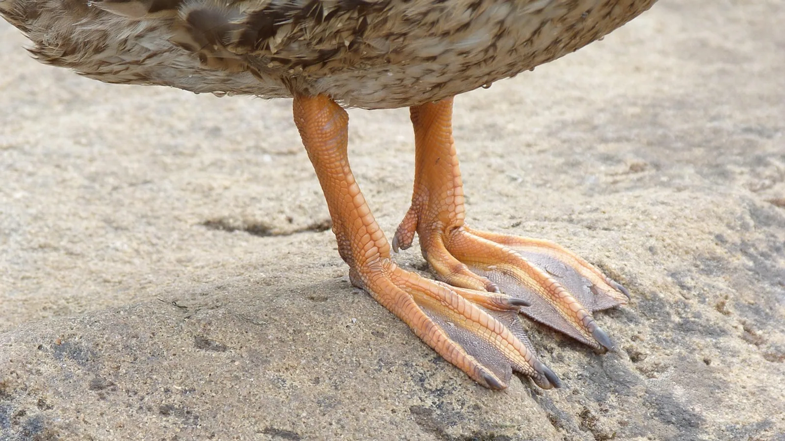
A duck's foot is a three-toed (trigid) propulsive paddle in which the second, third, and fourth digits are joined by a pliable interdigital web, a fold of skin rich in collagen and elastin rather than fused bone. The web is anchored to the phalanges by connective tissue and functions as a single hydrofoil surface during the power stroke, while the tibiotarsus, tarsometatarsus, and hypotarsus convert hindlimb muscle contraction into forward thrust.
This matters beyond trivia. Waterfowl foot anatomy explains why ducks are efficient swimmers and poor perchers, why pododermatitis and bumblefoot present where they do, why the plantar surface is thermally protected during winter foraging, and why a penetrating web wound can bleed heavily and heal slowly. For students, the duck foot is also the classic model for studying programmed cell death in the developing limb, because the same interdigital tissue that regresses in the chick persists in the duck [1][2].
This article is educational and is not a substitute for veterinary diagnosis or treatment.
Why the Duck Foot Is Not a Fused Hand
The single most common misconception is that the digits are fused. They are not. Each digit remains a separate skeletal unit with its own joints, tendons, and neurovascular supply. The web is a separate soft-tissue structure draped between the digits and continuous with the skin of the toe margins.
The difference is developmental. In birds with free digits, such as the chick, the interdigital mesenchyme undergoes apoptosis (programmed cell death) that removes the tissue between the toes. In the duck, that cell death is markedly reduced, and the surviving mesenchyme becomes fibroblastic connective tissue that forms the web [3]. The duck interdigital mesoderm also shows an attenuated response to bone morphogenetic protein (BMP)-induced apoptosis and to TGF-beta-induced chondrogenesis compared with free-digit species, accompanied by reduced expression of the msx-1 and msx-2 genes [2].
Fine-structural work on duck embryos showed that interdigital necrosis does occur, but it is less intense and shorter in duration than in the chick, and the accompanying changes (collagen deposition, basal lamina rupture, ectodermal cell detachment) are correspondingly milder [1]. The ectodermal surface changes that mark free-digit formation in the chick are largely absent or much less pronounced in the duck [4]. A final key difference is vascular: in the chick, interdigital blood vessels regress as the digits free themselves, but in the duck, the interdigital necrotic process is not followed by vascular regression, so the web retains its blood supply [5].
Digits, Phalanges, and the Web Fold
Ducks have three functional pedal digits (II, III, and IV, plus a reduced hallux or first digit that carries no web). Digit III is the longest and bears the largest number of phalanges. The interdigital web occupies the spaces between the three main digits and is thickest at the commissures (the hinge lines where web meets digit) and thinnest at its free distal margin.
Histologically, the web is a skin fold. It has an epidermis and dermis like the rest of the foot, but the dermal layer contains a dense, organized meshwork of collagen fibers interwoven with elastin. Collagen provides tensile strength, so the web resists stretching under the load of the power stroke. Elastin provides recoil, so the web passively springs back to its resting position during the recovery stroke, reducing the muscular work required to reset the foot for the next beat. This collagen-rich architecture is consistent with the collagenous deposition observed in the developing interdigital interface during web formation [1].
The web is not a rigid pane. During the power stroke it is spread taut by the water; during the recovery stroke it relaxes and folds slightly, reducing drag. This passive feathering behavior is a property of the tissue itself, not of active muscular contraction of the web.
The Osteology of the Swimming Hindlimb
Tibiotarsus
The tibiotarsus is the proximal long bone of the avian lower leg. It is formed by fusion of the distal tibia with proximal tarsal elements during development. In a duck it transmits the force from the strong hindlimb muscles of the thigh and shank down to the ankle joint. Its relatively long shaft and robust medullary cavity in diving and dabbling species support a substantial muscular envelope (the gastrocnemius and digital flexors). The tibiotarsus acts as the strut through which knee and hip extension is converted into an ankle-driven kick.
Tarsometatarsus
The tarsometatarsus is the distal long bone of the avian leg, formed by fusion of distal tarsal elements with the metatarsals. In a duck it is comparatively short and stout relative to the tibiotarsus, which gives the foot mechanical advantage for pushing water rather than for running. The three main digits articulate at its distal condyles. This bone is also the attachment site for the digital flexor tendons as they pass toward the toes, and its plantar surface supports the foot during standing. In ducks it is the structure that defines the width of the swimming foot, and its length relative to the tibiotarsus contributes to the characteristic leg silhouette of a swimming dabble.
Hypotarsus
The hypotarsus is a bony prominence on the plantar aspect of the proximal tarsometatarsus, and in ducks it is elaborated into a series of ridges and grooves. It acts as a pulley block for the flexor tendons. The deep digital flexor and the superficial digital flexor pass through these grooves, held in place by a retinaculum, and are redirected so that contraction of muscles in the shank produces flexion at the toe joints rather than lifting the foot. This pulley arrangement is what makes the duck foot an efficient paddle blade on the power stroke. It also lets the toes be folded compactly during the recovery stroke, reducing frontal area and drag.
The combination of a long tibiotarsus, a short stout tarsometatarsus, and a grooved hypotarsus gives the duck foot a favorable lever system for swimming without sacrificing the ability to stand or walk on land, though it does make long-distance terrestrial locomotion inefficient compared with cursorial birds.
The Digital Flexor Mechanism
The digital flexor mechanism is the tendon and pulley system that flexes the toes. Two key components are the superficial digital flexor and the deep digital flexor. Both originate from muscles in the shank, pass through the hypotarsal grooves, and insert on the phalanges of the three main digits, with the deep flexor inserting most distally. When these muscles contract, the toes are drawn into a curled, hook-like position, and the web is drawn back into a compact bundle. When the muscles relax during the power stroke, the toes remain extended and the web spreads.
The flexor tendons are enclosed in synovial sheaths along portions of their course, which reduces friction at the ankle and metatarsophalangeal joints. The hypotarsus is the principal redirection point for these tendons, and its grooved surface is why injuries to the plantar aspect of the proximal metatarsus can cause tendon luxation or rupture, a serious problem in waterfowl kept on abrasive flooring.
Vascular Supply and Heat Exchange in the Web
Cranial Tibial Artery
The cranial tibial artery supplies the dorsal and distal leg and contributes to the vascular network of the foot in birds. It is the principal named artery students should be able to trace from the popliteal region down the cranial aspect of the tibiotarsus and across the tarsal joint. Once in the foot, it ramifies into smaller branches that supply the digits, the interdigital web, and the plantar surface. The web itself is well vascularized, which is why laceration of a duck's webbed foot can bleed briskly.
The persistent vascularity of the interdigital tissue is developmentally significant. In the chick, interdigital vessels regress alongside the apoptotic mesh; in the duck they do not [5]. That retained vascular bed in the adult web is one substrate for the heat-exchange system described below.
Rete Mirabile and Countercurrent Heat Exchange
A rete mirabile (Latin for "wonderful net") is a complex of closely apposed, parallel arteries and veins that acts as a heat exchanger. In the duck's foot, the arterial blood traveling distally toward the webbed surface runs alongside venous blood returning proximally from the cold foot. The two vessels are adjacent and flow in opposite directions. Heat moves from the warm arterial blood to the cooler venous blood across the vessel walls.
The effect is that arterial blood arriving at the web is already cooled, and venous blood returning to the body core is already warmed. This is countercurrent heat exchange. The body core does not have to give up large amounts of heat to the environment, and the tissue of the web is exposed to a smaller thermal gradient between itself and the water or ice. Ducks standing on ice or swimming in near-freezing water lose less heat from the foot, and the risk of cold-induced tissue injury is reduced. The limb also functions as a thermal buffer, so the core temperature is defended when the periphery is cold.
This system is not unique to ducks. It appears in a range of birds and mammals that forage in cold environments, and it is one of the standard examples taught in comparative thermoregulation. The anatomy is important because it explains a counterintuitive observation: a duck's webbed foot can be quite cold to the touch while the bird is comfortable, and this is normal, not a sign of frostbite or hypothermia.
How the Foot Works During a Stroke
The sequence at the level of the foot is mechanical, not mysterious:
- During recovery (the leg swings forward), the digital flexors shorten, curling the toes and gathering the web into a compact bundle to reduce drag.
- At the top of the stroke, the flexors relax and the toes extend, spreading the web across the full span of the three digits.
- During the power stroke (the leg drives backward), the extended web reaches maximum surface area. Resistance from the water holds the web taut, and the tension is carried through collagen fibers in the web to the phalanges.
- Force from the tibiotarsus and tarsometatarsus is transferred through the ankle and metatarsophalangeal joints into the toes and then into the water.
- As the foot exits the water and begins the next recovery, the flexors re-engage, the web relaxes, and the cycle repeats.
The rete mirabile runs alongside this mechanical cycle continuously, moderating heat loss regardless of stroke phase.
Comparative Web Anatomy Across Waterbirds
Web structure varies across waterbirds because the developmental mechanisms that produce it vary. A comparative analysis of webbed foot types identified four categories: palmate, semipalmate, totipalmate, and lobate [6]. These differences reflect how long Gremlin1 (a BMP antagonist that inhibits interdigital cell death) remains expressed in the interdigital tissues and how the proliferating cell populations are distributed across the toes [6].
| Species group | Foot type | Web structure | Developmental note |
|---|---|---|---|
| Mallard-type duck (Anas platyrhynchos) | Palmate | Full web between digits II, III, and IV, three commissures, continuous with toe margins | Gremlin1 persists in interdigital tissue, keeping the web; attenuated msx-1/msx-2 response to BMP and TGF-beta compared with free-digit birds [2] |
| Geese (Anserini) | Palmate | Full web between digits II, III, and IV; broadly similar architecture to ducks, proportionally larger and heavier | Not directly analyzed in the cited comparative dataset |
| Gulls (Laridae) | Palmate | Full web between the three forward digits; anatomically similar to duck web in extent | Not directly analyzed in the cited comparative dataset |
| Great cormorant | Totipalmate | Web connects all four digits, including the hallux | Gremlin1 present in all interdigital tissues at St. 31, then lost except along the toes by St. 33, indicating a distinct developmental route from the palmate duck foot [6] |
| Common coot and little grebe | Lobate | Separate fleshy lobes along each digit, not a continuous interdigital membrane | Gremlin1 retained in interdigital tissue; lobate feet appear to have arisen independently in coot and grebe through distinct mechanisms, a case of convergent evolution [6] |
Two points from that comparison are worth emphasizing. First, palmate, semipalmate, totipalmate, and lobate are not a simple gradient of "more web." The lobate foot is a separate design, in which each toe bears an independent flap of skin rather than sharing an interdigital membrane. Second, the totipalmate cormorant foot and the palmate duck foot arose from different developmental programs, even though both produce a functioning paddle [6].
How the Web Is Studied in Practice
Researchers and anatomists investigate duck foot structure through several standard approaches:
- Embryological staging. Chick and duck limb buds are compared at matched developmental stages, and the timing of interdigital cell death is mapped by DNA fragmentation markers and electron microscopy [1][4].
- Gene expression analysis. In situ hybridization for Gremlin1, msx-1, msx-2, bmp genes, and fgf-8 in the developing autopod reveals which signals are attenuated or prolonged in webbed species [6][2].
- Vascular injection. Indian ink injection of the limb bud vasculature demonstrates whether interdigital vessels persist or regress, as in the chick-versus-duck comparison [5].
- Experimental ectoderm removal. Removing ectoderm from the duck leg bud can trigger ectopic cartilage formation, showing that the surviving web mesenchyme retains latent chondrogenic potential. Ectopic cartilages in the duck are rounded, smaller, and located more distally at the interdigital margin than the elongate cartilages seen in the chick, and extra digit formation is less frequent in the duck [3].
- Gross dissection and imaging. Adult foot anatomy is demonstrated by dissection of the tibiotarsus, tarsometatarsus, hypotarsus, flexor tendons, and cranial tibial artery, plus radiography or computed tomography to display the phalanges.
Students should be able to move between these levels. A web that persists in the adult duck is the direct consequence of reduced apoptotic signaling, retained interdigital vasculature, and a fibroblastic (not chondrogenic) fate for the surviving interdigital mesenchyme [2][3].
Clinical Relevance, Limitations and Common Mistakes
The web and the plantar surface of the foot are common sites of injury and disease in captive waterfowl. Pododermatitis (bumblefoot) typically involves the plantar metatarsal pad and can extend to the web and toe margins. Because the web has a rich vascular supply, penetrating wounds from wire, glass, or fishing tackle can cause significant hemorrhage and are prone to secondary infection. The rete mirabile and the foot's cold tolerance mean that a cool foot in a healthy duck is not itself alarming, but a foot that is cold and also swollen, discolored, or non-weight-bearing warrants examination.
Fractures of the tibiotarsus or tarsometatarsus, luxation of the flexor tendons at the hypotarsus, and avulsion injuries at the commissures are all described in waterfowl practice. A duck's short tarsometatarsus and long tibiotarsus are not adapted for high-impact landing on hard surfaces, so housing on concrete or wire without soft footing increases the risk of both soft tissue and skeletal injury.
Two common mistakes students make are worth correcting early. The first is describing the duck web as fused toes. The bones are separate and the joints are separate. The web is soft tissue only. The second is treating the duck foot as a scaled-up version of a free-digit bird's foot. The developmental pathway, the gene expression profile, and the retained interdigital vasculature are distinct [6][2][5].
Individual birds vary, and a diagnosis requires examination by a veterinarian. Anatomy knowledge supports clinical reasoning, but it does not replace it.
Quick Review
- A duck's foot is trigid (three main digits) with a palmate web between digits II, III, and IV.
- The web is a skin fold with a collagen and elastin-rich dermis, not fused bone.
- Web persistence results from reduced interdigital apoptosis, attenuated msx-1 and msx-2 signaling, and a fibroblastic fate for surviving mesenchyme [2][3].
- Interdigital blood vessels persist in the duck instead of regressing as they do in the chick [5].
- The tibiotarsus is the proximal leg strut, the tarsometatarsus is the distal fused bone, and the hypotarsus is the tendinous pulley on the plantar proximal metatarsus.
- The cranial tibial artery supplies the distal leg and foot, and the rete mirabile provides countercurrent heat exchange to limit heat loss.
- Palmate, semipalmate, totipalmate, and lobate feet are distinct morphologies with distinct developmental origins [6].
Frequently Asked Questions
Are a duck's toes fused together?
No. The digits are separate skeletal units with separate joints. The web is a fold of skin between them, not a fusion of bone.
Is the web made of muscle?
No. The web is a skin fold whose dermis is rich in collagen and elastin. Digits are moved by the digital flexor tendons, not by muscles inside the web itself.
Why do ducks not get frostbite on their feet in cold water?
The rete mirabile, a network of parallel arteries and veins, warms returning venous blood using heat from incoming arterial blood. This lowers the thermal gradient at the foot surface and protects the peripheral tissue.
What is the hypotarsus?
It is a bony prominence on the plantar proximal tarsometatarsus that acts as a pulley block, redirecting the digital flexor tendons so that shank muscle contraction flexes the toes.
Is the webbed foot unique to ducks?
No. Palmate feet occur in ducks, geese, and gulls. Totipalmate feet occur in cormorants, and lobate feet occur in coots and grebes. These are morphologically and developmentally distinct types [6].
Why does duck foot embryology matter in research?
The duck foot is a classic model of how reducing programmed cell death alters limb shape. Comparing duck and chick interdigital tissue revealed the roles of BMP signaling, Gremlin1, msx genes, and vascular regression in shaping the vertebrate limb [1][6][2][5].
Related Articles
- Avian Parasites of the Feet: Etiology, Diagnosis, and Management
- Dogs Licking Feet Diabetes
- Dog Licking Feet Obsessively
- Dog Keeps Licking My Feet
- Dog Licking Feet Video
- Dog Licking Feet Of Human
- Pekin Duck Breeding: Timing, Fertility and Care
- Ancona Duck: Breed Profile and Care Guide
Sources
- Fine structure of the interdigital membranes during the morphogenesis of the digits of the webbed foot of the duck embryo.
- Morphological diversity of the avian foot is related with the pattern of msx gene expression in the developing autopod.
- Interdigital chondrogenesis and extra digit formation in the duck leg bud subjected to local ectoderm removal.
- Surface changes in the embryonic interdigital epithelium during the formation of the free digits: a comparative study in the chick and duck foot.
- Vascular regression during the formation of the free digits in the avian limb bud: a comparative study in chick and duck embryos.
- Developmental mechanisms underlying webbed foot morphological diversity in waterbirds.