Dense Regular Connective Tissue: Structure and Function
By Dr. Zubair Khalid, DVM, MS, PhD ·
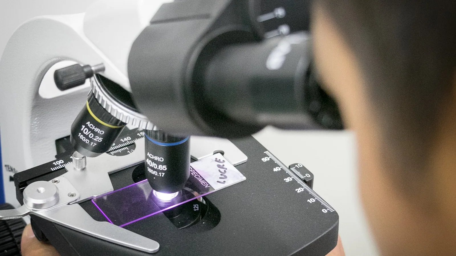
Dense regular connective tissue is a connective tissue in which collagen type I fibers are packed into parallel bundles, with sparse flattened fibroblasts (tenocytes) arranged in rows between the fiber bundles and very little ground substance. It forms the mechanical backbone of tendons and ligaments, which is why it is sometimes called dense fibrous connective tissue.
This tissue is the reason a horse can transmit several hundred kilograms of muscular force through a flexor tendon without the tissue tearing, and the reason a torn cruciate ligament in a dog does not knit back together on its own. Understanding what dense regular connective tissue is, how it is built, how it is identified under a microscope, and why it heals so poorly is foundational for veterinary anatomy, surgery, and rehabilitation. This article is educational and is not a substitute for veterinary diagnosis or treatment.
The Connective Tissue Map: Where Dense Regular Fits
Connective tissue proper is traditionally divided into loose and dense categories, and the dense category splits again into regular and irregular based on fiber direction. The classification rests on three variables: how much collagen is present, whether the collagen runs in one direction or many, and how much space is left for cells and ground substance.
Connective tissues are defined as specialized (bone and cartilage), dense regular (tendon and ligament), and dense irregular connective tissue [1]. That three-part framework is a useful starting point because it groups tendons and ligaments together and separates them from the skin dermis and joint capsule.
| Tissue | Predominant cell | Fiber arrangement | Representative locations | Primary function |
|---|---|---|---|---|
| Loose (areolar) connective tissue | Fibroblasts, plus macrophages, mast cells, adipocytes | Collagen and elastic fibers in a loose, random mesh | Beneath epithelia, around vessels and nerves, between muscle fibers | Support, diffusion, immune surveillance, filler |
| Dense irregular connective tissue | Fibroblasts | Collagen bundles woven in multiple directions | Skin dermis, joint capsule, fascia, organ capsules, periosteum | Resistance to multidirectional tension |
| Dense regular connective tissue | Tenocytes (specialized fibroblasts), flattened and aligned in rows | Collagen type I bundles parallel to the line of pull | Tendon, ligament, aponeurosis, some fascia | Uniaxial tensile strength and force transmission |
| Specialized connective tissue | Chondrocytes, osteocytes, osteoblasts | Cartilage matrix or mineralized collagen | Articular cartilage, bone | Load bearing, skeletal support |
A useful anchor for the whole system is the work on muscle connective tissue in the rat soleus, which described two distinct connective tissue compartments that meet only at the myotendinous junction. One compartment is areolar and lies between muscle fibers. The other is dense regular and lies beyond the ends of the muscle fibers in structures called microtendons, so the definitive tendon is effectively the accumulation of one microtendon per muscle fiber [2]. That study is a clean demonstration that dense regular connective tissue is not a bigger, tougher version of loose connective tissue. It is a different tissue with a different job at a different anatomical site.
Structure of Dense Regular Connective Tissue


Cells: Tenocytes in Rows
The resident cell of tendon and ligament is the tenocyte, a fibroblast lineage cell responsible for synthesizing and maintaining the collagen matrix. In dense regular connective tissue, tenocytes are sparse and flattened, with condensed nuclei and thin cytoplasmic extensions that wrap around the collagen bundles. They sit in longitudinal rows between the fiber bundles, and their nuclei appear as elongated basophilic streaks parallel to the collagen.
The shape is functional. Because the tissue is under near-constant uniaxial tension, cells that align with the axis of pull are positioned to sense strain and to deposit new collagen in the correct orientation. A tendon explant study of rat tenocytes found that cells migrating out of the tendon gradually aggregate and align in parallel rows with the tendon's long axis as culture time extends to 12 days, with accompanying remodeling of the collagen bundles [3]. Alignment is not incidental. It is part of how the tissue maintains its architecture.
Collagen orientation also shapes cell behavior from the outside in. When human dermal fibroblasts and tenocytes were seeded onto multilamellar collagen I membranes, the membrane with oriented fibers supported better cell proliferation and adhesion and provided an instructive pattern for cell growth and alignment [4]. Dense regular connective tissue therefore works as a two-way system. Cells build the aligned matrix, and the aligned matrix tells cells how to behave.
Fibers: Parallel Collagen Type I
Collagen type I makes up the overwhelming majority of the dry weight of tendon and ligament. The fibers are arranged in a hierarchical plan. Tropocollagen molecules assemble into fibrils, fibrils group into fibers, fibers group into bundles, and bundles run parallel to the tendon axis. Collagen composition and organization determine ligament integrity, with type I collagen providing tensile strength and type III collagen supporting remodeling [5].
At the level of the individual fibril, the structure is not perfectly straight. Tendons show a crimp pattern, meaning the collagen fibrils run in aligned bundles with a regular undulating wave. Crimp is established during embryonic development and functions as a shock absorber during loading [6]. In a 3D culture model, contraction of tendon fibroblasts was the critical factor driving the buckling of collagen fibrils that produces the crimp, and the resulting superstructure created the characteristic toe region of the stress-strain curve [6]. The toe region is the initial low-stiffness phase of tendon loading, when the crimp is being straightened before the fibers take up load directly. This is why tendon does not behave like a rigid beam at low forces.
Ground Substance and Water
Dense regular connective tissue contains very little ground substance compared with loose connective tissue or cartilage. The matrix is dominated by collagen, with a small amount of proteoglycan and water bound within and between the fibrils. The low proportion of ground substance is one reason the tissue is stiff and has limited capacity for diffusion-based nutrition. The high fiber-to-cell ratio is a defining feature that also explains the tissue's low metabolic activity and limited regeneration potential [7].
Vasculature and Nerves
Dense regular connective tissue is poorly vascularized. Tendons receive blood from the surrounding paratenon, from the musculotendinous junction, and from the osseous insertion, with vessels passing through the endotenon septa that separate fiber bundles. The mid-substance of a tendon has the weakest blood supply, and intrasynovial segments of flexor tendons are especially dependent on synovial fluid diffusion.
A cadaveric comparison of two human lateral ankle ligaments illustrates how much vascularity varies within the same region. The anterior talofibular ligament showed loosely arranged collagen fibers, lower fiber density, and higher cellularity and vascularity, which indicated increased remodeling but reduced structural strength. The calcaneofibular ligament showed dense, well-organized parallel collagen bundles with lower cellularity, consistent with greater mechanical stability [5]. Both tissues are dense regular connective tissue by category, but their fiber organization and vascularity differ in ways that track with their injury patterns.
Nerve endings in and around tendon and ligament are not uniform. The architectural arrangement of connective tissue in the musculoskeletal system has functional consequences for proprioception, and the connective tissue matrix should be treated as an integrating structure rather than as passive force-guiding material [8].
How to Identify Dense Regular Connective Tissue Under the Microscope
Standard hematoxylin and eosin staining is usually sufficient for orientation, with special stains for fiber typing.
- Start at low magnification. Look for long, sweeping, wavy bands of eosinophilic (pink) material running in one direction across the entire field. This is the parallel collagen pattern.
- Find the nuclei. They should be sparse, flattened, and elongated, with the long axis parallel to the collagen bundles. Fibroblast nuclei appear as small dark streaks in rows between the collagen, not as plump round cells scattered randomly.
- Assess cellularity. If the field is crowded with nuclei, you are probably looking at loose connective tissue or a healing tendon, not mature dense regular connective tissue.
- Assess vascularity. Mature tendon and ligament have few visible blood vessels in the mid-substance. Prominent vessels usually indicate the paratenon, a healing response, or an inflamed synovial environment.
- Confirm with stains. Picrosirius red or polarized light microscopy highlights collagen birefringence and fiber orientation. Immunostaining for type I and type III collagen can distinguish mature from remodeling matrix. Alcian blue or toluidine blue will show whether any cartilage-like matrix is present, which matters when assessing insertional regions.
The practical value of this exercise is that it lets you predict behavior. A tissue with parallel collagen, sparse flattened tenocytes, minimal ground substance, and few vessels will be strong in tension and slow to heal. That prediction holds across species.
Tendon as the Classic Example
Tendon is the standard example of dense regular connective tissue because it connects muscle to bone and transmits force along a single axis. Tendons are dense regular connective tissue structures defined by their anatomical position connecting muscle to bone, and despite that common feature, tendons from different locations vary substantially in morphological, molecular, and mechanical properties related to their specialized function [9].
That variation has direct functional meaning. Tendons used in a spring-like manner, such as the equine superficial digital flexor tendon or the human Achilles tendon, are subjected to high strains during locomotion and store and return elastic energy. Positional tendons, such as those that simply hold a joint in position, experience much lower strains. The matrix composition differs between these types, and the ability of the resident cells to synthesize and degrade matrix also varies, which appears to relate to the magnitude of strain the tendon experiences during normal activity [9].
Tendon development itself is instructive. In an in vitro model, human tendon fibroblasts produced tendon-like constructs with large elongated cells aligned along the axis and collagen fibrils that increased in diameter by about 50 percent between day 14 and day 35 of culture, approaching the fibril size seen in adult human tendon. That increase in fibril diameter was accompanied by a fivefold increase in mechanical strength, from roughly 0.9 MPa to 4.9 MPa, and a rise in Young's modulus from about 5.8 MPa to 32.3 MPa, while the maximal strain at failure stayed constant at around 16 percent [10]. The lesson is that fibril diameter, not just collagen quantity, drives tendon strength.
Comparable findings come from engineered tendon in a porcine model, where parallel collagen alignment was observed at both ends of a repaired flexor tendon defect at 6 weeks, but not in the middle, and the repair tissue was more cellular than native tendon [11]. Early repair tissue is hypercellular and disorganized. Maturation is the process of removing cells and pulling collagen into alignment.
Ligament: Similar Tissue, Different Fiber Mix
Ligament connects bone to bone and is structurally similar to tendon, with the same parallel collagen type I bundles and sparse tenocytes. The main differences are more variable fiber orientation, a somewhat higher content of elastic fibers, and a different mechanical role. Ligaments must resist tension while allowing controlled joint motion, so they typically have slightly more extensibility than tendons in the same animal.
Molecular analysis of intraarticular and extraarticular connective tissues has shown that these differences are real at the gene expression level. Comparing the anterior cruciate ligament, posterior cruciate ligament, and medial collateral ligament with the patellar tendon, Achilles tendon, and synovium, researchers identified distinct clusters of genes that separate intraarticular connective tissues from extraarticular ones, along with a new marker of tendons and ligaments [12]. The intraarticular ligaments differ from extraarticular ligaments and tendons in biochemical, biomechanical, and viscoelastic properties, and most importantly in their ability to heal after surgical repair [12]. That differential healing capacity is one of the most clinically consequential facts in veterinary orthopedics.
Elastic fiber content is another point of structural variation. In subcutaneous fascia, both loose and dense regions contain hierarchical collagen fiber bundles associated with elastic fibers. In loose fascia, the elastic fibers sit mainly along the surface of the flattened collagen bundles, while in dense fascia they are found on the surface and within the bundles, consistent with repeated hierarchical organization. Elastase treatment altered bundle architecture and increased passive expansion [13]. Elastic fibers therefore contribute to how dense collagen bundles behave under stretch, even though they are a minor component by mass.
Comparative Clinical Relevance: Why Healing Is Slow
The Fiber-to-Cell Problem
Healing depends on cells. Dense regular connective tissue has very few cells relative to its matrix volume, and those cells have low metabolic activity. The high fiber-to-cell ratio and low metabolic activity and regeneration potential explain why healing frequently requires surgical implantation or reconstruction, with a high risk of reinjury [7]. A tendon cannot simply grow new tissue quickly, because there are not enough cells present to do the work at speed.
The Vascularity Problem
Tendon and ligament rely on diffusion from synovial fluid or from vessels in the paratenon and endotenon. The mid-substance of a tendon is at the end of a long diffusion distance. Low perfusion limits the delivery of oxygen, nutrients, growth factors, and inflammatory cells to the injury site. The anterior talofibular ligament's higher cellularity and vascularity suggest greater remodeling capacity than the denser calcaneofibular ligament, but that same remodeling capacity comes with reduced structural strength [5]. More vessels and cells do not automatically mean better mechanics.
The Scar Problem and the Adhesion Problem
Repair tissue rarely reproduces native architecture. In dog flexor tendon grafting studies, intrasynovial tendon grafts remained viable when transferred into the synovial space and appeared to heal through an intrinsic process with preservation of the gliding surface and better functional characteristics. Extrasynovial tendon grafts instead acted as scaffolding for early ingrowth of vessels and cells, with early cellular necrosis followed by ingrowth of fibrovascular adhesions from the periphery. Those dense peripheral adhesions obliterated the gliding surface, reduced tendon excursion, and limited joint rotation [14]. The distinction between intrinsic and extrinsic healing is a central concept in tendon surgery. Adhesions are the price of bringing a blood supply into a tissue that needs to glide.
A similar pattern appears in ligament reconstruction. When rat medial collateral ligaments were replaced with autogenous fascia lata, ligament, or patellar tendon grafts, the early stage was marked by transplant necrosis and acute inflammation. During the intermediate stage, host cells migrated in and collagen was rebuilt. Loose, extensible tissues such as fascia lata were fully colonized by fibroblasts, while dense strong tissues such as patellar tendon were destroyed and recolonized at the periphery early, with the center destroyed and recolonized later and more irregularly. In the late consolidation phase, a dense collagen network was rebuilt and reoriented, homogeneous with parallel bundles in fascia lata transplants but not homogeneous in tendon transplants, which retained a nodular periphery and well-oriented bundles separated by calcified or granulomatous foci centrally [15]. Graft architecture and graft tissue type determine how repair proceeds.
The Matrix Turnover Problem
Even in uninjured tendon, matrix turnover is slow. Tendon cells may be preprogrammed during embryological development for the strain they will encounter in life, or may simply respond to the particular strain environment they experience [9]. This matters for rehabilitation. Loading drives matrix maintenance. Complete immobilization removes the mechanical signal that tells tenocytes to maintain collagen. Controlled loading during healing is a strategy to supply that signal without exceeding tissue strength.
Nutritional and pharmacological support for collagen synthesis is an active area. In a rat model of complete Achilles tendon rupture, high-dose vitamin C given once every 2 days produced more evident angiogenesis on day 3, a significant difference in type I collagen production on day 10, and repair tissue that was almost regular dense connective tissue in structure by day 21. Mean collagen fiber diameter was higher and active fibroblast counts were slightly elevated in the vitamin C group throughout healing [16]. This is an experimental finding in healthy rats, not a clinical dosing recommendation, and it should be read as evidence that collagen synthesis is a rate-limiting step in tendon repair.
Tissue Engineering and the Architecture Lesson
The persistent difficulty of getting dense regular connective tissue to regenerate has driven a large body of scaffold work. Decellularization aims to remove cells from donor tendon while preserving the native molecular structure of the extracellular matrix, because proteins, lipids, nucleic acids, and other extracellular molecules are involved in differentiation, proliferation, vascularization, and collagen deposition. There is no standardized decellularization protocol for dense regular connective tissue, and many modifications to bioscaffold structure, shape, and composition have been described to improve therapeutic outcomes [7].
The recurring design principle in this field is alignment. Scaffolds that mimic the hierarchical structure of tendon, with aligned fibers and appropriate stiffness, give tenocytes the physical cues that maintain the tenogenic phenotype. Mechanical stimulation can enhance tenocyte proliferation and help maintain that phenotype, and cell/scaffold constructs grown under dynamic culture show improved mechanical properties in vivo [17]. Aligned nanofiber scaffolds with sustained release of growth factor also enhance tenocyte proliferation, upregulate tenogenic gene expression, and increase tendon-specific protein synthesis [18]. Human neo-tendon tissue has been generated in vitro from dermal fibroblasts under static mechanical strain, with longitudinally aligned collagen fibers and spindle-shaped cells, and collagen fibril diameter and tensile strength increased with time, peaking at 14 weeks. In contrast, the same cells in a tension-free condition formed disorganized fibrous tissue with significantly weaker strength and poor collagen fiber formation [19]. Tension and alignment are the two non-negotiable inputs.
Dermal fibroblasts are attractive as a cell source because they are easily accessible and do not create a major donor site defect. In a porcine model, dermal fibroblast engineered tendon and tenocyte engineered tendon were similar to each other in gross appearance, histology, and tensile strength [11]. This suggests the matrix environment, not just the cell type, dictates the outcome.
Clinical Relevance, Limitations and Common Mistakes
The single most important clinical consequence of dense regular connective tissue structure is that injury to tendon and ligament heals slowly and incompletely, and some lesions never heal without surgery. The anterior cruciate ligament in dogs is the classic example. Its intraarticular environment, poor vascularity, and limited intrinsic healing capacity mean that conservative management rarely restores normal joint stability, and surgical stabilization is the standard approach.
Common student mistakes in this topic include the following.
Confusing dense regular with dense irregular. The distinction is fiber direction and function. Dermis, joint capsule, and organ capsules resist tension from multiple directions and are dense irregular. Tendon and ligament resist tension along one axis and are dense regular.
Treating all dense connective tissue as equivalent. The lateral ankle ligament comparison shows that even two ligaments in the same joint can differ in collagen organization, cellularity, and vascularity, with corresponding differences in strength and remodeling capacity [5].
Assuming more cells and vessels mean stronger tissue. Healing tendon is hypercellular and more vascular than native tendon, and it is weaker. The repair tissue at 6 weeks after porcine flexor tendon repair had more cellular components than natural tendon and lacked mid-substance collagen alignment [11].
Forgetting that tendon is not uniform along its length. The rat soleus model shows that areolar and dense regular connective tissue meet only at the myotendinous junction, and collagen fibril diameter distributions change as microtendon collagen blends into tendon collagen [2].
Assuming collagen is a single molecule. Ligament integrity depends on the balance between type I collagen for tensile strength and type III collagen for remodeling, and the ratio shifts with injury and repair [5]. Type III collagen is smaller and less stiff, which is why scar tissue is mechanically inferior.
Overlooking the role of elastic fibers. In dense fascia, elastic fibers sit both on the surface of and within collagen bundles, and elastase treatment changes bundle architecture and increases passive expansion [13]. Elastic fibers matter for tissues that must recoil.
Ignoring the fascia and the wider architectural context. The connective tissue matrix connects structures and transmits force across joints, and an architectural view of how muscle and connective tissue are organized in series is more appropriate than treating ligaments as isolated passive straps [8]. For a veterinary student, this means thinking about the whole myotendinous unit and its surrounding sheaths, not just the tendon proper.
Practical clinical implications follow from the histology. Suture placement in tendon should engage the collagen bundles rather than sit in loose areolar tissue. Postoperative protocols balance the need for controlled loading against the risk of gap formation, because the repair tissue has few cells and little initial tensile strength. Vitamin C and other cofactors for collagen synthesis are relevant to healing biology, but experimental animal findings do not translate directly into clinical protocols for every species or patient. Cases involving tendon or ligament injury require individual assessment by a veterinarian.
Quick Review
- Dense regular connective tissue has parallel collagen type I bundles, sparse flattened tenocytes in rows between the fibers, minimal ground substance, and poor vascularity.
- Tendon is the classic example, ligament is similar but with more variable fiber orientation and more elastic fibers.
- Dense irregular connective tissue (dermis, joint capsule) has woven collagen and resists multidirectional tension. Loose areolar connective tissue has more cells, more ground substance, and a random fiber mesh.
- The high fiber-to-cell ratio, low metabolic activity, and poor blood supply explain slow tendon and ligament healing.
- Collagen crimp, established during development by tenocyte contraction, gives tendon its toe region on the stress-strain curve and acts as a shock absorber.
- Intrasynovial and extrasynovial tendons heal differently, and extrinsic healing with adhesions damages the gliding surface.
- Fibril diameter correlates with tendon strength, not just total collagen content.
Frequently Asked Questions
What is dense regular connective tissue?
Dense regular connective tissue is connective tissue with parallel collagen type I bundles, sparse flattened tenocytes arranged in rows between the bundles, and very little ground substance. Tendon and ligament are the principal examples.
How does dense regular connective tissue differ from dense irregular connective tissue?
The difference is fiber direction. Dense regular connective tissue has collagen running in one direction to resist uniaxial tension, while dense irregular connective tissue has collagen woven in multiple directions to resist tension from many directions, as in skin dermis and joint capsule.
Why does dense regular connective tissue heal so slowly?
It has few cells relative to its matrix volume, low metabolic activity, and poor vascularity, which limits the delivery of cells and nutrients to an injury site. This combination explains the frequent need for surgical repair and the high risk of reinjury.
Is a ligament the same as a tendon?
No. Both are dense regular connective tissue, but tendon connects muscle to bone and ligament connects bone to bone. Ligaments generally have more variable fiber orientation and a somewhat higher content of elastic fibers, and intraarticular ligaments have notably poorer healing capacity than tendons.
What is collagen crimp in tendon?
Crimp is a regular undulating wave pattern in the aligned collagen fibrils of tendon. It forms during development through tenocyte contraction and acts as a shock absorber, producing the toe region at the start of the tendon stress-strain curve.
Is dense regular connective tissue the same as dense fibrous connective tissue?
Dense fibrous connective tissue is a broad term for dense connective tissue that is rich in collagen fibers, and dense regular connective tissue is one specific subtype of it. Tendon and ligament are the classic dense fibrous connective tissues of the musculoskeletal system.
Related Articles
- G-Quadruplex Telomere Structure and Function
- Equine Hoof Anatomy: Structures and Function
- Plasmid in Bacteria: Structure, Types, and Functions
- Histone Variants: Structure, Function, and Implications
- Chromatin in a Cell: Structure, Function, and Dynamics
- Nucleosome Protein: Structure, Function, and Assembly
- Connective Tissue Types: Loose, Dense, and More
- Muscle Tissue: Types, Structure, and Function
Sources
- Non-myogenic Contribution to Muscle Development and Homeostasis: The Role of Connective Tissues.
- The dual connective tissue system of rat soleus muscle.
- Nuclear image changes in cells originating from tenocytes that migrated from tendon explants.
- Collagen I membranes for tendon repair: effect of collagen fiber orientation on cell behavior.
- A Histological Evaluation of Lateral Ankle Ligaments in Human Cadavers: Implications for Connective Tissue Integrity.
- Tenocyte contraction induces crimp formation in tendon-like tissue.
- Decellularization of Dense Regular Connective Tissue-Cellular and Molecular Modification with Applications in Regenerative Medicine.
- The architecture of the connective tissue in the musculoskeletal system-an often overlooked functional parameter as to proprioception in the locomotor apparatus.
- Tendon matrix composition and turnover in relation to functional requirements.
- In vitro tendon tissue development from human fibroblasts demonstrates collagen fibril diameter growth associated with a rise in mechanical strength.
- Repair of tendon defect with dermal fibroblast engineered tendon in a porcine model.
- Genome-wide expression analysis of intra- and extraarticular connective tissue.
- Elastic fiber-mediated hierarchical bundling of collagen fibers in subcutaneous fascia.
- The Marshall R. Urist Young Investigator Award. Autogenous flexor tendon grafts. Biologic mechanisms for incorporation.
- [[Reconstruction of the medial collateral ligament of the knee in rats using a free autogeneic transplant of fascia lata, ligament or tendon].](https://pubmed.ncbi.nlm.nih.gov/8171976/)
- High-dose vitamin C supplementation accelerates the Achilles tendon healing in healthy rats.
- Hierarchical scaffolds amalgamating bFGF-immobilized fibrous yarn bundles with cryogel tube for tendon tissue engineering.
- Development of high resilience spiral wound suture-embedded gelatin/PCL/heparin nanofiber membrane scaffolds for tendon tissue engineering.
- Engineering human neo-tendon tissue in vitro with human dermal fibroblasts under static mechanical strain.