Muscle Tissue: Types, Structure, and Function
By Dr. Zubair Khalid, DVM, MS, PhD ·
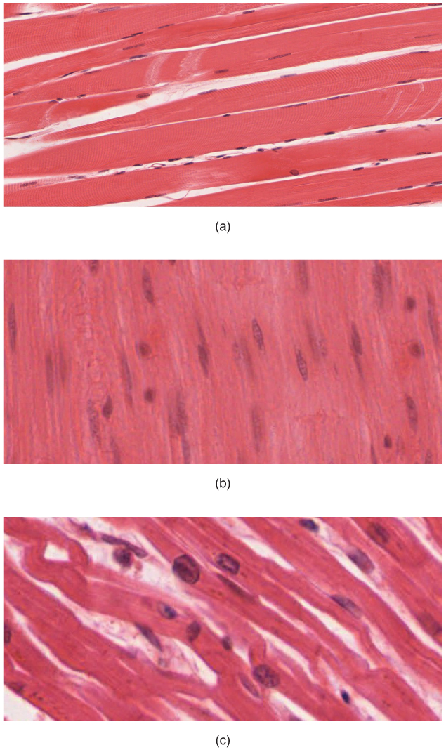
Muscle and muscle tissue come in three distinct types in domestic animals: skeletal, cardiac, and smooth. Skeletal muscle is striated, multinucleate, and under voluntary control, cardiac muscle is striated, mononucleate, branched, joined by intercalated discs and gap junctions, and involuntary, and smooth muscle is non-striated, spindle-shaped, mononucleate, and involuntary.
That single sentence is the backbone of every comparative question on muscle tissue in veterinary anatomy and physiology. The rest of this article unpacks how each type is built, how the sarcomere generates force, and what those differences mean in a live dog, cat, horse, or cow.
This article is educational and is not a substitute for veterinary diagnosis or treatment.
The Three Types of Muscle Tissue at a Glance

Muscle is one of the four basic tissue types, alongside epithelium, connective tissue, and nervous tissue. Muscular tissue is specialized for contraction, and contraction converts chemical energy from ATP into mechanical work. In animals, that work moves the skeleton, pumps blood, and pushes ingesta through the gut.
The three muscle types share the same core machinery. All three use actin and myosin as their contractile proteins, and all three switch contraction on and off by changing the concentration of calcium ions in the cytoplasm. What differs is the arrangement of that machinery, the way calcium is delivered, and the way the cells are wired together.
Skeletal muscle
Skeletal muscle attaches to bone through tendons and produces voluntary movement, posture, and heat. Its cells are long, cylindrical, and multinucleate, meaning each fiber contains many nuclei. During development, many precursor cells fuse into a single fiber, and each contributing nucleus stays inside the shared cytoplasm. That is why a single skeletal muscle fiber can be several centimeters long in a horse and still contain hundreds of nuclei.
Skeletal muscle is striated. Striations are the alternating light and dark bands visible under a light microscope, and they arise from the precise, repeating alignment of sarcomeres along the length of the fiber. This is the muscle striation pattern that gives striated muscle its name.
Cardiac muscle
Cardiac muscle forms the myocardium, the muscular wall of the heart. Its cells are striated like skeletal fibers, but they are mononucleate and branched. Branches let the cells interlock into a three-dimensional mesh rather than running in parallel bundles.
Cardiac muscle cells meet end to end at intercalated discs. These are specialized junctional complexes that contain desmosomes for mechanical adhesion and gap junctions for electrical coupling. Gap junctions let ions pass directly from cell to cell, so an electrical signal spreads through the myocardium as a coordinated wave. The heart therefore behaves as a functional syncytium even though each cell is a separate unit. Cardiac muscle is involuntary, meaning it contracts without conscious input.
Cardiac muscle is also the most aerobic of the three types. It depends on oxidative metabolism, contains abundant mitochondria, and is rich in myoglobin, the oxygen-binding pigment that gives myocardium its deep red color. That design makes the heart resistant to fatigue, which matters because it never rests.
Smooth muscle
Smooth muscle lines the walls of hollow organs and tubes. It is found in the gastrointestinal tract, blood vessels, airways, bladder, uterus, and many other structures. Smooth muscle cells are non-striated, spindle-shaped, and mononucleate, with a single centrally placed nucleus. Because the contractile filaments are not aligned into sarcomeres, no striations appear under the microscope.
Smooth muscle is involuntary. It contracts slowly and can sustain contraction for long periods with low energy cost. In the gut, smooth muscle produces peristalsis. In arteries and arterioles, it sets vascular tone and therefore controls blood pressure and local blood flow. In the bladder, it empties urine. Smooth muscle cells can act as isolated units or as organized syncytia, and the organization of the smooth muscle contractile cytoskeleton is still less well understood than that of striated muscle [1].
Comparison Table: Skeletal, Cardiac, and Smooth Muscle
| Feature | Skeletal muscle | Cardiac muscle | Smooth muscle |
|---|---|---|---|
| Striations | Present (striated) | Present (striated) | Absent (non-striated) |
| Nuclei per cell | Many (multinucleate) | One (mononucleate) | One (mononucleate) |
| Cell shape | Long, cylindrical, unbranched | Branched, short, cylindrical | Spindle-shaped, tapering |
| Control | Voluntary (somatic motor neurons) | Involuntary (autonomic plus intrinsic conduction) | Involuntary (autonomic, hormonal, local) |
| Junctions | No intercalated discs | Intercalated discs with desmosomes and gap junctions | Gap junctions in unitary smooth muscle |
| Regeneration capacity | Limited, satellite cells support repair | Essentially none, scar replaces lost myocardium | Good, can proliferate and hypertrophy |
| Typical locations | Limb, trunk, diaphragm, tongue, eye muscles | Myocardium of atria and ventricles | Gut, blood vessels, airways, bladder, uterus |
| Mitochondrial density | Variable by fiber type | Very high | Low to moderate |
| Fatigue resistance | Low in fast fibers, high in slow fibers | Very high | High |
The Sarcomere: Functional Unit of Striated Muscle
The sarcomere is the functional unit of contraction in striated muscle, and it is bounded at each end by a Z-line (also called a Z-disc). A sarcomere is the segment of a myofibril between two adjacent Z-lines. Hundreds to thousands of sarcomeres in series give a muscle fiber its length, and their simultaneous shortening gives the fiber its contraction.
Each sarcomere has a defined banding pattern that is visible on histology.
- The A-band is the dark band. It corresponds to the full length of the thick filaments, which are made of myosin.
- The I-band is the light band. It contains the portions of the thin filaments, made of actin, that do not overlap thick filaments. The Z-line runs through the middle of the I-band.
- The H-zone is the lighter region in the center of the A-band where only thick filaments are present and thin filaments have not reached.
- The M-line runs down the center of the H-zone and links adjacent thick filaments.
During contraction, the A-band stays the same length. The I-band and H-zone both shorten. That observation is the classic evidence for the sliding-filament mechanism.
The sliding-filament mechanism
The sliding-filament mechanism states that thin filaments slide past thick filaments, pulling the Z-lines closer together, without the filaments themselves changing length. Myosin heads on the thick filament bind to actin on the thin filament, pivot, and release in a repeated cycle called the cross-bridge cycle. Each cycle consumes ATP and produces a small amount of force. Millions of cross-bridges acting together produce the force of a whole muscle.
Contraction is switched on by calcium. When calcium binds to troponin C on the thin filament, tropomyosin shifts and exposes myosin-binding sites on actin, allowing cross-bridges to form [2]. When calcium is pumped back into the sarcoplasmic reticulum, the sites are covered again and the muscle relaxes.
Relaxation is not simply the absence of calcium. Recent work using time-resolved small-angle X-ray diffraction in mouse extensor digitorum longus muscle showed that reducing load at high calcium is more effective at switching off both thick and thin filaments than reducing calcium at high load [3]. In other words, both filaments have their own regulatory states, and relaxation depends on the interplay between them. This dual-filament regulation helps explain how muscles relax quickly and efficiently during movements such as rapid eye movements and postural adjustments.
How sarcomere length tunes force
Striated muscle has a length-tension relationship. Stretch a sarcomere modestly and it generates more force at the same calcium concentration. This property is called length-dependent activation, and in the heart it underlies the Frank-Starling mechanism, the rule that the heart pumps more forcefully when it is filled more fully [4].
The molecular basis of length-dependent activation is still being mapped. Work on skinned rat myocardial fibers indicates that sarcomere stretch increases the calcium sensitivity of troponin through interactions with myosin heads in a relaxed, disordered state rather than through strong force-generating cross-bridges [4]. A related study implicates the N-terminal domains of myosin binding protein C in this stretch-sensing mechanism [5]. Myosin binding protein C is a thick-filament protein, and its role in tuning contraction is one reason mutations in this protein matter clinically in both animals and people.
Fiber types in skeletal muscle
Skeletal muscle fibers are not all the same. They are classified by myosin heavy chain isoform into Type I (slow-twitch, oxidative) and Type II (fast-twitch, glycolytic, with IIA and IIX subtypes). Type I fibers resist fatigue and support posture and endurance. Type II fibers contract quickly and powerfully but tire fast.
A meta-analysis of 110 biopsy studies found that men had greater cross-sectional areas for all fiber types and greater distribution percentages of Type II fibers, while women had greater Type I distribution percentages [6]. The same meta-analysis reported that most biopsies came from the vastus lateralis in healthy adults aged 18 to 59. These findings describe human muscle, but the underlying fiber-type biology is conserved across mammals, and the same slow versus fast design logic applies to canine, equine, and bovine muscle.
How Muscle Tissue Is Organized
Muscle tissue is built in nested layers of connective tissue. In skeletal muscle, the endomysium wraps individual fibers, the perimysium bundles fibers into fascicles, and the epimysium surrounds the whole muscle. These layers merge with the tendon at the muscle-tendon junction and transmit force to bone.
Blood supply matters as much as the contractile cells. A study using fluorescently labeled lectins to perfuse mouse skeletal muscle found that wheat germ agglutinin labeled the entire vascular network and labeled capillaries more brightly than other markers, while isolectin GS-IB4 labeled arterioles and only the early capillary segments [7]. The practical point is that the microvasculature of skeletal muscle is dense and highly organized, and it adapts to the metabolic demands of the fibers it serves.
Mitochondrial content varies by muscle type and by fiber type. A study in rats compared mitochondria isolated from red skeletal muscle (soleus), mixed skeletal muscle (pectoralis major and rectus abdominis), and white skeletal muscle (biceps brachii and gastrocnemius), measuring quantity, size, respiratory complex activity, and ATP content [8]. This line of work matters for veterinary research because skeletal muscle is a rich mitochondrial source and mitochondrial function is central to muscle health.
Comparative Notes Across Domestic Species
The three muscle types are conserved across mammals, but their proportions and properties differ by species and by function.
Dogs and cats. Skeletal muscle mass is large relative to body weight, and fiber-type composition varies by breed and by the job the muscle performs. Sighthounds have a high proportion of fast-twitch fibers in limb muscles, while working and herding breeds have more oxidative fibers suited to sustained activity. Cardiac muscle in dogs and cats follows the standard mammalian pattern of striated, mononucleate, branched cells with intercalated discs. Cats are notable for a high reliance on anaerobic glycolysis during sprint activity and a corresponding sensitivity to disorders of muscle energy metabolism.
Horses. The horse is the classic endurance and power athlete. Equine skeletal muscle shows pronounced fiber-type plasticity with training, and the gluteal and semitendinosus muscles are common biopsy sites. Cardiac muscle in the horse has a very high mitochondrial density, consistent with the heart's continuous aerobic workload. Smooth muscle is prominent in the large intestinal wall, where it drives the segmented and propulsive motility that keeps the hindgut moving.
Cattle and sheep. Ruminant skeletal muscle is organized into distinct fiber-type groups, and the proportion of red oxidative fibers differs between breeds selected for meat yield and breeds selected for milk or wool. Smooth muscle in the ruminant forestomach is unusual in that it must accommodate large volumes of ingesta while maintaining tone, and the smooth muscle of the abomasum and intestine follows the standard pattern.
Pigs and poultry. Pigs have a high proportion of fast-twitch glycolytic fibers in the major muscle groups, which contributes to pale, soft meat when stress before slaughter depletes energy stores. Poultry have a striking split between red oxidative fibers in dark meat (legs) and white glycolytic fibers in light meat (breast), which is one of the clearest demonstrations of fiber-type specialization in any domestic species.
What Goes Wrong When Muscle Tissue Fails
Muscle tissue is affected by a wide range of conditions in animals, and the three types are not equally vulnerable.
Skeletal muscle can atrophy, as shown in mouse models where elevated phosphate levels reduced skeletal muscle mass and function [9]. Skeletal muscle also participates in systemic signaling. A study in mice found that skeletal muscle deficiency of the mitochondrial protein SIRT3 contributed to pulmonary vascular remodeling in pulmonary hypertension associated with heart failure with preserved ejection fraction, and elevated circulating LOXL2 was detected in plasma and skeletal muscle of affected animals [10]. This is an example of muscle acting as an endocrine organ rather than a purely mechanical one.
Cardiac muscle is vulnerable to microvascular and metabolic disease. In a rat model of diabetic cardiomyopathy, microvascular dysfunction appeared in skeletal muscle before myocardial vascular changes, and the timing differed by sex, with exercise tolerance declining earlier in males [11]. Cardiac muscle has essentially no regenerative capacity, so injury is repaired with scar tissue rather than new contractile cells.
Smooth muscle is central to vascular and hollow-organ disease. Dystrophin, best known for its role in skeletal and cardiac muscle, is also expressed in vascular smooth muscle cells, and its deficiency alters vascular smooth muscle cell plasticity and mitochondrial dynamics [12]. Vascular smooth muscle cells also switch between a contractile phenotype and a proliferative, inflammatory phenotype, and this switch is controlled epigenetically. The chromatin remodeling subunit BAF60c preserves the contractile phenotype of vascular smooth muscle cells, and its loss aggravates abdominal aortic aneurysm formation in mice [13]. Smooth muscle contractile tone is also regulated by myosin light chain phosphorylation, and disruption of this pathway causes gastrointestinal dysmotility in a mouse model of myotonic dystrophy [14].
Clinical Relevance, Limitations and Common Mistakes
The most common mistake is confusing striation with voluntary control. Striation simply means the contractile filaments are organized into sarcomeres. Cardiac muscle is striated but involuntary, and smooth muscle is non-striated and involuntary. A second common mistake is assuming cardiac muscle can regenerate like skeletal muscle. It cannot, and lost myocardium is replaced by fibrous scar. A third mistake is treating all skeletal muscle as one tissue. Fiber-type composition varies by muscle, by breed, and by training status, and that variation explains why some muscles fatigue quickly and others do not.
Practical implications for owners and keepers follow from these facts. Skeletal muscle responds to exercise with measurable changes in fiber type, mitochondrial content, and capillary density, so conditioning programs work. Cardiac muscle depends on continuous aerobic metabolism, so anything that compromises coronary perfusion or oxygen delivery hits the heart hard. Smooth muscle function in the gut and vasculature is influenced by hydration, electrolyte balance, and autonomic tone, which is why dehydration and stress can both slow gut motility and alter blood pressure.
Individual animals vary, and any specific diagnosis or treatment plan needs a veterinarian who can examine the animal and interpret laboratory and imaging findings in context.
Frequently Asked Questions
What are the three types of muscle tissue?
Skeletal, cardiac, and smooth muscle. Skeletal muscle is striated, multinucleate, and voluntary, cardiac muscle is striated, mononucleate, branched, and involuntary, and smooth muscle is non-striated, spindle-shaped, mononucleate, and involuntary.
What does striated muscle mean?
Striated muscle has visible light and dark bands under the microscope. The bands come from the regular alignment of sarcomeres, the repeating contractile units bounded by Z-lines. Both skeletal and cardiac muscle are striated.
Why is cardiac muscle involuntary?
Cardiac muscle contracts in response to its own intrinsic conduction system and to autonomic input, not to conscious commands. Gap junctions at intercalated discs let the electrical signal spread from cell to cell so the heart contracts as a coordinated unit.
How does the sarcomere shorten?
The sarcomere shortens when thin actin filaments slide past thick myosin filaments. Myosin heads bind actin, pivot, and release in the cross-bridge cycle, pulling the Z-lines closer together while the filaments themselves keep their length.
Which muscle type has the most mitochondria?
Cardiac muscle. It depends on aerobic metabolism, contains abundant mitochondria, and is rich in myoglobin, which is why myocardium is deep red and highly fatigue resistant.
Can smooth muscle regenerate?
Yes. Smooth muscle cells can proliferate and hypertrophy, which is why the uterus enlarges during pregnancy and why vascular smooth muscle can remodel after injury. Cardiac muscle has essentially no regenerative capacity, and skeletal muscle regeneration is limited and depends on satellite cells.
What is the difference between the A-band and the I-band?
The A-band is the dark band and spans the full length of the thick myosin filaments. The I-band is the light band and contains the parts of the thin actin filaments that do not overlap thick filaments. The Z-line sits in the middle of the I-band.
Do all domestic species have the same muscle types?
Yes. Skeletal, cardiac, and smooth muscle are present in all domestic mammals and birds. What differs between species is the proportion of fiber types within skeletal muscle and the metabolic specialization of those fibers, not the fundamental tissue types.
Related Articles
- Equine Muscular System: Major Muscle Groups and Function
- Equine Muscular Physiology: Energy Metabolism and Fatigue
- Plasmid in Bacteria: Structure, Types, and Functions
- Plasmid Definition: Structure, Types, and Functions in Genetics
- What Is True About RNA: Structure, Types, and Functions
- Nucleotide Molecule: Structure, Types, and Functions in DNA and RNA
- Muscle Cell: Structure, Types, and Function
- Fibrous Cartilage: Structure, Types, and Locations
- Nerve Tissue: Neurons, Glia, and Structure Explained
Sources
- Smooth muscle contractile cytoskeleton in health and disease.
- Temperature dependence of activation of thick and thin filaments in skeletal and cardiac muscles.
- Dual-filament regulation of relaxation in mammalian fast skeletal muscle.
- Myosin-actin crossbridge independent sarcomere length induced Ca(2+) sensitivity changes in skinned myocardial fibers: Role of myosin heads.
- Ca(2+) sensitivity changes in skinned myocardial fibers induced by myosin-actin crossbridge-independent sarcomere stretch: Role of N-domain of MyBP-C.
- Sex differences in skeletal muscle fiber types: A meta-analysis.
- Perfusion Staining Methods for Visualization of Intact Microvascular Networks in Whole Mount Skeletal Muscle Preparations.
- Comparative Analysis of the Bioactivity and Anti-Inflammatory Effects Against Endotoxin in Mitochondria for Transplantation: Impact of Muscle Origin in Rats.
- Hyperphosphatemia Contributes to Skeletal Muscle Atrophy in Mice.
- Skeletal Muscle SIRT3 Deficiency Contributes to Pulmonary Vascular Remodeling in Pulmonary Hypertension Due to Heart Failure With Preserved Ejection Fraction.
- Microvascular Dysfunction in Skeletal Muscle Precedes Myocardial Vascular Changes in Diabetic Cardiomyopathy: Sex-Dependent Differences.
- Impact of dystrophin deficiency on vascular smooth muscle cell.
- BAF60c prevents abdominal aortic aneurysm formation through epigenetic control of vascular smooth muscle cell homeostasis.
- MBNL loss of function in smooth muscle as a model for myotonic dystrophy associated gastrointestinal dysmotility.