Coronoid Process Anatomy: Attachments and Function
By Dr. Zubair Khalid, DVM, MS, PhD ·
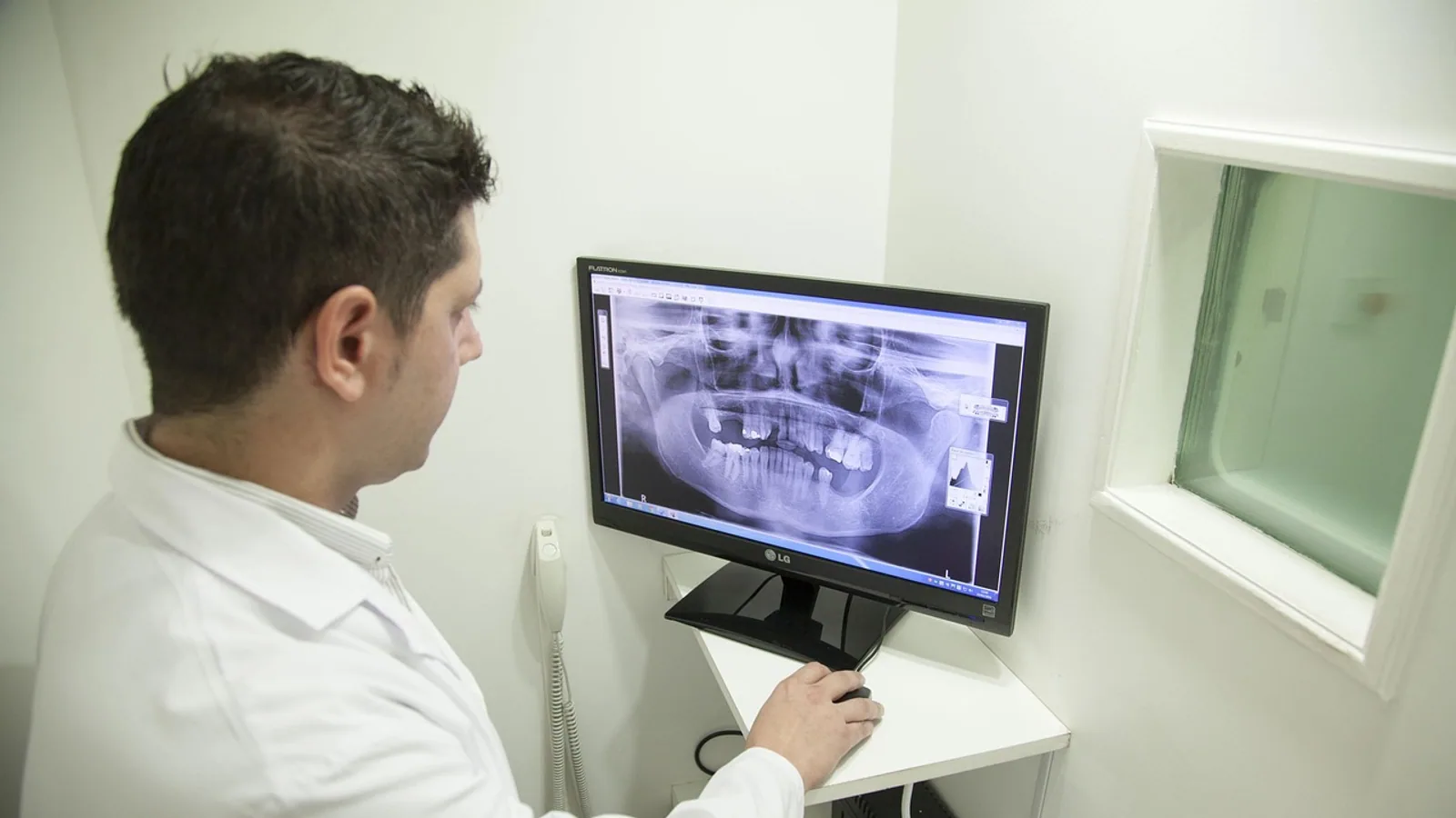
The coronoid process of the mandible is a thin, pointed bony projection that rises from the rostral edge of the mandibular ramus and serves as the primary insertion site for the temporalis muscle. A second, structurally distinct coronoid process projects from the proximal ulna and contributes to elbow joint congruity, and the two structures share a name but nothing else.
Students lose marks on this topic for one predictable reason. They read "coronoid process" in a question about the jaw and answer with the elbow, or the reverse. The confusion is understandable because both are named for the same Greek root, korone, meaning crow's beak, and both are pointed projections that anchor soft tissue. In clinical practice the two structures rarely appear in the same case, so the mix-up is a study problem rather than a diagnostic one. Once you separate them by bone, by joint, and by muscle attachment, the anatomy becomes straightforward.
This article covers the mandibular coronoid process in dogs, cats, horses, and cattle, the ulnar coronoid process in dogs, the temporalis insertion, comparative size differences between carnivores and herbivores, and the two clinical scenarios that bring each structure into the operating room.
What the Coronoid Process Is and Where It Sits


The coronoid process is a dorsally directed projection of bone at the rostral border of the mandibular ramus. It sits between the mandibular notch caudally and the alveolar margin of the last cheek teeth rostrally, and it forms the rostral wall of the temporal fossa when the jaw is closed. Its medial surface faces the pterygoid muscles, and its lateral surface faces the masseter and the zygomatic arch.
The process is not a static shelf. It is a stress-bearing enthesis, meaning a site where tendon meets bone and transmits load. Micro- and nano-level analysis of the coronoid process apex shows a fibrocartilaginous layer at the tendon-bone junction that acts as a stretching brake to control stress concentration [1]. That detail matters because it explains why the process remodels under load and why abnormal loading, as in a dysplastic process, produces measurable downstream effects on the muscle itself.
Developmentally, the process does not self-differentiate. In human embryos and fetuses the temporal and masseter muscle anlagen appear before the skeleton they attach to, and the coronoid process differentiates as a discrete entity inside the mass of the temporal muscle anlage at roughly 7 to 7.5 weeks of fertilization age [2]. The process then unites with the main portion of the ramus at about eight weeks [2]. The sequence is muscle first, bone second, which is the opposite of what many students assume and which explains why anything that disrupts the temporalis during development also disrupts the process.
The Two Coronoid Processes Are Not Related
The mandibular coronoid process belongs to the skull. The ulnar coronoid process belongs to the forelimb. They differ in bone, joint, muscle attachment, and clinical behavior. The only shared feature is shape.
| Feature | Mandibular coronoid process | Ulnar coronoid process |
|---|---|---|
| Bone | Mandible (ramus) | Ulna (proximal) |
| Joint | Temporomandibular joint region | Elbow joint |
| Main attachment | Temporalis muscle | Joint capsule and collateral ligament complex |
| Primary function | Leverage for jaw closure | Elbow congruity and rotational stability |
| Comparative note | Large in carnivores, reduced in herbivores | Prominent in dogs, clinically important |
| Clinical condition | Coronoidectomy, coronoid dysplasia | Fragmented medial coronoid process |
Temporalis Attachment: The Core Function
The temporalis muscle inserts on the mandibular coronoid process. This is the single most examinable fact in the topic, and it is also the fact most often stated too simply.
The classic description is a single muscle layer originating at the temporal line, converging to a tendon, and inserting onto a narrow site of the process. Dissection work shows a more complex picture. The temporalis ends at two distinct terminal tendons with wider insertion sites than textbooks present, and it separates into two parts that act as a single structural unit [3]. The superficial part is the large fan-shaped muscle most people picture. It converges inferomedially to form the superficial tendon and the lateral boundary of the retromolar triangle. The deep part is a narrow, vertically oriented rectangular muscle that converges posterolaterally [3].
Additional dissection has identified three parts in cadaveric specimens: the classically recognized superficial part, a zygomatic part, and a complex deep part [4]. The deep temporalis contains muscle bundles that insert into the internal aspect of the coronoid process and the retromolar triangle, and these interdigitate with the buccinator, mylohyoid, and superior constrictor muscles [4]. The zygomatic part originates from the zygomatic arch and inserts into the superficial part of the temporalis as that part inserts into the lateral surface of the coronoid process [4].
A systematic review of distal temporalis insertions across dissection, histology, and imaging identified three bony insertion patterns: on or through the coronoid, and additional insertions beyond the process [5]. The practical takeaway is that the temporalis footprint on the coronoid process is larger and more layered than a single tendon line.
What the Temporalis Does Once It Is Attached
The temporalis is a jaw-closing muscle. Its fibers run from the temporal fossa downward and forward to the coronoid process, so contraction pulls the process dorsocaudally and closes the mouth. Because the process sits rostral to the temporomandibular joint, the muscle has a mechanical advantage for elevation of the mandible and for retraction.
The relationship between muscle and bone is bidirectional. Functional pressure from the temporalis affects bone quality and strength at the process, and the orientation of biological apatite crystallites differs by site, being unidirectional in the mesiodistal direction at the apex and oriented vertical to the occlusal plane elsewhere [1]. Young's modulus varies with that crystallite orientation [1]. In other words, the bone is built to match the direction of pull.
The relationship is also developmental. In a transgenic mouse model with a dysplastic coronoid process, the temporalis developed normally up to the point of attachment, then showed altered orientation and a reduced cross-sectional area from that point onward, persisting to weaning [6]. The mutant mice acquired long-face morphology with anterior open bite during postnatal growth [6]. A process that does not form correctly changes the muscle that pulls on it, and that changed muscle changes the growing face.
Accessory Attachments and Surgical Anatomy
The temporalis has accessory attachments beyond the coronoid process. Cadaveric dissection found multiple accessory attachments to adjacent structures, including the masseter and pterygoid muscles, which explains why sliding the muscle free is more difficult than the textbook insertion suggests [7].
Variation occurs in the other direction as well. A case report described unduly extensive temporalis attachment in which extra fleshy fibers completely occupied the mandibular notch and descended to insert on the coronoid process plus the external and internal surfaces of the ramus [8]. The mandibular notch in that specimen was 3.1 cm wide [8]. Such variations matter for flap design, for radiographic interpretation, and for any procedure that releases the muscle.
Comparative Anatomy Across Species
The relative size of the coronoid process tracks diet and jaw mechanics. Carnivores have a large process because they need a powerful temporalis for a snapping bite and for holding struggling prey. Herbivores have a reduced process because their jaw mechanics favor a wide grinding stroke driven more by the masseter and pterygoid muscles, with less demand for a tall coronoid lever.
Dog
The dog has a well-developed coronoid process with a broad base and a strong temporalis insertion. The process is tall relative to the ramus, which gives the temporalis a long lever arm for jaw closure. The dog also has a prominent ulnar coronoid process at the elbow, described below.
Cat
The cat follows the carnivore pattern with a large, sharply pointed coronoid process and a dominant temporalis. The feline jaw is built for a rapid, forceful bite, and the coronoid lever reflects that. The process is proportionally similar to the dog's, though the skull is shorter and the temporal fossa is correspondingly compressed.
Horse
The horse has a reduced coronoid process relative to body size. The equine mandible is long and the ramus is deep, but the coronoid projection is modest because the masticatory pattern is a lateral grinding stroke dominated by the masseter and the pterygoid muscles. The temporalis is present and functional but does not drive the jaw the way it does in a carnivore.
Cow
The cow shows the herbivore pattern even more clearly. The coronoid process is small and the temporalis is correspondingly reduced. Ruminant mastication relies on a wide circular grinding motion, and the muscle mass sits in the masseter and pterygoid groups. The coronoid process still anchors the temporalis, but it is not a dominant landmark on the bovine mandible.
Summary of Species Differences
| Species | Coronoid process size | Dominant jaw muscle pattern | Clinical note |
|---|---|---|---|
| Dog | Large, broad base | Temporalis-dominant closure | Ulnar coronoid process clinically important at elbow |
| Cat | Large, sharply pointed | Temporalis-dominant closure | Carnivore pattern |
| Horse | Reduced | Masseter and pterygoid grinding | Herbivore pattern |
| Cow | Small | Masseter and pterygoid grinding | Herbivore pattern |
The Ulnar Coronoid Process in Dogs
The ulnar coronoid process is a separate structure on the proximal ulna. In dogs it is a prominent medial projection that articulates with the humeral condyle and contributes to elbow congruity. It sits adjacent to the radial head and the trochlear notch, and it is part of the load-bearing surface of the elbow.
Its role is mechanical. The medial coronoid process helps maintain congruent contact between the ulna and the humerus through the range of motion, and it shares load with the radial head. When the process is intact and properly seated, the elbow glides. When it is not, the joint loses congruity and the cartilage pays the price.
The clinical condition tied to this structure is fragmented medial coronoid process, usually abbreviated FMCP. The process separates or fissures, and the resulting fragment irritates the joint, causes pain, and leads to osteoarthritis. It is one of the components of elbow dysplasia in dogs. The condition is diagnosed by imaging and confirmed at surgery or arthroscopy, and treatment ranges from fragment removal to more extensive procedures depending on the degree of joint change.
Students should keep the two processes separate in their notes. The mandibular coronoid process anchors the temporalis and is involved in jaw surgery. The ulnar coronoid process maintains elbow congruity and is involved in lameness workups. A dog with a fragmented medial coronoid process has an elbow problem, not a jaw problem.
How the Anatomy Is Observed and Tested
Anatomical study of the mandibular coronoid process relies on dissection, histology, and imaging. Cadaveric dissection with careful sketching against bony landmarks is the standard method for mapping the temporalis tendon footprint [3]. Histology adds the tendon-bone interface detail, including the fibrocartilaginous layer at the apex [1]. Imaging, including CT and MRI, allows the muscle parts and their insertions to be visualized in living subjects and has been used to confirm the confluence of deep temporalis fibers with the buccinator [4].
For the ulnar coronoid process, radiographic and cross-sectional imaging of the elbow is the practical route. Survey radiographs, CT, and arthroscopy each contribute, and the choice depends on what the clinician needs to see and what the patient can tolerate.
In the gross anatomy lab, the fastest way to keep the two structures straight is to identify the bone first. If the specimen is a skull or hemimandible, the process you are looking at is mandibular. If the specimen is a forelimb, it is ulnar. Then identify the joint. The mandibular process sits at the temporomandibular joint region. The ulnar process sits at the elbow.
Clinical Relevance, Limitations and Common Mistakes
Coronoidectomy in Oral Surgery
Coronoidectomy is surgical removal of the mandibular coronoid process. It is performed for several reasons, and the shared logic is that releasing the temporalis insertion frees the muscle.
Temporalis muscle transfer for facial reanimation depends on this release. In lengthening temporalis myoplasty, the coronoid tendinous insertions are transferred to the lip and nasolabial fold, and the freed tendon is 4 to 6 cm wide [9]. Orthodromic transfer techniques release the insertion from the coronoid process and suture it to fascial strips anchored at the commissure and paralyzed hemilip [10]. A related technique exposes the coronoid process through a preauricular incision, transects it with a saw, and anchors fascia lata strips to the nasolabial fold musculature [11]. A further refinement mobilizes the coronoid process in continuity with the temporalis insertion through a nasolabial fold approach [12].
Coronoidectomy also extends the reach of a temporalis muscle flap. Releasing the muscle insertion from the coronoid process increases flap length and arc of rotation, adding at least 2 cm of length, which is enough to cover defects crossing the midline and can make a unilateral flap sufficient where bilateral flaps were previously needed [13]. The rotation point shifts from the level of the zygomatic arch to the lower part of the mandibular neck [13].
In neurosurgery, splitting the temporalis vertically in the direction of its coronoid insertion up to the zygomatic arch reduces obstruction during subtemporal approaches, and the technique preserved muscle function and bulk in the reported cases [14].
In temporomandibular joint replacement, the coronoid process is often resected because it carries the temporalis insertion. In a prospective study of unilateral total alloplastic joint replacement, the coronoid process was resected in 11 of 14 patients, and postoperative surface electromyography showed relative symmetry above 72 percent in both the masseter and anterior temporalis muscles [15]. Detaching the muscle did not abolish its activity.
A less common application is intraoral transpositional plastic surgery of the coronoid process for recurrent temporomandibular joint luxation. The procedure shifts the temporalis insertion so that extreme opening provokes an early stretch reflex and limits mouth opening [16].
Fragmented Medial Coronoid Process in Dogs
FMCP is the canine clinical condition tied to the ulnar coronoid process. It presents as forelimb lameness, often in young dogs of predisposed breeds, and it is part of the elbow dysplasia complex. Diagnosis uses imaging and arthroscopy. Treatment aims to remove or address the fragment and manage the secondary osteoarthritis. Because the process is a load-bearing part of the elbow, losing it changes joint mechanics, and long-term management often includes weight control and activity modification alongside surgical intervention.
Common Mistakes
The first mistake is mixing up the two coronoid processes. Anchor each to its bone and joint and the problem disappears.
The second is treating the temporalis insertion as a single point. The footprint is wide, layered, and sometimes extends beyond the process, with accessory attachments to adjacent muscles [7][8]. Surgical planning that assumes a simple tendon line will underestimate the release needed.
The third is assuming the muscle and bone develop independently. The process forms within the temporalis anlage, and disrupting one disrupts the other [2][6].
The fourth is assuming that detaching the temporalis eliminates its function. Postoperative electromyography after coronoid resection showed preserved and relatively symmetric activity [15].
The fifth is reading a "coronoid" finding on a radiograph without checking which joint was imaged.
This article is educational and is not a substitute for veterinary diagnosis or treatment.
Quick Review
- The mandibular coronoid process is a projection of the mandibular ramus and the insertion site of the temporalis muscle.
- The ulnar coronoid process is a separate structure on the proximal ulna that contributes to elbow congruity in dogs.
- The temporalis insertion is wider and more layered than the classic single-tendon description, with superficial, deep, and zygomatic parts.
- The coronoid process is large in carnivores such as dogs and cats and reduced in herbivores such as horses and cattle.
- The process develops within the temporalis anlage, so muscle and bone influence each other during growth.
- Coronoidectomy is used in facial reanimation, temporalis flap mobilization, and temporomandibular joint surgery.
- Fragmented medial coronoid process is a canine elbow condition, not a jaw condition.
Frequently Asked Questions
What muscle attaches to the coronoid process of the mandible?
The temporalis muscle inserts on the mandibular coronoid process. Its fibers converge from the temporal fossa onto the process, and the insertion includes superficial and deep components with a wider footprint than most textbook descriptions show.
Is the coronoid process the same in the jaw and the elbow?
No. The mandibular coronoid process is part of the jaw and anchors the temporalis. The ulnar coronoid process is part of the proximal ulna and contributes to elbow congruity. They share a name and a pointed shape and nothing else.
Why is the coronoid process large in dogs and small in horses?
Size tracks jaw mechanics. Carnivores need a powerful temporalis for a forceful bite, so the process is tall and the lever arm is long. Herbivores rely on a grinding stroke driven by the masseter and pterygoid muscles, so the temporalis and its bony attachment are reduced.
What is fragmented medial coronoid process in dogs?
It is a condition in which the medial coronoid process of the ulna separates or fissures, causing elbow pain and osteoarthritis. It is part of the elbow dysplasia complex and is managed with imaging, surgery, and long-term joint care.
Why would a surgeon remove the coronoid process?
Removing the process releases the temporalis insertion. Surgeons do this to transfer the muscle for facial reanimation, to extend the reach of a temporalis muscle flap, and to clear the field during temporomandibular joint replacement.
Does removing the coronoid process stop the temporalis from working?
No. A prospective study of unilateral temporomandibular joint replacement, in which the coronoid process was resected in most patients, found preserved and relatively symmetric masseter and anterior temporalis activity after surgery.
Related Articles
- Equine Larynx Anatomy and Function in Respiration
- Canine Lymphatic System: Anatomy and Function
- Equine Hoof Anatomy: Structures and Function
- Fish Gills: Anatomy, Function, and Common Health Issues
- Equine Muscular System: Major Muscle Groups and Function
- Ruminant Digestive Anatomy and Physiology: A Clinical Reference
- Coracoid Process: Anatomy and Comparative Notes
- How Roosters Fertilize Eggs: Step-by-Step Process
- Greater Trochanter: Anatomy, Attachments, and Role
Sources
- Micro- and nano-bone analyses of the human mandible coronoid process and tendon-bone entheses.
- The morphogenetic relationship of the temporal muscle to the coronoid process in human embryos and fetuses.
- Morphology of the temporalis muscle focusing on the tendinous attachment onto the coronoid process.
- The human temporalis muscle: superficial, deep, and zygomatic parts comprise one structural unit.
- Towards an integrative understanding of the human temporalis muscle: Anatomical, histological, and imaging evidence.
- Association of Dysplastic Coronoid Process with Long-Face Morphology.
- The temporalis muscle and its relationship to the accessory attachments and the main pedicle-a cadaveric study.
- Unduly extensive mandibular insertion of temporalis muscle: A case report.
- Anatomic study of the tendinous insertion lamina of the temporalis muscle.
- Facial reanimation utilizing combined orthodromic temporalis muscle flap and end-to-side cross-face nerve grafts.
- Orthodromic transfer of the temporalis muscle in incomplete facial nerve palsy.
- Temporalis muscle transfer for facial paralysis: a further refinement.
- New method for maximum mobilization of temporalis muscle flap.
- Splitting of temporalis muscle for basal exposure--technical note.
- How Does a Unilateral Temporomandibular Joint Replacement Affect Bilateral Masseter and Temporalis Muscle Activity?-A Prospective Study.
- [[Intraoral transpositional plastic surgery of coronoid process in treatment of recurrent TMJ luxation. Preliminary report].](https://pubmed.ncbi.nlm.nih.gov/1818633/)