Cerebral Peduncles: Brain Anatomy Explained
By Dr. Zubair Khalid, DVM, MS, PhD ·
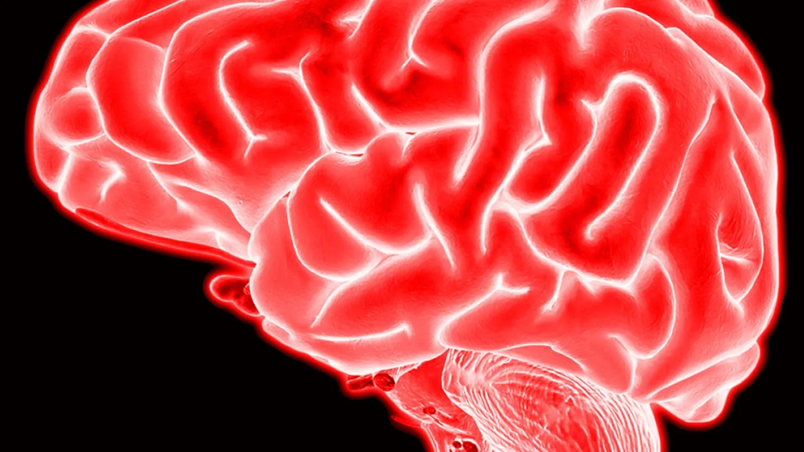
The cerebral peduncles are the paired, pillar-like structures on the ventral surface of the midbrain. Each cerebral peduncle (Latin: pedunculus cerebri) contains a ventral crus cerebri packed with descending motor fibers and a dorsal tegmentum that carries ascending sensory and integrative pathways.
They are not the same as the cerebellar peduncles. The cerebellar peduncles are three pairs of stalks (superior, middle, inferior) that connect the cerebellum to the brainstem. The cerebral peduncles belong to the midbrain and connect the forebrain to the hindbrain. Confusing the two is the single most common naming error in neuroanatomy, and it matters because the two structures carry completely different tracts and fail in different ways.
This article explains what the cerebral peduncles are, what runs through them, how they look across domestic species, and why a veterinarian or biomedical student should care about a structure roughly the size of a pencil eraser.
This article is educational and is not a substitute for veterinary diagnosis or treatment.
Where the Cerebral Peduncles Sit in the Brain
The brainstem has three parts stacked from top to bottom: midbrain (mesencephalon), pons, and medulla oblongata. The cerebral peduncles form the most ventral part of the midbrain. They look like two thick columns that emerge from under the thalamus and plunge downward toward the pons.
Each peduncle has two functional floors:
- Crus cerebri (also called the basis pedunculi). The ventral, superficially visible surface. This is a solid highway of descending cortical fibers.
- Tegmentum. The dorsal region, hidden behind the crus. It holds ascending tracts, cranial nerve nuclei, the red nucleus, and the reticular formation.
The substantia nigra sits between them, a pigmented band of dopamine-producing neurons that separates the crus cerebri from the tegmentum. In fresh tissue the substantia nigra is visibly dark because of neuromelanin pigment. It belongs to neither the crus nor the tegmentum in a strict functional sense, but anatomically it forms the boundary between the two.
Between the two cerebral peduncles on the ventral surface is a deep, diamond-shaped depression called the interpeduncular fossa. The floor of this fossa is the posterior perforated substance, pierced by small penetrating arteries. The oculomotor nerve (cranial nerve III) exits the midbrain on the medial edge of each cerebral peduncle, in the wall of the interpeduncular fossa. This is a high-yield landmark. A lesion at the medial peduncle can produce an ipsilateral oculomotor palsy plus contralateral hemiparesis, a classic brainstem syndrome.
The Crus Cerebri: Descending Motor Highways
The crus cerebri is the ventral third of each peduncle. It is made almost entirely of descending axons from the cerebral cortex. Three fiber systems run through it, arranged in a consistent medial-to-lateral order:
1. Frontopontine fibers (medial)
These originate from the frontal lobe and terminate in the pontine nuclei. They are part of the corticopontine system, which feeds the cerebellum with information about intended movement. In the crus they occupy the most medial fifth.
2. Corticospinal and corticobulbar fibers (middle)
This is the pyramidal tract. Corticospinal fibers travel from the motor cortex down to the spinal cord. Corticobulbar fibers (also called corticonuclear fibers) end on cranial nerve motor nuclei in the brainstem. In the crus cerebri they occupy the middle three-fifths. This is the single most clinically important occupant of the cerebral peduncle.
The corticospinal tract (CST) is the main voluntary motor pathway. Its integrity can be measured with diffusion tensor imaging (DTI), which calculates fractional anisotropy (FA), a number between 0 and 1 that reflects how directionally organized the white matter is. Higher FA means healthier, more coherent axons. In human stroke research, FA measured at the cerebral peduncle correlates with motor function and walking ability. One study of 40 subacute stroke patients found that the FA ratio at the cerebral peduncle correlated with lower limb motor scores and walking ability [1]. Another study of 36 chronic stroke patients used cerebral peduncle area asymmetry as one of three imaging markers of CST integrity [2]. A third study of 124 patients with intracerebral hemorrhage found that FA asymmetry restricted to the cerebral peduncle was associated with poor ambulation at 3 months [3].
These human studies matter to veterinary readers because the anatomy is conserved. The dog, cat, and horse all have a corticospinal tract that passes through the crus cerebri, though the relative size and functional importance differ by species.
3. Temporopontine, parietopontine, and occipitopontine fibers (lateral)
These corticopontine fibers arise from the temporal, parietal, and occipital lobes and end in the pontine nuclei. They occupy the lateral fifth of the crus. Together with the frontopontine fibers, they form the corticopontocerebellar pathway, which is how the cortex tells the cerebellum what movement it intends to make.
The Tegmentum: Ascending and Integrative Territory
The tegmentum is the dorsal part of each cerebral peduncle, sitting behind the substantia nigra. It is more complex than the crus because it contains multiple systems:
- Ascending sensory tracts. The medial lemniscus (touch and proprioception), spinothalamic tracts (pain and temperature), and trigeminothalamic tracts pass through the tegmentum on their way to the thalamus.
- Red nucleus. A large motor relay that receives input from the cerebellum and motor cortex and gives rise to the rubrospinal tract.
- Cranial nerve nuclei. The oculomotor (III) and trochlear (IV) nuclei sit in the midbrain tegmentum.
- Reticular formation. The midbrain reticular formation contributes to arousal and motor tone.
- Substantia nigra. Technically between crus and tegmentum, but functionally part of the midbrain motor system. The pars compacta produces dopamine and projects to the basal ganglia. The pars reticulata is a GABAergic output nucleus.
The tegmentum is where the midbrain integrates movement, sensation, and arousal. Damage here produces a different clinical picture than damage to the crus. A crus lesion causes weakness. A tegmental lesion causes sensory loss, eye movement deficits, or movement disorders.
Cerebral Peduncles vs Cerebellar Peduncles
This distinction deserves its own section because students and clinicians mix them up constantly.
| Feature | Cerebral peduncles | Cerebellar peduncles |
|---|---|---|
| Location | Ventral midbrain | Between cerebellum and brainstem |
| Number | One pair | Three pairs (superior, middle, inferior) |
| Main contents | Corticospinal, corticobulbar, corticopontine fibers (crus); ascending tracts and nuclei (tegmentum) | Superior: mostly efferent from cerebellum. Middle: massive corticopontocerebellar input. Inferior: afferent from spinal cord and medulla |
| Clinical signature | Contralateral hemiparesis, oculomotor palsy | Cerebellar ataxia, intention tremor, dysmetria |
| Key landmark | Interpeduncular fossa, oculomotor nerve exit | Fourth ventricle, cerebellar hemispheres |
The cerebellar peduncles are named for what they connect: the cerebellum. The cerebral peduncles are named for what they connect: the cerebrum to the brainstem. Both are "peduncles" in the sense of a stalk or stem, but they are in different parts of the brain and carry different traffic.
A useful memory hook: cerebral peduncles are in the midbrain and carry the pyramidal tract. Cerebellar peduncles are at the back of the brainstem and carry cerebellar input and output.
Species Differences in the Cerebral Peduncles
The basic plan is conserved across mammals, but the size and prominence of the cerebral peduncles vary with the size of the corticospinal tract, which in turn varies with the animal's reliance on fine distal limb control.
Dog
The dog has a well-developed corticospinal tract, though it is smaller relative to body size than in primates. The cerebral peduncles are prominent and easy to identify on ventral brainstem dissection. The crus cerebri carries corticospinal fibers that contribute to voluntary limb movement, but the dog relies heavily on brainstem and cerebellar pathways for locomotion. The red nucleus and rubrospinal tract are relatively more important in dogs than in humans.
Cat
The cat's corticospinal tract is smaller than the dog's. The cerebral peduncles are still clearly visible but less bulky. Cats have excellent postural and righting reflexes that depend on brainstem and cerebellar circuits rather than the pyramidal tract. The tegmentum is well developed because of the importance of the red nucleus and reticular formation in feline motor control.
Horse
The horse has a large brainstem and prominent cerebral peduncles. The corticospinal tract is present but relatively small compared to the size of the animal. Horses depend on brainstem and cerebellar control for gait and posture. The interpeduncular fossa and oculomotor nerve exit are consistent landmarks. The equine midbrain is large enough that a skilled anatomist can dissect the crus cerebri and tegmentum separately.
Human
The human cerebral peduncles are proportionally large because the corticospinal tract is massive. The crus cerebri is the dominant feature, and the corticospinal fibers within it are the main target of stroke imaging research. The human literature on cerebral peduncle lesions is extensive, including case reports of inflammatory lesions presenting as painful tonic spasms [4] and multiple sclerosis lesions in the lateral cerebral peduncle [5]. Human studies also use cerebral peduncle FA as a biomarker for motor recovery after stroke, hemorrhage, and tumor surgery [6][1][2][3][7].
Comparative summary table
| Component | Main tract or nucleus | Function | Dog | Cat | Horse | Human |
|---|---|---|---|---|---|---|
| Crus cerebri (medial) | Frontopontine fibers | Cortical input to pons | Present | Present | Present | Prominent |
| Crus cerebri (middle) | Corticospinal, corticobulbar | Voluntary motor output | Well developed | Moderate | Small relative to size | Massive |
| Crus cerebri (lateral) | Temporo/parieto/occipitopontine | Cortical input to pons | Present | Present | Present | Prominent |
| Substantia nigra | Pars compacta, pars reticulata | Dopamine output, movement modulation | Present | Present | Present | Present |
| Tegmentum | Medial lemniscus, spinothalamic, red nucleus, CN III/IV nuclei | Sensory relay, motor integration, eye movement | Well developed | Well developed | Well developed | Well developed |
| Interpeduncular fossa | Oculomotor nerve exit | CN III emerges | Present | Present | Present | Present |
What the Cerebral Peduncles Do: Function by Component
Corticospinal tract
The CST carries voluntary motor commands from the motor cortex to the spinal cord. It passes through the corona radiata, internal capsule, crus cerebri, pons, and medulla, where most fibers cross the midline (pyramidal decussation) and descend in the lateral corticospinal tract. A smaller uncrossed portion forms the anterior corticospinal tract. In humans, CST damage causes contralateral weakness. In animals, the deficit is less severe because of redundancy in brainstem motor pathways.
DTI studies in humans show that CST integrity at the cerebral peduncle predicts motor recovery. A study of 81 patients with brain tumors found that the FA ratio at the cerebral peduncle was significantly higher in patients who recovered motor function after surgery than in those who did not [7]. Another study of 69 chronic stroke patients found that FA in the cerebral peduncle was reduced in the most impaired gait clusters [8]. A longitudinal study of basal ganglia infarction found that FA in the cerebral peduncle decreased progressively over 90 days and that the 7-day cerebral peduncle FA ratio correlated with 180-day motor scores [9].
Corticobulbar tract
Corticobulbar fibers end on cranial nerve motor nuclei. They control facial expression, mastication, swallowing, and tongue movement. In the crus cerebri they travel with corticospinal fibers. A lesion in the crus can cause both limb weakness and cranial nerve deficits, depending on the exact location.
Corticopontine tract
Corticopontine fibers end in the pontine nuclei, which project to the cerebellum via the middle cerebellar peduncle. This pathway lets the cortex inform the cerebellum about planned movements. It is essential for smooth, coordinated motor activity. The frontopontine fibers occupy the medial crus, and the temporo/parieto/occipitopontine fibers occupy the lateral crus.
Tegmental systems
The tegmentum carries ascending sensory information and houses motor nuclei. The medial lemniscus carries fine touch and proprioception from the contralateral side of the body. The spinothalamic tract carries pain and temperature. The red nucleus coordinates motor output from the cerebellum and cortex. The oculomotor and trochlear nuclei control eye movements. Damage to the tegmentum can cause sensory loss, ataxia, or diplopia.
Clinical Relevance, Limitations and Common Mistakes
Why the cerebral peduncles matter clinically
The cerebral peduncles are a bottleneck. All corticospinal and corticobulbar fibers pass through the crus cerebri on their way from the cortex to the brainstem and spinal cord. A small lesion here can cause disproportionately large motor deficits. This is why the cerebral peduncle is a key landmark in stroke imaging and surgical planning.
In humans, cerebral peduncle lesions have been reported in inflammatory conditions. A case report described a post-COVID inflammatory lesion in the right cerebral peduncle that presented as painful tonic spasms [4]. Another reported a lateral cerebral peduncle lesion in a patient with multiple sclerosis, also presenting with painful tonic spasms [5]. A series of three patients with autoimmune GFAP astrocytopathy showed delayed bilateral cerebral peduncle lesions on follow-up MRI [10]. These cases show that the cerebral peduncle can be a target of immune-mediated injury, not just vascular disease.
In veterinary medicine, cerebral peduncle lesions are less commonly reported but can occur with trauma, neoplasia, inflammatory disease, and vascular events. The clinical signs depend on which part of the peduncle is affected. Crus lesions cause motor deficits. Tegmental lesions cause sensory or cranial nerve deficits.
Common mistakes
Mistake 1: Confusing cerebral and cerebellar peduncles. This is the most frequent error. The cerebral peduncles are in the midbrain. The cerebellar peduncles connect the cerebellum to the brainstem. They are not the same structure and do not carry the same tracts.
Mistake 2: Assuming the cerebral peduncle is only a motor structure. The crus cerebri is motor, but the tegmentum carries sensory and integrative pathways. A lesion in the tegmentum can cause sensory loss or eye movement deficits.
Mistake 3: Forgetting the substantia nigra. The substantia nigra sits between the crus and the tegmentum. It is not part of either, but it is part of the cerebral peduncle region. Dopaminergic loss here causes Parkinson's disease in humans and has been linked to movement disorders in animals.
Mistake 4: Overlooking the oculomotor nerve. The oculomotor nerve exits between the cerebral peduncles in the interpeduncular fossa. A lesion at the medial peduncle can compress the nerve and cause ipsilateral pupil dilation and eye movement palsy. This is a clinical clue that the lesion is at the midbrain level.
Mistake 5: Assuming animal and human motor deficits are identical. Animals have more redundant brainstem motor pathways. A dog with a partial corticospinal lesion may still walk, while a human with the same lesion may have severe hemiparesis. Species differences in the size and importance of the corticospinal tract explain this.
Limitations
This article covers the gross and functional anatomy of the cerebral peduncles. It does not cover embryological development in detail or human-specific stroke syndromes. Individual cases require a veterinarian for diagnosis and treatment. Imaging findings such as FA values are research tools and are not a substitute for clinical examination.
Practical Implications for Owners and Keepers
If your animal has a neurological deficit that localizes to the brainstem, the cerebral peduncles may be involved. Signs to watch for include:
- Weakness on one side of the body (hemiparesis)
- Difficulty with eye movement or a drooping eyelid
- Facial weakness or difficulty swallowing
- Changes in gait or coordination
- Abnormal postural reactions
These signs are not specific to the cerebral peduncles. They can occur with lesions anywhere in the brainstem or cortex. A veterinarian will use neurological examination and imaging to localize the problem.
If your animal is diagnosed with a midbrain lesion, the prognosis depends on the cause. Inflammatory lesions may respond to immunosuppressive treatment. Vascular lesions may stabilize or improve with supportive care. Tumors may require surgery or radiation. The cerebral peduncle itself is not a diagnosis. It is a location.
For students and technicians, the cerebral peduncles are a high-yield anatomy topic. Learn the crus cerebri, the tegmentum, the substantia nigra, and the interpeduncular fossa. Know the three fiber systems in the crus and their medial-to-lateral arrangement. Know that the oculomotor nerve exits medially. These facts will serve you in neurology, surgery, and imaging.
What Is Still Uncertain
The role of the cerebral peduncle in veterinary motor recovery is not well studied. Most DTI research on the cerebral peduncle comes from human stroke and tumor patients [6][1][2][3][8][7][9]. We do not have equivalent large-scale studies in dogs, cats, or horses. The anatomy is conserved, but the functional recovery patterns may differ because of differences in corticospinal tract size and reliance on brainstem pathways.
The significance of cerebral peduncle lesions in inflammatory diseases is also an active area. Human case reports describe lesions in COVID-related inflammation [4], multiple sclerosis [5], and GFAP astrocytopathy [10]. Whether similar lesions occur in animals with inflammatory brain disease is not well documented.
Finally, the exact contribution of the corticopontine fibers to motor learning and coordination is still being mapped. The cerebral peduncle is a conduit, but how the brain uses the information that passes through it is a question for ongoing research.
Frequently Asked Questions
What are the cerebral peduncles?
The cerebral peduncles are paired ventral midbrain structures. Each contains a crus cerebri with descending motor fibers and a tegmentum with ascending sensory and integrative pathways.
Are cerebral peduncles the same as cerebellar peduncles?
No. Cerebral peduncles are in the midbrain and carry corticospinal, corticobulbar, and corticopontine fibers. Cerebellar peduncles connect the cerebellum to the brainstem and carry cerebellar input and output.
What tracts run through the crus cerebri?
The crus cerebri carries frontopontine fibers medially, corticospinal and corticobulbar fibers in the middle, and temporo/parieto/occipitopontine fibers laterally.
What is the substantia nigra?
The substantia nigra is a pigmented band of dopamine-producing neurons between the crus cerebri and the tegmentum. It modulates movement through projections to the basal ganglia.
Where does the oculomotor nerve exit?
The oculomotor nerve exits the midbrain on the medial edge of each cerebral peduncle, in the wall of the interpeduncular fossa.
Do dogs and cats have cerebral peduncles?
Yes. All mammals have cerebral peduncles. The size and prominence vary with the size of the corticospinal tract, which is larger in humans and smaller in cats and horses.
What happens if the cerebral peduncle is damaged?
Damage to the crus cerebri causes contralateral motor weakness. Damage to the tegmentum causes sensory loss, eye movement deficits, or movement disorders. The exact signs depend on the location and extent of the lesion.
Can the cerebral peduncle recover after injury?
Recovery depends on the cause and severity. Human studies show that corticospinal tract integrity at the cerebral peduncle correlates with motor recovery after stroke and surgery [6][7]. The same principles likely apply in animals, but species-specific data are limited.
Related Articles
- Bovine Neurological Anatomy: Cranial Nerves and Brainstem
- Feline Toxoplasmosis: Neurological Manifestations and Cerebral Involvement
- Dog Shaking Head Brain Tumor
- Brain Single Cell Rna Seq
- Toxoplasmosis in Cats and the Risk of Brain Infection in Humans
- Why Is My Dog walking in circles? Vestibular Disease vs Brain Tumor
- Brain Ventricles: Anatomy and Function of the Ventricular System
- Falx Cerebri: Anatomy, Function, and Dural Reflections
Sources
- The relationship between corticospinal tract and the affected lower limb function and walking ability in subacute stroke patients: a preliminary study.
- Asymmetry in fractional anisotropy of the corticospinal tract correlates with measures of body structure/function and activity in the chronic phase after stroke.
- Association of Corticospinal Tract Asymmetry With Ambulatory Ability After Intracerebral Hemorrhage.
- Post-coronavirus inflammatory cerebral peduncle lesion presenting as painful tonic spasms.
- Lacosamide-Responsive Paroxysmal Tonic Spasms Associated With a New Lateral Cerebral Peduncle Lesion in Multiple Sclerosis: A Case Report.
- Rehabilitation with hybrid assistive limb improves upper limb paralysis in patients with cerebral hemorrhage by repairing axonal injury of the corticospinal tract.
- Prediction of Motor Recovery Using Diffusion Tensor Imaging and Regional Cerebral Blood Flow in Postoperative Brain Tumors.
- Motor tract integrity in gait pattern clusters of patients with chronic Stroke: A diffusion tensor imaging and motion capture study.
- Secondary Degeneration of White Matter Tract following Basal Ganglia Infarction: A Longitudinal Diffusion Tensor Imaging Study.
- Delayed appearance of bilateral cerebral peduncle lesions in autoimmune glial fibrillary acidic protein astrocytopathy: A serial MRI case series.