Brain Ventricles: Anatomy and Function of the Ventricular System
By Dr. Zubair Khalid, DVM, MS, PhD ·
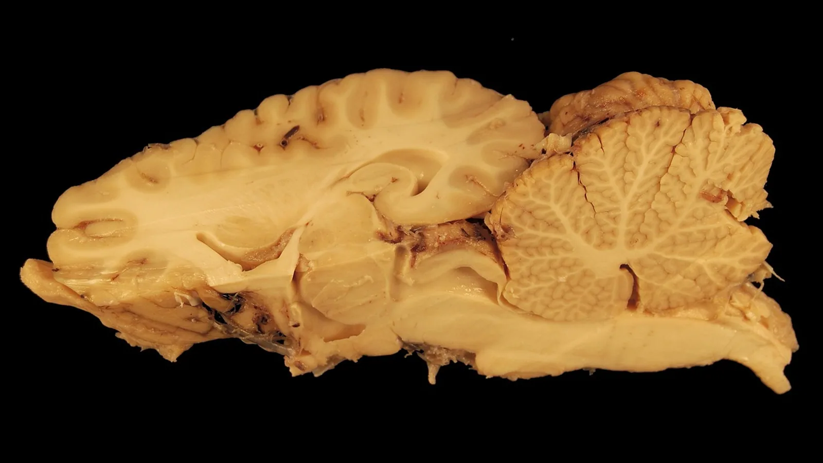
The brain ventricles are four interconnected, fluid-filled cavities inside the brain: two lateral ventricles, a third ventricle, and a fourth ventricle. Together they form the ventricular system of the brain, a continuous channel that produces cerebrospinal fluid (CSF), moves it through the central nervous system, and connects it to the subarachnoid space that bathes the outer surface of the brain and spinal cord.
This guide covers the anatomy of each ventricle, the narrow channels that link them, the flow path of CSF from its production sites to its exit points, and the core functions of the system: buoyancy, mechanical protection, and waste clearance. It also covers the biology behind these functions, the questions people ask most often, and what remains uncertain in current research.
What Are the Brain Ventricles?
The cerebral ventricles are cavities within the brain parenchyma, the functional tissue of the brain. They are lined by ependyma, a specialized ciliated epithelium that regulates local CSF flow and the exchange of substances between CSF and brain tissue [1]. The ventricles are not empty space. They are filled with CSF, a clear fluid that is produced continuously and circulates through the ventricular system and the subarachnoid space.
The ventricular system of the brain is often described as the body's "third circulation," alongside the blood circulation and the lymphatic system. That framing has been revised in recent years. Current models treat CSF dynamics as a broader system that includes the glymphatic pathway, in which CSF enters periarterial spaces and exchanges with interstitial fluid through aquaporin-4 channels on astrocytic endfeet, and meningeal lymphatic drainage, which carries waste-laden fluid out of the cranium [2].
The Four Ventricles of the Brain
Lateral Ventricles
The two lateral ventricles are the largest cavities in the ventricular system. Each sits in one cerebral hemisphere and has a characteristic C shape that follows the curve of the surrounding cortex. Anatomists divide each lateral ventricle into a frontal horn, a body, an occipital horn, and a temporal horn. The lateral ventricles are the origin point of the CSF circulation pathway, which makes them the target for intraventricular drug delivery and tracer studies [3].
Third Ventricle
The third ventricle is a narrow, midline cavity situated between the two halves of the diencephalon, the region that contains the thalamus and hypothalamus. It connects to each lateral ventricle through an interventricular foramen, also called the foramen of Monro. The third ventricle is a site where CSF flow patterns can become abnormal. In communicating hydrocephalus, researchers have observed rapid CSF flow and abnormal vortices in the third ventricle using 4-dimensional flow MRI [4].
Fourth Ventricle
The fourth ventricle is a diamond-shaped cavity located between the brainstem and the cerebellum. It receives CSF from the third ventricle through the cerebral aqueduct, a narrow channel that runs through the midbrain. CSF exits the fourth ventricle into the subarachnoid space through three openings: the median aperture (foramen of Magendie) and the two lateral apertures (foramina of Luschka).
Function Table: Ventricles of the Brain
| Ventricle | Location | Connections | Key Function |
|---|---|---|---|
| Lateral ventricles (two) | Cerebral hemispheres, one per side | Interventricular foramina of Monro to third ventricle | Largest CSF compartment, origin of the CSF circulation pathway, main site of intraventricular access [3] |
| Third ventricle | Midline, between the thalami | Foramina of Monro from lateral ventricles, cerebral aqueduct to fourth ventricle | Midline conduit, site of normal and abnormal CSF flow patterns [4] |
| Fourth ventricle | Between brainstem and cerebellum | Cerebral aqueduct from third ventricle, median and lateral apertures to subarachnoid space | Exit point of ventricular CSF into the subarachnoid space |
| Connecting channels | Foramina of Monro, cerebral aqueduct, median and lateral apertures | Link all four ventricles to the subarachnoid space | Maintain a continuous CSF pathway and set resistance to flow [5] |
How CSF Flows Through the Ventricular System
CSF flow follows a defined path. Fluid produced in the lateral ventricles passes through the interventricular foramina of Monro into the third ventricle. From there it moves through the cerebral aqueduct into the fourth ventricle. It then leaves the ventricular system through the median aperture and the two lateral apertures, entering the subarachnoid space that surrounds the brain and spinal cord.
The flow does not depend on a single pump. Vascular pulsations help propel CSF through the ventricular system and subarachnoid space, and the coordinated beating of ependymal cilia contributes locally [6]. Recent imaging work has shown that subarachnoid CSF flow in humans is brain-wide and spatiotemporally coherent, with strong cardiac-driven oscillations. Flow velocities are higher and responses come earlier in subarachnoid spaces near major arteries, and the pattern propagates outward to more distal spaces [7].
The diagram below traces the main CSF flow path from production to the subarachnoid space.
flowchart TD
    A[Choroid plexus produces CSF] --> B[Lateral ventricles]
    B --> C[Foramina of Monro]
    C --> D[Third ventricle]
    D --> E[Cerebral aqueduct]
    E --> F[Fourth ventricle]
    F --> G[Median aperture]
    F --> H[Lateral apertures]
    G --> I[Subarachnoid space]
    H --> I
    I --> J[Glymphatic exchange]
    I --> K[Meningeal lymphatic drainage]
CSF Production and Turnover
CSF is produced mainly by the choroid plexus, a vascular structure found in the walls of the ventricles. In humans, the choroid plexus produces roughly 500 mL of CSF per day. The total volume of CSF in the body at any moment is about 150 mL. That means the entire CSF volume turns over several times daily, roughly three to four times under normal conditions.
This high turnover matters for two reasons. First, it means the composition of CSF can be refreshed quickly, which supports stable ion concentrations around the brain. Second, it means that any substance introduced into the CSF, whether a drug, a tracer, or a waste product, is subject to continuous dilution and transport.
The choroid plexus is not a passive filter. Its epithelial cells are multiciliated and respond to mechanical stimuli from CSF flow. The mechanically activated ion channel Piezo1 is expressed on the apical and lateral membranes of mature choroid plexus epithelial cells, where it mediates calcium influx in response to fluid flow. Either knockout or overexpression of Piezo1 in these cells causes severe hydrocephalus in mice, reduces CSF flow, and disrupts ciliary maintenance [8]. This shows that CSF production is actively regulated, not just a fixed secretion rate.
Ion regulation is part of that control. The inwardly rectifying potassium channel Kir7.1 is prominently expressed at the apical membrane of choroid plexus epithelial cells and helps set the concentration of potassium in CSF. Mice carrying a pathological mutation in this channel show reduced potassium conductance and significantly decreased CSF potassium concentration, while CSF secretion rates remain unchanged [9]. The system regulates what is in the fluid, not only how much fluid is made.
Functions of the Ventricular System
Buoyancy
The brain is a soft organ that weighs roughly 1,400 grams in adults. Suspended in CSF, its effective weight drops dramatically. This buoyancy reduces the mechanical load on the brain's own tissue and on the structures that anchor it, and it allows the brain to maintain its shape without resting its full weight on the skull base.
Protection
CSF acts as a hydraulic cushion. Because the fluid surrounds the brain and fills the ventricles, mechanical forces applied to the head are distributed and damped rather than concentrated on one region of cortex. The ventricular system also provides a route for pressure to equalize across compartments, which is why obstruction at a narrow point such as the cerebral aqueduct can produce region-specific changes in ventricular size.
Waste Clearance
CSF is a clearance medium. In the glymphatic pathway, CSF enters periarterial spaces and exchanges with interstitial fluid through aquaporin-4 channels on astrocytic endfeet, which promotes the removal of waste from brain parenchyma. Waste-laden fluid then exits along perivenous routes and drains out of the cranium through meningeal lymphatics [2]. This pathway links the ventricular system to the broader question of how the brain handles metabolic byproducts.
Imaging studies support the connection between CSF movement and waste transport. Higher regional CSF motility, measured as mean pseudo-diffusivity on low b-value diffusion MRI, is consistently associated with higher CSF concentrations of amyloid-beta 40, and CSF motility measures collectively explained about 18 percent of the variance in CSF amyloid-beta 40 across regions [10]. That is a correlation, not proof of causation, but it points to fluid motion as a meaningful variable in how soluble proteins are handled.
The Ventricular System and Brain Development
The CSF-ventricular system does not develop separately from the cortex. Genetic and epigenetic programs coordinate the two "hand in glove," according to a multiomic study of about 2,700 trio-based exomes from patients with congenital cerebral ventriculomegaly. About 25 percent of cases were associated with rare, damaging de novo variants in mutation-intolerant genes, and 35 exome-wide significant genes converged on pathways involved in chromatin remodeling, histone methylation, and phosphoinositide 3-kinase signaling. Knockout of selected genes in mouse models caused ventriculomegaly by impairing both CSF dynamics and cortical cytoarchitecture [11].
Microglia, the brain's resident immune cells, also participate in ventriculogenesis. Recurrent damaging variants in the adaptor protein GRB2 have been identified in patients with congenital hydrocephalus. Mice with conditional deletion of Grb2 in microglia show congenital absence of microglia and severe communicating hydrocephalus, along with depletion of cortical neurons and impaired glia-lymphatic CSF flow [12]. These findings place the ventricular system at the center of early brain development, not at its margins.
Ventricular Borders as Active Tissue
The ependyma and choroid plexus are often described as barriers. Current research treats them as active participants in brain function. The choroid plexus can undergo vascular, stromal, and epithelial remodeling that turns it into a gateway for immune cell entry and a source of cytokines and chemokines that change CSF composition. The ependyma, normally a ciliated barrier regulating local CSF flow and CSF-brain exchange, can undergo transcriptional and structural changes that compromise ventricular barrier integrity and propagate inflammation into periventricular tissue [1].
This matters for understanding why periventricular regions are vulnerable in conditions such as multiple sclerosis, where diffuse pathology appears in a surface-in gradient near CSF-filled spaces [1]. The ventricular border is not a passive wall. It shapes what reaches the surrounding parenchyma.
Variability in CSF Circulation
CSF production, pressure, flow, and absorption are tightly linked to brain homeostasis, and all four vary between people and within the same person over time. Phase contrast MRI has shown that CSF circulation is affected by circadian rhythm, age, molecular factors, and disease states [6]. Aqueductal resistance, the resistance to flow through the cerebral aqueduct, varies with heart rate and brain size, which complicates comparisons between individuals [5].
This variability has practical consequences. When a drug or tracer is delivered into the lateral ventricle, its distribution depends on the anatomic configuration of the CSF pathways, the local contribution of ependymal cilia, and the pulsatility of the vascular system [6]. Two patients with the same diagnosis can have very different CSF transport profiles.
Common Mistakes and Limitations
A few misunderstandings come up repeatedly when people read about the ventricular system.
Treating the ventricles as empty space. The ventricles are filled with CSF and lined by active ependymal tissue. Their size and shape reflect the balance of CSF production, flow, and absorption, not the absence of brain tissue.
Assuming CSF flow is driven by one pump. Flow depends on vascular pulsations, ependymal cilia, and the geometry of the pathways. Removing any one contributor changes the system but does not stop it entirely [6].
Confusing ventricular volume with ventricular function. A larger ventricle is not automatically a dysfunctional one. Ventricular size must be interpreted alongside flow measurements and clinical context. In healthy young adults, researchers have worked to establish ratio-based indices precisely because raw size varies with body and brain dimensions [5].
Reading animal data as human data. Much of what is known about ventriculogenesis and CSF regulation comes from mouse models, including the Piezo1, Kir7.1, and GRB2 studies [12][9][8]. These models are valuable, but species differences in brain size, CSF turnover, and anatomy limit direct translation.
Assuming a single imaging measurement tells the whole story. CSF motility, ventricular volume, choroid plexus volume, and aqueductal resistance each capture a different part of the system. Studies in Alzheimer's disease, for example, have found that choroid plexus volume, CSF volume, and glymphatic function interact, with mediation analysis showing interdependent relationships among them [13][14].
Individual cases require clinical evaluation. A veterinarian or physician interprets imaging and flow data in the context of symptoms, history, and other findings. No single number stands alone.
Frequently Asked Questions
How many ventricles are in the brain?
There are four: two lateral ventricles, one third ventricle, and one fourth ventricle. They are connected in series by the interventricular foramina of Monro, the cerebral aqueduct, and the median and lateral apertures.
What is the main function of the ventricles of the brain?
The ventricles produce and circulate cerebrospinal fluid, which provides buoyancy, cushions the brain against mechanical forces, and supports waste clearance through the glymphatic and meningeal lymphatic pathways.
How much CSF is produced each day?
The choroid plexus produces roughly 500 mL per day in humans. Because total CSF volume is about 150 mL, the fluid turns over several times daily.
Where does CSF go after the fourth ventricle?
It exits through the median aperture and the two lateral apertures into the subarachnoid space. From there it can enter periarterial spaces for glymphatic exchange or drain through meningeal lymphatics.
What connects the lateral ventricles to the third ventricle?
The interventricular foramina of Monro. Each lateral ventricle has its own foramen connecting it to the midline third ventricle.
Does the choroid plexus only produce CSF?
No. Choroid plexus epithelial cells regulate CSF ion concentrations, respond to mechanical flow signals through Piezo1, and can act as a gateway for immune cell entry under inflammatory conditions.
Can CSF flow be measured in a living person?
Yes. Phase contrast MRI and newer techniques such as slow-flow-sensitized phase-contrast imaging can quantify CSF flow velocity and direction in humans, including ultra-slow flow on the order of 100 micrometers per second.
Why does ventricular size vary between people?
Ventricular size varies with age, brain size, heart rate, and disease state. Researchers use ratio-based indices rather than raw measurements to compare individuals more reliably.
Related Articles
- Canine Lymphatic System: Anatomy and Function
- Equine Muscular System: Major Muscle Groups and Function
- Ruminant Digestive Anatomy and Physiology: A Clinical Reference
- Bovine Neurological Anatomy: Cranial Nerves and Brainstem
- Equine Larynx Anatomy and Function in Respiration
- Equine Hoof Anatomy: Structures and Function
- Cerebral Peduncles: Brain Anatomy Explained
- Falx Cerebri: Anatomy, Function, and Dural Reflections
Sources
- Margins of control: ventricular brain borders as architects in central nervous system autoimmunity.
- [[Concepts of Cerebrospinal Fluid Management Based on a Paradigm Shift in CSF Dynamics:From the Third Circulation to the Glymphatic System and Meningeal Lymphatic Drainage].](https://pubmed.ncbi.nlm.nih.gov/42220197/)
- Imaging Cerebrospinal Fluid Transport by Contrast-Enhanced Magnetic Resonance Imaging via Intraventricular Infusion in Rodents.
- Cerebrospinal Fluid Flow and Vorticity in Hydrocephalus on 4-Dimensional Flow MRI.
- Relationships Between Lateral Ventricle Size, Cerebrospinal Fluid Dynamics, and Aqueductal Resistance in Young Healthy Adults.
- Variability in the circulation of cerebrospinal fluid: causes and clinical implications for intraventricular drug delivery.
- Mapping subarachnoid cerebrospinal fluid circulation in the human brain.
- Piezo1-mediated mechanotransduction in choroid plexus epithelial cells governs ciliogenesis and cerebrospinal fluid homeostasis.
- Pathological mutation L241P in Kir7.1 channel inhibits its activity in the choroid plexus epithelium and decreases cerebrospinal fluid K(+) concentration in the mouse.
- Regional Cerebrospinal Fluid Motility as a Key Determinant of Soluble Amyloid-β Levels and Kinetics.
- Developmental genetic determinants of the human cerebrospinal fluid-ventricular system.
- Microglial GRB2 is essential for brain ventriculogenesis and CSF homeostasis.
- Multimodal MRI reveals impaired glymphatic function with choroid plexus enlargement and cerebrospinal fluid expansion in alzheimer's disease.
- Choroid plexus-glymphatic axis disruption in Alzheimer's disease: Cerebrospinal fluid expansion as a mediator of metabolic dysfunction and cognitive decline.