Body Planes and Sections: Anatomical Terminology
By Dr. Zubair Khalid, DVM, MS, PhD ·
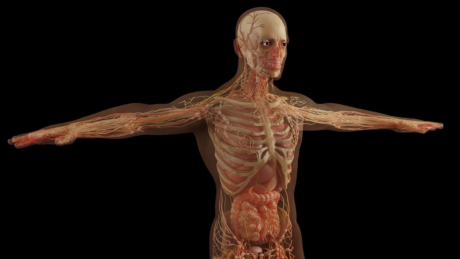
A body plane is an imaginary flat surface that passes through the body and divides it into portions, and a section is the actual cut surface produced when the body or a structure is sliced along that plane. The three cardinal planes are the sagittal plane, the coronal (or frontal) plane, and the transverse plane, and every directional term used in veterinary anatomy is defined relative to them.
These terms matter because they are the shared language of surgery, imaging, and dissection. A surgeon plans an osteotomy in the coronal plane, a radiologist reads a transverse CT slice of a cat's thorax, and a student describes a muscle's fiber direction relative to the sagittal plane. When the vocabulary is loose, the anatomy is loose with it. Veterinary students who arrive at dissection with a firm grasp of basic directional terms engage more effectively with the laboratory work [1], and the same foundation carries into clinical practice.
The Three Cardinal Planes of the Body
Sagittal Plane
A sagittal plane is any vertical plane that runs parallel to the long axis of the body and divides it into left and right portions. The word comes from the Latin sagitta, meaning arrow, a reference to the direction of the sagittal suture of the skull.
The midsagittal plane (also called the median plane) is the single sagittal plane that passes exactly through the midline of the body and divides it into equal left and right halves. Any sagittal plane off the midline is called parasagittal. A parasagittal section through the thorax of a dog, for example, might pass through the right lung fields without touching the heart.
Because the sagittal plane is parallel to the long axis, sections taken in this plane show the body in profile. Sagittal views are the standard way to assess patellar tracking, spinal curvature in the craniocaudal direction, and the anterior-to-posterior insertion footprint of ligaments. A cadaveric study of the medial patellofemoral ligament in children noted that the coronal-plane anatomy of the patellar insertion had been well described, but the sagittal-plane anatomy had not been widely studied, so the investigators measured the sagittal insertion footprint directly [2]. That is the kind of gap that plane terminology exposes.
Coronal (Frontal) Plane
A coronal plane, also called the coronal/frontal plane or simply the frontal plane, is any vertical plane that runs parallel to the long axis of the body and divides it into dorsal and ventral portions. In human anatomy the same plane divides the body into anterior and posterior portions, which is why the term "coronal frontal plane" appears so often in the literature. The two names describe the same geometric plane.
The name comes from corona, a crown, because in humans the coronal suture of the skull lies roughly in this plane. In a standing quadruped, a coronal section through the thorax separates the dorsal half of the chest from the ventral half. A coronal section through the skull separates the dorsal calvaria from the ventral facial structures.
Coronal-plane alignment is a major surgical variable. In periacetabular osteotomy for hip dysplasia, computational modeling found that the coronal plane was the principal rotational plane and had the strongest effect on joint contact pressure compared with the sagittal and transverse planes [3]. In total knee arthroplasty, a retrospective study of 310 robotic-assisted cases examined alignment beyond the coronal plane, noting that true personalization must extend into the sagittal and axial planes as well [4]. The coronal plane is where most alignment work begins, but it is rarely where it ends.
Transverse Plane
A transverse plane (also called the horizontal plane or axial plane) is any plane that runs perpendicular to the long axis of the body and divides it into cranial and caudal portions. In human anatomy the same plane divides the body into superior and inferior portions. A transverse section through the abdomen of a dog produces a slice that shows the cross-sectional arrangement of the viscera at that level.
The transverse plane is the workhorse of computed tomography. A pilot study of the thorax of Shirazi cats used CT to display the thoracic cage, trachea, bronchi, lungs, heart, vessels, mediastinum, and pleural cavity on sagittal and coronal planes and, exceptionally, on a transverse plane, providing detail that radiographs could not [5]. Transverse slices are how modern imaging resolves structures that overlap on a plain film.
Dorsal Plane: A Veterinary Preference
In veterinary anatomy, the term dorsal plane is often preferred over transverse plane when describing sections of the trunk of quadrupeds. The dorsal plane is a plane parallel to the back (the dorsal surface) that divides the body into dorsal and ventral parts, which is geometrically the same as a coronal plane. The preference exists because "dorsal" is unambiguous in a quadruped whose back is horizontal, while "transverse" can be confused with the transverse plane of imaging, which is perpendicular to the long axis.
The practical rule for students: in the thorax and abdomen of a dog or cat, a section that separates dorsal from ventral structures may be called a dorsal plane or a coronal plane, and both are correct. A section that separates cranial from caudal structures is a transverse plane. The terminology shifts with the region and with the imaging convention, so always confirm which plane a colleague or a report means.
Orientation: Why Quadrupeds Change the Vocabulary
The single most common source of confusion for students entering veterinary anatomy is that the human directional vocabulary assumes a vertical trunk, while the veterinary vocabulary assumes a horizontal trunk. In a standing human, the long axis runs from head to foot, so "superior" and "inferior" describe position along that axis. In a standing dog, the long axis runs from head to tail, so "cranial" and "caudal" replace them.
The same shift applies to the dorsoventral axis. In a human, "anterior" and "posterior" describe the front and back of the trunk. In a quadruped, "ventral" and "dorsal" do the same job, and they remain accurate regardless of whether the animal is standing, recumbent, or suspended for surgery. This is why veterinary anatomists prefer dorsal and ventral: the terms are tied to the animal's own body, not to gravity.
Two additional terms apply only to quadrupeds. Rostral means toward the nose, and it is used for structures on the head, particularly the skull and face, where "cranial" would be ambiguous. The term derives from rostrum, meaning beak or snout. Palmar means the aspect of the forelimb distal to the carpus that contacts the ground, and plantar means the equivalent aspect of the hindlimb distal to the tarsus. In a dog, the palmar surface of the paw and the plantar surface of the paw are both ventral-facing, but they are named separately because the limbs are structured differently.
A related pair is proximal and distal, which describe position along a limb relative to the point of attachment to the trunk. Proximal means closer to the trunk, distal means farther from it. These terms are used in both human and veterinary anatomy and do not change with body orientation.
Summary Table: Human Versus Veterinary Directional Terms
| Human Term | Veterinary Term | Meaning |
|---|---|---|
| Superior | Cranial | Toward the head |
| Inferior | Caudal | Toward the tail |
| Anterior | Ventral | Toward the belly |
| Posterior | Dorsal | Toward the back |
| Anterior (on head) | Rostral | Toward the nose |
| Palmar surface (hand) | Palmar surface (forepaw) | Ground-contact aspect of the forelimb distal to the carpus |
| Plantar surface (foot) | Plantar surface (hindpaw) | Ground-contact aspect of the hindlimb distal to the tarsus |
| Proximal | Proximal | Closer to the trunk |
| Distal | Distal | Farther from the trunk |
| Medial | Medial | Toward the midline |
| Lateral | Lateral | Away from the midline |
Note that medial and lateral, proximal and distal, and superficial and deep are unchanged between the two systems. Only the terms tied to the head-to-tail and back-to-belly axes shift.
The Coronal Axis Versus the Coronal Plane
A frequent error in student writing and even in clinical notes is to use "coronal axis" when the correct term is "coronal plane." The two are not interchangeable.
A plane is a two-dimensional surface that extends infinitely in two directions. A coronal plane is a flat surface that divides the body into dorsal and ventral portions. An axis is a line about which rotation occurs. The coronal axis, also called the frontal axis or the transverse axis, is a line that runs from side to side through the body, perpendicular to the sagittal plane. Rotation about the coronal axis produces flexion and extension, which are movements in the sagittal plane.
The distinction matters because alignment studies and surgical planning documents refer to both. A study of lower extremity alignment and recurrent patella dislocation assessed alignment in the sagittal, coronal, and transverse planes and found that increased external tibial rotation in the transverse plane was associated with recurrent dislocation, while sagittal and coronal alignment showed no such relationship [6]. The transverse plane is where the rotational abnormality lived. If a reader confused the coronal plane with the coronal axis, the finding would be misread as a flexion-extension problem rather than a rotational one.
A simple memory rule: planes divide, axes rotate. A coronal section is a slice. A coronal axis is a hinge.
How Planes Are Used in Practice
Imaging
Sagittal, coronal, and transverse planes are the standard orthogonal planes of cross-sectional imaging. A study of the vidian canal used axial, coronal, and sagittal CT slices to measure canal length, transverse diameter, and angulation relative to the sagittal plane, finding that the angle between the canal and the sagittal plane differed between males and females [7]. A study of cervical pedicle morphology measured pedicle and vertebral artery diameters in sagittal and coronal planes to assess the feasibility of pedicle screw placement [8]. These are not abstract geometric exercises. The plane determines which structure is visible and which is obscured.
Three-dimensional imaging adds a layer of complexity. A study of craniofacial angular measurement found that measurements performed on standard sagittal, coronal, and transverse sections may not reflect true spatial geometry because of projection-related distortion, and proposed a reoriented plane protocol to align the measurement plane with the true spatial configuration of landmarks [9]. The lesson is that standard planes are conventions, not laws of nature, and there are times when a nonstandard plane gives a truer answer.
Surgery
Surgical planning is plane-specific. In spinal deformity correction, the deformity is a three-dimensional alteration in the coronal, sagittal, and transverse planes, and surgical techniques aim to restore coronal and sagittal alignment while managing the rotational component in the transverse plane [10]. A surgeon who plans only in the coronal plane will miss the rotational deformity.
In pediatric supracondylar humerus fractures, a biomechanical study compared three pin configurations and found that pins diverging in both the coronal and sagittal planes (the 3LDD configuration) provided more stable fixation than pins diverging only in the coronal plane (the 3LDP configuration) or crossed pins [11]. The sagittal plane spread was the variable that mattered. This is a concrete example of how a plane-specific decision changes a clinical outcome.
Dissection and Teaching
In the dissection laboratory, students learn to identify structures by their position relative to planes. A muscle that lies medial to the sagittal plane is on the medial side of the limb. A vessel that crosses the coronal plane from dorsal to ventral is passing from the back to the belly. Veterinary anatomy courses increasingly use pre-matriculation materials on basic directional terms to prepare students before they enter the laboratory, because students who are fluent in the vocabulary engage more effectively with dissection [1].
Clinical Relevance, Limitations and Common Mistakes
The most common mistake is applying human directional terms to quadrupeds. Saying "superior" when you mean "cranial" or "anterior" when you mean "ventral" creates ambiguity in a species whose trunk is horizontal. The fix is to use the veterinary terms consistently, even in casual conversation, until they become automatic.
The second mistake is confusing the coronal plane with the coronal axis, as discussed above. Planes divide, axes rotate.
The third mistake is assuming that "transverse" always means the same thing. In imaging, the transverse plane is perpendicular to the long axis. In some veterinary contexts, the dorsal plane is preferred for sections of the trunk that separate dorsal from ventral structures. Always confirm the convention in use.
The fourth mistake is treating the midsagittal plane as interchangeable with any sagittal plane. Only the midsagittal plane divides the body into equal halves. A parasagittal section does not.
The fifth mistake is forgetting that planes are defined relative to the body's long axis, not to the ground. A recumbent dog has the same sagittal, coronal, and transverse planes as a standing dog. Gravity does not change the anatomy.
This article is educational and is not a substitute for veterinary diagnosis or treatment. Individual patients require evaluation by a licensed veterinarian.
Quick Review
- A sagittal plane runs parallel to the long axis and divides the body into left and right portions. The midsagittal plane divides it into equal halves.
- A coronal (frontal) plane runs parallel to the long axis and divides the body into dorsal and ventral portions in quadrupeds, or anterior and posterior portions in humans.
- A transverse plane runs perpendicular to the long axis and divides the body into cranial and caudal portions in quadrupeds, or superior and inferior portions in humans.
- In veterinary anatomy, the dorsal plane is often preferred over transverse for trunk sections that separate dorsal from ventral structures.
- Quadrupeds use cranial, caudal, dorsal, ventral, rostral, palmar, and plantar in place of the human superior, inferior, anterior, and posterior.
- A coronal plane is a surface. A coronal axis is a line of rotation. Do not confuse them.
- Medial, lateral, proximal, distal, superficial, and deep are unchanged between human and veterinary anatomy.
Frequently Asked Questions
What is the difference between a sagittal and a midsagittal plane?
A sagittal plane is any vertical plane parallel to the long axis that divides the body into left and right portions. The midsagittal plane is the specific sagittal plane that passes through the midline and divides the body into equal left and right halves.
Why do veterinarians say "cranial" instead of "superior"?
Because a quadruped's trunk is horizontal, not vertical. Cranial means toward the head and caudal means toward the tail, which are unambiguous regardless of the animal's posture. Superior and inferior assume a vertical trunk and do not translate cleanly to dogs, cats, horses, or cattle.
Is the coronal plane the same as the frontal plane?
Yes. Coronal plane and frontal plane are two names for the same geometric plane, the one that divides the body into dorsal and ventral portions in quadrupeds or anterior and posterior portions in humans. The combined term coronal/frontal plane is used when both names need to be acknowledged.
What does "transverse plane" mean in a CT scan?
In CT imaging, the transverse plane is the plane perpendicular to the long axis of the body. A transverse slice shows a cross-section of the body at that level, which is why it is also called an axial slice.
What is the coronal axis?
The coronal axis is a line that runs from side to side through the body, perpendicular to the sagittal plane. Rotation about the coronal axis produces flexion and extension. It is not the same thing as the coronal plane, which is a surface.
Why is the dorsal plane preferred over the transverse plane in some veterinary contexts?
Because "dorsal" is unambiguous in a quadruped whose back is horizontal. The dorsal plane separates dorsal from ventral structures, which is the same geometric result as a coronal plane. The preference is a matter of convention and clarity, not a difference in geometry.
Related Articles
- Veterinary Terminology: A Guide for Students and Professionals
- Statistical Synonyms: A Guide to Terminology in Statistics
- Poultry Terminology: A Glossary for Farmers and Enthusiasts
- Predictor vs. Covariate: Clarifying Terminology in Research
- Body Composition Analysis
- Laboratory Terminology for Beginners: Aliquot, Blank, Control, Replicate, and Standard
- Body Regions: Anatomical Terminology Guide
- Midsagittal Plane: Definition, Examples, and Body Planes
- Coronal Cut: Definition and Anatomical Planes
Sources
- Improving Veterinary Students' Interest and Engagement During Anatomy Dissection Sessions: Approaches Related to Flow Theory.
- A Cadaveric Study of the Sagittal Patellar Insertion of the Medial Patellofemoral Ligament in Children: Implications for Reconstruction
- Computer-Assisted Optimization of the Acetabular Rotation in Periacetabular Osteotomy Using Patient's Anatomy-Specific Finite Element Analysis
- Beyond the coronal plane in robotic total knee arthroplasty-Part 2: Combined flexion does not affect outcomes
- Comparative pilot study of radiography and computed tomography for the thorax of Shirazi cat
- Alignment in the transverse plane, but not sagittal or coronal plane, affects the risk of recurrent patella dislocation
- Imaging anatomy of the vidian canal and its clinical significance
- Morphometric Analysis of the Cervical Pedicle and Transverse Foramen with Vertebral Artery: A Donor-Based, Dry Bone and Radiologic Study.
- Geometric plane alignment as a methodological requirement for accurate angular measurement in three-dimensional craniofacial imaging.
- Biomechanical Principles of Spinal Deformity Correction in the Thoracolumbar Spine.
- Sagittal plane alignment affects the strength of pin fixation in supracondylar humerus fractures