Axon Hillock: Structure, Function, and Action Potential Initiation
By Dr. Zubair Khalid, DVM, MS, PhD ·
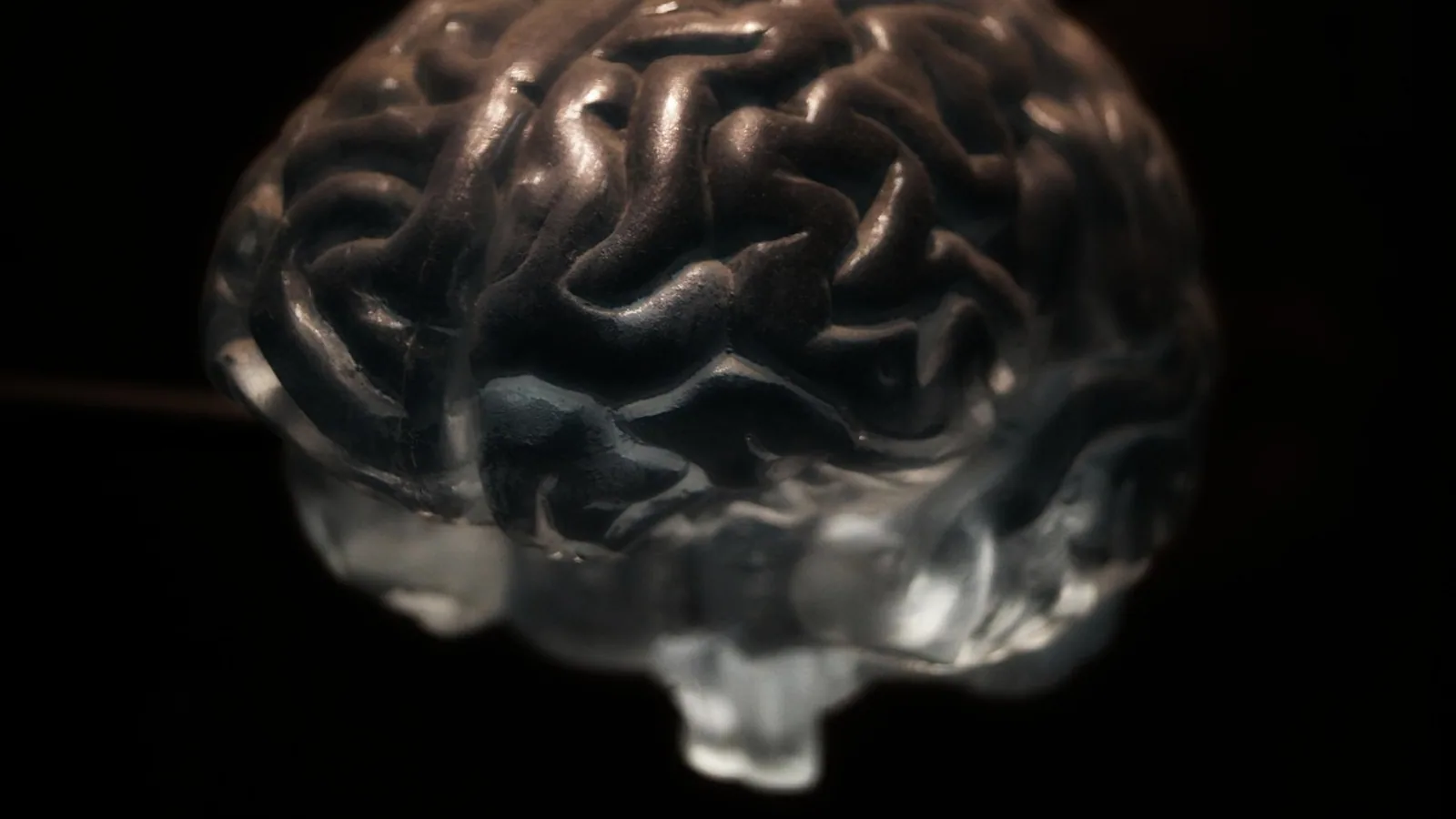
The axon hillock is the tapered, cone-shaped junction where the cell body (soma) narrows into the axon. The axon initial segment (AIS), a short specialized stretch of axon just distal to the hillock, is the true trigger zone in most neurons because it carries the highest density of voltage-gated sodium channels and the lowest threshold for firing.
Why the Axon Hillock Matters
Every action potential a neuron sends is a decision made at one small patch of membrane. The axon and axon hillock sit at the end of the somatodendritic compartment, where hundreds or thousands of excitatory and inhibitory synaptic inputs converge. Those inputs arrive as graded potentials that fade with distance and time. The hillock and AIS are the place where the sum of those fading signals is compared against a threshold. If the summed depolarization is large enough, the neuron fires an all-or-none action potential. If it is not, nothing leaves the cell.
This arrangement explains a core feature of nervous system function. Synaptic integration is analog and graded, while axonal output is digital and all-or-none. The axon hillock and AIS are the converter between the two. In veterinary physiology, this matters for understanding everything from spinal reflex arcs to the effects of local anesthetics, which block the sodium channels concentrated at the AIS.
Structure of the Axon Hillock and Axon Initial Segment
The Hillock Proper
The axon hillock is a region of the soma, not a separate organelle. Under light microscopy it appears as a pale, cone-shaped zone where the cytoplasm of the cell body funnels into the axon. It contains no Nissl bodies (the rough endoplasmic reticulum granules that fill the rest of the soma), which is why it looks paler than surrounding cytoplasm. Microtubules and neurofilaments bundle together here and continue into the axon as the axonal cytoskeleton.
The hillock is best understood as a structural transition zone. It is the last part of the somatodendritic membrane and the first part of the axonal membrane. Its membrane can support graded potentials, but it is not the main site of spike initiation.
The Axon Initial Segment
The AIS is the specialized domain immediately distal to the hillock. In most mammalian neurons it is roughly 20 to 60 micrometers long, though length varies by cell type and can change with activity. The AIS is defined by three features.
First, it has a very high density of voltage-gated sodium channels, mainly NaV1.6 and NaV1.2 subtypes. These channels open at relatively negative voltages, which gives the AIS a low threshold for spike initiation, near -55 to -50 mV in many neurons. The exact threshold depends on the cell type and on the mix of channels present.
Second, the AIS has a distinctive cytoskeleton. Ankyrin-G (AnkG) is the master scaffolding protein that anchors sodium channels to the membrane. BetaIV-spectrin links ankyrin-G to the actin cytoskeleton. Together they form a dense, stable submembrane lattice that clusters sodium channels and holds them in place. Disrupting ankyrin-G or betaIV-spectrin disperses the channels and impairs spike initiation [1][2].
Third, the AIS is a diffusion barrier. It separates the somatodendritic membrane from the axonal membrane, keeping lipids and proteins in their proper compartments. This barrier is part of how neurons maintain polarity, the structural difference between dendrites and axon [2].
Myelination begins just beyond the AIS. The first myelin segment and its paranode sit next to the distal AIS and can influence the threshold for action potentials [3]. The AIS itself is unmyelinated.
The Trigger Zone in Context
Textbooks often call the axon hillock the trigger zone. The more precise statement is that the trigger zone is the AIS, with the hillock serving as the structural gateway. The two are adjacent and functionally linked, which is why the terms are sometimes used loosely. For a student, the key point is that the hillock is where the soma ends and the axon begins, and the AIS is where the spike actually starts.
How Action Potentials Are Initiated: Step by Step
The initiation sequence follows a predictable order. Each step depends on the one before it.
- Synaptic inputs arrive on dendrites and the soma. Excitatory inputs produce excitatory postsynaptic potentials (EPSPs), which are small depolarizations. Inhibitory inputs produce inhibitory postsynaptic potentials (IPSPs), which are small hyperpolarizations or shunts.
- Graded potentials spread passively toward the hillock and AIS. They decay with distance and time, so inputs near the trigger zone have more influence than distant ones.
- EPSPs and IPSPs summate at the hillock and AIS. Summation can be temporal (inputs arriving close together in time) or spatial (inputs arriving at different places at once). The result is a single membrane potential at the trigger zone.
- If the summed depolarization reaches threshold, near -55 to -50 mV, voltage-gated sodium channels in the AIS open. Because the AIS has a high density of these channels, a small depolarization opens enough of them to start a regenerative inward sodium current.
- The regenerative current drives the membrane potential rapidly toward the sodium equilibrium potential. This is the upstroke of the action potential. The spike is all-or-none because once the regenerative current starts, it no longer depends on the size of the original stimulus.
- The action potential propagates in two directions. It travels forward along the axon toward the terminals, and it backpropagates into the soma and dendrites. Backpropagation can influence synaptic plasticity.
- After the spike, sodium channels inactivate and potassium channels open. The membrane repolarizes and then returns to rest. The AIS is ready for the next cycle.
The critical distinction for students is this. EPSPs and IPSPs are graded and can be any size. The action potential is all-or-none and always has roughly the same amplitude in a given cell. The AIS is where the graded signal is converted into the all-or-none output.
Channel Types and Thresholds Across Neuron Regions
Different parts of the neuron have different channel complements and different roles. The table below summarizes the main comparison.
| Region | Dominant channels | Threshold | Role |
|---|---|---|---|
| Dendrite | Ligand-gated channels, some NaV and Kv | High or none | Receives synaptic input, produces graded EPSPs and IPSPs |
| Soma | Ligand-gated channels, NaV, Kv, CaV | High | Integrates inputs, supports backpropagating spikes |
| Axon hillock | Mixed, transitional | Intermediate | Structural gateway from soma to axon |
| Axon initial segment | High-density NaV1.6 and NaV1.2, Kv1, KCNQ | Low, near -55 to -50 mV | Spike initiation, main trigger zone |
| Node of Ranvier | High-density NaV, some Kv | Low | Regenerates the action potential during saltatory conduction |
The AIS and the node of Ranvier both have high sodium channel density, but they serve different purposes. The AIS initiates the spike. The node regenerates it as it travels along a myelinated axon.
Comparative Notes Across Species and Fiber Types
Giant Squid Axon
The giant squid axon is the classic preparation in which Hodgkin and Huxley worked out the ionic basis of the action potential. It is a large, unmyelinated fiber, and its spike initiation zone is distributed along a long stretch of membrane rather than concentrated in a short AIS. The squid axon does not have a compact AIS of the mammalian type. Its sodium channels are spread over a broad region, and the threshold is set by the overall channel density and the passive properties of the fiber. This is why the squid axon is a model for the general mechanism of the action potential but not for the precise geometry of the mammalian trigger zone.
Mammalian Motor Neurons
Mammalian motor neurons have a well-defined AIS with high sodium channel density. The AIS of spinal motor neurons is plastic. In adult rats, successful H-reflex up-conditioning was associated with greater AIS length and greater distance from the soma, and greater length correlated with greater reflex increase. Down-conditioning was associated with more GABAergic terminals on the AIS, weaker ankyrin-G immunoreactivity, and a positive shift in firing threshold [4]. These findings show that the AIS of motor neurons is not fixed. It changes with learning and with altered activity.
Invertebrate Unmyelinated Fibers
Many invertebrate neurons have unmyelinated axons and a less sharply defined initiation zone. In some invertebrate systems, spike initiation occurs at a region that resembles the AIS but lacks the full ankyrin-G and betaIV-spectrin scaffold of vertebrates. The general principle still holds. The spike starts where sodium channel density is highest and threshold is lowest. The anatomical specialization is less pronounced.
Sensory Neurons
Myelinated dorsal root ganglion neurons assemble an AIS in the proximal region of their stem axon, enriched in NaV1.1 and NaV1.7. In a model of neuropathic pain, NaV1.7 channels at this AIS were associated with spontaneous activity, and the AIS lowered the voltage threshold for spontaneous discharges [5]. This is a useful reminder that the AIS is not only a normal trigger zone. It can also be a source of pathological spontaneous firing.
How the Axon Hillock and AIS Are Studied
Several methods are used to study the hillock and AIS in research and teaching settings.
Immunofluorescence is the standard method for visualizing the AIS. Antibodies against ankyrin-G, betaIV-spectrin, NaV1.6, and NaV1.2 label the AIS as a distinct band at the proximal axon. This approach is used to measure AIS length, position, and channel density [1][4][6].
Whole-cell patch clamp recording measures the threshold and firing properties of the neuron. The recording electrode is placed on the soma, and the experimenter injects current to find the threshold for spike initiation. Combined with pharmacology, this method can isolate the contribution of specific channels [7][8].
Computational modeling is used to test how changes in AIS length, channel density, and channel kinetics affect excitability. Models of pyramidal cells and fast-spiking interneurons have shown that the spatial distribution of sodium channel subtypes along the AIS affects both spike initiation and backpropagation [9].
Two-photon imaging allows longitudinal tracking of AIS structure in living animals. This has been used to show that the AIS remodels during associative fear learning and extinction [10].
Serial section electron microscopy provides three-dimensional views of the AIS and its relationship to myelin. This method revealed that the paranode of the first myelin segment undergoes age-dependent remodeling near the AIS [3].
Clinical Relevance, Limitations and Common Mistakes
The AIS is a site of plasticity, and changes there can alter excitability in ways that matter clinically. In a mouse model of autism spectrum disorder, pyramidal neurons in the medial prefrontal cortex had shortened AIS and reduced excitability, and a circuit-specific intervention normalized AIS structure and rescued social behaviors [11]. In a model of diabetic brain complications, endoplasmic reticulum stress was linked to AIS shortening in cortical cultures [12]. In the sub-acute phase after ischemic stroke, AIS structure and intrinsic excitability were altered in peri-infarct neurons [6]. These findings are from animal and cell models, and they do not translate directly into clinical recommendations for patients.
Several common mistakes appear in student work.
Confusing the hillock with the AIS. The hillock is the structural junction. The AIS is the functional trigger zone. They are adjacent but not the same.
Treating the trigger zone as a single point. The AIS is a region, roughly 20 to 60 micrometers long, and its properties vary along its length. The proximal and distal AIS can have different sodium channel subtypes and different gating properties [9].
Assuming the threshold is fixed. Threshold depends on the mix of channels, the recent activity of the cell, and the position of the AIS relative to the soma. Changes in axial resistance between the soma and AIS can shift the somatic threshold by several millivolts [13].
Forgetting that the AIS is unmyelinated. Myelination begins beyond the AIS. The first myelin segment and its paranode are adjacent to the distal AIS and can influence threshold [3].
Treating graded potentials and action potentials as the same kind of signal. EPSPs and IPSPs are graded and decremental. Action potentials are all-or-none and regenerative. The AIS is where the conversion happens.
This article is educational and is not a substitute for veterinary diagnosis or treatment. Individual animals require evaluation by a veterinarian.
Quick Review
- The axon hillock is the tapered junction where the soma narrows into the axon.
- The axon initial segment (AIS) is the true trigger zone in most neurons.
- The AIS is roughly 20 to 60 micrometers long and has high densities of NaV1.6 and NaV1.2.
- The threshold for spike initiation is near -55 to -50 mV in many neurons.
- Ankyrin-G and betaIV-spectrin form the cytoskeletal scaffold that clusters sodium channels at the AIS.
- EPSPs and IPSPs are graded and summate at the hillock and AIS. The action potential is all-or-none.
- Myelination begins just beyond the AIS.
Frequently Asked Questions
What is the difference between the axon hillock and the axon initial segment?
The axon hillock is the tapered structural junction where the soma meets the axon. The axon initial segment is the specialized stretch of axon just distal to the hillock, and it is the main site of action potential initiation.
Why is the axon initial segment called the trigger zone?
It has the highest density of voltage-gated sodium channels and the lowest threshold for firing, so the action potential starts there in most neurons.
What is the threshold for action potential initiation at the AIS?
In many neurons the threshold is near -55 to -50 mV, though the exact value depends on the cell type and the channels present.
Do graded potentials and action potentials occur at the same place?
No. Graded EPSPs and IPSPs summate at the hillock and AIS, and the action potential is initiated at the AIS. The AIS converts the graded input into an all-or-none output.
What holds the sodium channels at the AIS in place?
Ankyrin-G and betaIV-spectrin form a submembrane scaffold that clusters the channels and anchors them to the cytoskeleton.
Does myelination cover the axon initial segment?
No. The AIS is unmyelinated. Myelination begins just beyond it, and the first myelin segment can influence the threshold for action potentials.
Related Articles
- TATA Box DNA: Structure, Function, and Role in Transcription Initiation
- Circular RNA in Cancer: Biogenesis, Functions, and Clinical Potential
- G-Quadruplex Telomere Structure and Function
- The Role of Histone Chaperones in Cancer: A Review of Their Function and Therapeutic Potential
- Equine Hoof Anatomy: Structures and Function
- Plasmid in Bacteria: Structure, Types, and Functions
- Action Potential: Phases, Ions, and Diagram
- Condyloid Joint: Structure and Movement
- Papillary Dermis: Structure and Function Explained
Sources
- Tsc1 deletion in Purkinje neurons disrupts the axon initial segment, impairing excitability and cerebellar function.
- Axon Initial Segment: Structure, Biological Functions, Diseases, and Therapeutic Targets.
- Evidence for glia-mediated, age-dependent remodeling of myelin at the axon initial segment.
- Motor learning changes the axon initial segment of the spinal motoneuron.
- Sensory neurons have an axon initial segment that initiates spontaneous activity in neuropathic pain.
- Characterisation of the axon initial segment and intrinsic excitability in the sub-acute phase post-ischaemic stroke.
- Activation of Dopamine D1 Receptors at the Axon Initial Segment-Like Process of Retinal AII Amacrine Cells Modulates Action Potential Firing.
- Axon Initial Segment GABA Inhibits Action Potential Generation throughout Periadolescent Development.
- Impact on backpropagation of the spatial heterogeneity of sodium channel kinetics in the axon initial segment.
- Axon initial segment dynamics during associative fear learning.
- Restoration of axon initial segment plasticity via chemogenetic activation rescues autism-related behaviors.
- Endoplasmic Reticulum Stress Mediates Axon Initial Segment Shortening: Implications for Diabetic Brain Complications.
- Neural excitability increases with axonal resistance between soma and axon initial segment.