Dog Ultrasound Sedation: Is It Needed and Is It Safe?
By Dr. Zubair Khalid, DVM, MS, PhD ·
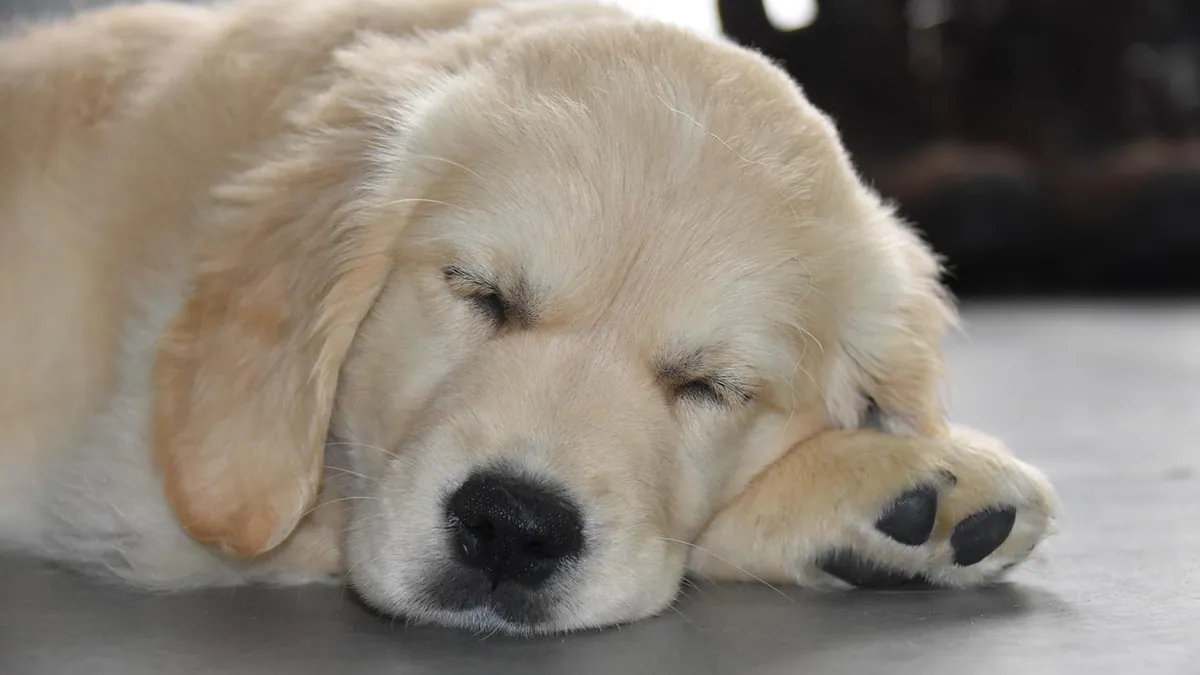
Most dogs do not need sedation for an ultrasound. The procedure is painless, and many dogs lie still with gentle handling and a calm room. Sedation is used when a dog is anxious, painful, or unable to stay still long enough to get clear images, and the drugs used are generally safe when a veterinarian tailors the dose to your dog.
This article is educational and is not a substitute for veterinary diagnosis or treatment.
Quick Answer
- Ultrasound uses sound waves, not radiation, and the transducer is simply moved across the skin, so the exam itself does not hurt [1].
- Many dogs complete an abdominal or thyroid ultrasound fully awake, especially with gentle restraint and a quiet setting [2].
- Sedation is most often chosen for anxious dogs, painful dogs, or exams that require a very still patient, such as echocardiography or contrast studies [3][4].
- Common sedation drugs include dexmedetomidine, butorphanol, and midazolam, and your veterinarian calculates the dose from your dog's weight and health status [3][5][4].
- Sedation wears off, but reversal drugs such as atipamezole can speed recovery when dexmedetomidine is used [3].
What a Dog Ultrasound Is and Why It Is Done
An ultrasound machine sends high-frequency sound waves into the body through a handheld probe called a transducer. When those waves hit tissue boundaries, some bounce back, and the machine turns those returning echoes into a real-time picture of your dog's organs [1]. Because the image updates many times per second, the veterinarian can watch organs move and can freeze or record clips for later review [1].
Ultrasound is used to look at many body areas. Abdominal ultrasound helps assess organs such as the pancreas, liver, kidneys, and bladder [6][7]. Thyroid ultrasound examines the thyroid lobes beside the trachea [2]. Heart ultrasound, called echocardiography, looks at chamber size and pumping function [3]. Ultrasound can also guide a needle for sampling a mass or draining fluid, and contrast-enhanced ultrasound can help identify dead tissue inside a mass before a sample is taken [7].
If your veterinarian is deciding between imaging tests, a guide like Radiography, Ultrasound, CT, and MRI in Veterinary Medicine: Imaging Selection by Clinical Question can help you understand why ultrasound was chosen.
Does a Dog Ultrasound Require Sedation?
Usually, no. The exam is noninvasive, and the probe only touches the skin. For thyroid scans, most dogs and cats are successfully examined without sedation [2]. For many abdominal scans, a cooperative dog can be scanned while lying on its side or back with a helper holding gently.
Sedation becomes useful in specific situations:
- Your dog is anxious, wiggly, or reactive to handling.
- Your dog is in pain, such as with pancreatitis or a painful mass.
- The exam needs a very still patient, such as echocardiography or a contrast study [3][4].
- The area is hard to reach, or the dog will not tolerate the needed position.
- A procedure is combined with the scan, such as a fluid drain or a nerve block.
If your dog needs a procedure alongside imaging, sedation or anesthesia may be part of the plan. You can read more about procedure-based imaging in Ultrasound-Guided Pericardiocentesis for Therapeutic Drainage in Dogs and Ultrasound-Guided Celiac and Mesenteric Nerve Block in Dogs.
Common Sedation Drugs for Dog Ultrasound Sedation
Veterinarians choose drugs based on your dog's age, heart health, pain level, and the type of scan. The dose is always calculated for your individual dog. Never give human sedatives or leftover pet medications at home.
Dexmedetomidine
Dexmedetomidine is a common sedative in veterinary imaging. In one study, healthy dogs received 10 µg/kg of dexmedetomidine intravenously with 0.2 mg/kg of butorphanol for echocardiography [3]. A separate cardiac imaging study used a lower dose of 0.5 to 1 µg/kg intravenously with butorphanol at 0.1 to 0.3 mg/kg [4]. These doses show how much the plan can vary. Your veterinarian sets the dose for your dog, and you should not compare your dog's dose to these numbers.
Dexmedetomidine can be reversed with atipamezole given into the muscle, which shortens recovery [3].
Butorphanol
Butorphanol is an opioid-like pain reliever often paired with dexmedetomidine [3][5][4]. It helps with comfort and mild sedation. It is frequently combined with other drugs instead of used alone.
Midazolam
Midazolam is a benzodiazepine that can be added to a sedation plan. In one study, a combination of dexmedetomidine, butorphanol, and midazolam scored better for ease of the procedure than some other protocols [5].
Ketamine and Propofol
These are stronger drugs used when deeper sedation or general anesthesia is needed. In one study, a dexmedetomidine and ketamine protocol was linked to a seizure in one dog, and dexmedetomidine with propofol was also compared [5]. This does not mean these drugs are unsafe in general. It means the choice matters and should match your dog's health.
Reversal Agents
Atipamezole reverses dexmedetomidine and is given into the muscle [3]. Reversal can bring your dog back to normal more quickly, which is helpful for same-day discharge.
How to Prepare Your Dog for an Ultrasound
Preparation depends on the area being scanned and whether sedation is planned.
- Food: Many abdominal ultrasounds work best on an empty stomach. Your veterinarian will tell you how many hours to withhold food. Do not withhold food unless you are told to.
- Water: Ask whether water should be limited. Some scans, such as ureteral exams, may involve a diuretic like furosemide to improve visibility, and that changes the plan [8].
- Medications: Tell the veterinarian about every medication, supplement, and previous reaction to sedation.
- Health history: Share any heart, liver, kidney, or seizure history. This affects drug choice [5].
- Fur: The scan area is often clipped and cleaned with alcohol, then coated with coupling gel [2]. Do not be surprised by a small shaved patch.
- Transport: If sedation is planned, bring a leash and consider a second person to help you carry or steady your dog afterward.
What Happens During the Ultrasound
Most exams follow a similar flow.
- Check-in and exam. The veterinarian or technician weighs your dog and may do a brief physical exam.
- Positioning. For thyroid scans, dogs are often placed on their back, though other positions can work [2]. Abdominal scans usually use side or back lying.
- Clipping and gel. The fur over the area is clipped, alcohol is applied, and coupling gel is spread on the skin [2].
- Scanning. The probe is moved slowly across the skin. The veterinarian watches a live image and may freeze or record clips [1].
- Sedation if needed. If sedation is used, it is usually given by injection. Your dog becomes relaxed and sleepy but is monitored throughout.
- Recovery. If a reversal drug is used, your dog wakes faster [3]. Otherwise, sedation wears off on its own.
For heart scans, sedation can change some measurements. In one study, sedation affected all measured echocardiographic variables, though most dogs stayed within normal reference ranges for most values except ejection fraction and fractional shortening [3]. This is why your veterinarian may repeat certain measurements after your dog is fully awake. You can learn more in Echocardiography in Dogs and Cats: Indications and Basic Interpretation.
Recovery Timeline and Home Care
Recovery depends on the drugs used and whether a reversal agent was given. The table below gives general guidance. Your veterinarian will give you specific instructions.
| Time After Procedure | What to Expect | What to Do |
|---|---|---|
| 0 to 30 minutes | Sleepy, relaxed, may be wobbly | Keep your dog warm, quiet, and on the floor or a low bed |
| 30 minutes to 2 hours | Gradually more alert, may still be unsteady | Offer a small amount of water, no food until fully awake |
| 2 to 6 hours | Most dogs are close to normal | Short leash walks only, no stairs if wobbly |
| 6 to 24 hours | Full alertness in most dogs | Normal food and water if your veterinarian approves |
| 24 to 48 hours | Any mild grogginess should be gone | Call if your dog is still dull, not eating, or vomiting |
If atipamezole was used, recovery may be faster than the timeline above [3]. If a longer-acting drug was used, recovery may take longer. Do not leave a sedated dog alone on furniture or near stairs.
Possible Complications and Warning Signs
Serious problems from ultrasound sedation are uncommon, but they can happen. In one study of dogs receiving sedation for stress radiography, temperature, heart rate, and respiratory rate stayed within normal ranges for all protocols, and one dog had a seizure after a dexmedetomidine and ketamine combination [5]. In a cardiac CT study using low-dose dexmedetomidine and butorphanol, heart rate ranged from 37 to 141 beats per minute, mean arterial pressure ranged from 58 to 117 mmHg, mild low blood pressure occurred in two dogs, and no deaths occurred within 48 hours [4]. These findings show that monitoring matters and that drug choice affects the cardiovascular system.
Watch for these warning signs after sedation:
- Trouble breathing, noisy breathing, or blue gums
- Collapse, severe weakness, or inability to stand after a few hours
- Repeated vomiting or vomiting while sleepy
- A seizure or unusual twitching
- A very slow or very fast heart rate that you can feel
- Pale or white gums
- Extreme sleepiness that does not improve
Call your veterinarian right away if you see any of these. If your dog is struggling to breathe or cannot be woken, treat it as an emergency.
Questions to Ask Your Veterinarian
Before the appointment, ask:
- Does my dog need sedation for this ultrasound, or can we try without it?
- Which drug or drugs will you use, and why are they right for my dog?
- What are the risks for my dog based on age, heart, liver, kidney, and seizure history?
- Will you use a reversal drug, and how fast will my dog wake up?
- How long should I withhold food and water before the visit?
- What should I watch for at home, and who do I call after hours?
- Will sedation change the results, and do we need to repeat any measurements later [3]?
Limitations and When to Contact a Veterinarian
Ultrasound findings can vary between observers. In one study of pancreatic ultrasound, agreement between different veterinary radiologists was poor, while agreement by the same radiologist was moderate [6]. This means the person scanning and the quality of the images matter. Sedation can also change some measurements, so timing of the scan matters [3].
Contact your veterinarian the same day if your dog is still very sleepy several hours after the appointment, refuses food, vomits more than once, or seems painful.
Go to an emergency clinic now if your dog has trouble breathing, collapses, has a seizure, has pale gums, or cannot be woken.
Schedule a routine recheck if your dog is eating and acting normally but you have questions about the results or want to review the images.
Every dog is different, and only your veterinarian can decide what is safe and appropriate for your pet.
Frequently Asked Questions
Will my dog be asleep for an ultrasound?
Most dogs are awake for an ultrasound. The exam is painless, and many dogs lie still with gentle handling [2]. Sedation is reserved for anxious, painful, or wiggly dogs, or for exams that need a very still patient [3][4].
Is sedation safe for older dogs?
Sedation can be safe for older dogs when the drug and dose are chosen for their health. Your veterinarian will review heart, liver, kidney, and seizure history first [5][4]. Monitoring during and after the procedure lowers risk.
Can I stay with my dog during the ultrasound?
Many clinics allow owners to stay for the scan, but policies vary. If sedation is used, some clinics prefer owners to wait in the lobby to keep the room calm. Ask your clinic what they allow.
How long does sedation last after an ultrasound?
It depends on the drug. Dexmedetomidine can be reversed with atipamezole, which speeds recovery [3]. Without reversal, most dogs are close to normal within a few hours, though some remain groggy longer.
What if my dog is too anxious for an ultrasound without sedation?
Tell your veterinarian ahead of time. They can plan light sedation, adjust the appointment time, or use a quieter room. Trying to force a scan on a stressed dog can lead to poor images and a bad experience.
Related Articles
- Ultrasound-Guided Pericardiocentesis for Therapeutic Drainage in Dogs
- Echocardiography in Dogs and Cats: Indications and Basic Interpretation
- Ultrasound-Guided Celiac and Mesenteric Nerve Block in Dogs
- Ultrasound-Guided Injection of Stifle Joint in Dogs for Therapeutic Purposes
- Radiography, Ultrasound, CT, and MRI in Veterinary Medicine: Imaging Selection by Clinical Question
- Ultrasound-Guided Aspiration of Cystic Lesions in Small Animals
- Dog Ultrasound: How the Procedure Works and What It Shows
- Dog Sedation Medications: Types, Uses, and Safety
- Dog Sedation vs Anesthesia: What's the Difference?
- Point-of-Care Ultrasound (POCUS) in Veterinary Emergency Triage: Protocols and Interpretation
References
- Ultrasonography in Animals - Clinical Pathology and Procedures. Merck Veterinary Manual.
- Thyroid ultrasound scanning technique in dogs and cats. Journal of the American Veterinary Medical Association, 2025.
- Same-day echocardiography in dogs sedated with intravenous dexmedetomidine followed by reversal with intramuscular atipamezole. Journal of the American Veterinary Medical Association, 2026.
- Anaesthetic Management Including Low-Dose Dexmedetomidine and Butorphanol in Dogs Undergoing ECG-Gated Cardiac Computed Tomography: A Retrospective Case Series. Veterinary sciences, 2026.
- Comparison of Four Sedation and Anaesthesia Protocols during Stress Radiography of the Coxofemoral Joint Using a Distension Device Modified for Force Measurements. Veterinary and comparative orthopaedics and traumatology : V.C.O.T, 2026.
- Survey on Inter- and Intra-Observer Variations of the Ultrasound Assessment of Dog Pancreases. Veterinary radiology & ultrasound : the official journal of the American College of Veterinary Radiology and the International Veterinary Radiology Association, 2026.
- Added Value of Contrast-Enhanced Ultrasound for Cytological Sample Collection of Hepatic and Pulmonary Masses in Dogs. Animals : an open access journal from MDPI, 2026.
- Furosemide Administration Enhances Diagnostic Confidence in the Evaluation of Ureteral Ectopia in Dogs Using Ultrasound. Veterinary radiology & ultrasound : the official journal of the American College of Veterinary Radiology and the International Veterinary Radiology Association, 2026.