Lead Poisoning in Dogs and Cats: Sources, Symptoms, Treatment
By Dr. Zubair Khalid, DVM, MS, PhD ·
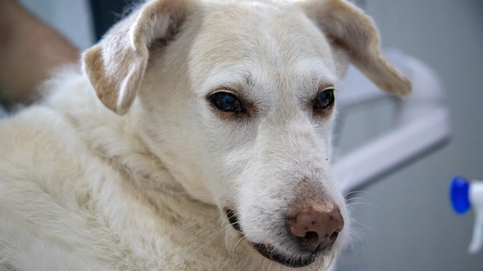
Lead poisoning in dogs and cats is a preventable toxicosis that still occurs in American homes, especially during renovation of older buildings. The most common source in cats is old lead-based paint disturbed by chipping, sanding, or scraping during home renovation, and paint is also the single most frequently identified source in dogs [1][2]. Pets can absorb lead by licking contaminated dust from their coats and paws, chewing painted surfaces, swallowing fishing weights or curtain weights, or eating shot and contaminated soil. Gastrointestinal signs such as vomiting and anorexia and neurologic signs such as seizures are the classic presentation, but the signs are often vague, which is why lead toxicosis is likely underdiagnosed in cats [1].
Owner triage summary. If you saw your pet eat paint chips, a fishing weight, a curtain weight, or shotgun pellets, call your veterinarian or a pet poison hotline immediately, even if your pet looks normal. If your pet is having a seizure, is wobbly, is vomiting repeatedly, or has stopped eating, treat it as an emergency and go to a veterinary hospital now. Do not try to make your pet vomit at home unless a veterinarian instructs you to do so.
This article is educational and is not a substitute for veterinary diagnosis or treatment.
At a Glance: Lead Poisoning in Dogs and Cats
| Feature | Dogs | Cats |
|---|---|---|
| Most common source | Paint, then other lead objects [2] | Old paint from home renovation in 84% of reviewed cases [1] |
| Typical signs | Gastrointestinal and neurologic signs predominate [2] | Anorexia, vomiting, seizures [1] |
| Age pattern | Median age 1.5 years in a case series [2] | Younger cats more likely to show central nervous system signs [1] |
| Key blood clue | Metarubricytosis, basophilic stippling, anemia [3] | Macrocytic regenerative anemia in some cases [4] |
| Confirmatory test | Blood lead concentration [1][3] | Blood lead concentration is the gold standard [1] |
| First-line chelator | Succimer orally for 10 days [5] | Succimer, calcium EDTA [6][4] |
| Sentinel value | Dogs and cats can signal lead exposure in children in the same home [7] | Same [7] |
Why Lead Is Toxic to Pets
Lead is a heavy metal that interferes with several enzyme systems in the body. Two systems matter most for how the disease looks in a dog or cat.
The first is heme synthesis, the pathway that builds hemoglobin inside red blood cells. Lead blocks enzymes in that pathway, including delta-aminolevulinic acid dehydratase, and the result is anemia plus a buildup of precursor molecules. That buildup is what produces the abnormal red blood cell shapes and the stippling seen on a blood smear. In a study of dogs from different habitats, delta-aminolevulinic acid dehydratase activity was measured as a marker of lead exposure, and blood lead concentrations were significantly higher in dogs from industrial areas than in controls [8].
The second system is the nervous system. Lead damages neurons and the myelin sheath that insulates nerve fibers. In cats, characteristic histological changes in the central nervous system include neuronal necrosis and demyelination [1]. That damage explains seizures, behavioral change, wobbliness, and the swallowing and voice problems reported in some affected cats.
Lead also accumulates in bone, where it is stored long term and released slowly. A German Shepherd Dog with multiple fractures and delayed bone healing had high lead concentration in bone mineral even though the serum lead concentration was normal [9]. That case shows why a single blood test does not always capture the full body burden, and why lead can keep affecting a pet long after the exposure stops.
The kidneys can be injured as well. Two dogs diagnosed with lead toxicity had glucosuria and proteinuria on urinalysis consistent with damage to the proximal renal tubules, and one had metarubricytosis on the complete blood count [3].
Pets as Sentinels for Children in the Same Home
Dogs and cats share the environment with the people in the house, so they can be exposed to the same toxicants. A case report from a farm documented elevated blood lead concentrations in cattle, a dog, a cat, and a pregnant woman living on the farm, with the woman at 37.3 µg/dL and the animals ranging from 8.42 µg/dL in the cat to 85.41 µg/dL in a calf [7]. Clinical signs of lead poisoning were not apparent in those animals, and further testing pointed to paint in the barn and home as the likely source [7].
That case is the practical reason veterinarians treat a lead diagnosis in a pet as a household event. If a dog or cat has an elevated blood lead level, children in the same home may also be exposed, and the child's pediatrician should be told. Dogs have also been proposed as biomonitors of environmental lead exposure in industrial areas, where blood lead concentrations were significantly higher than in control dogs [8]. A pet with unexplained lead exposure is a signal to check the whole household.
Risk Factors
Age is a clear factor. In a retrospective study of 85 small companion animals, ages ranged from 0.3 to 48 years with a median of 1.5 years [2]. Young animals explore with their mouths, chew more, and groom more, so they ingest more dust and debris. In cats, younger individuals seem more likely to show central nervous system signs [1].
Environment is the other major factor. Pets in older housing under renovation, pets in industrial or mining areas, and pets in households with occupational lead exposure carry the highest risk. In the Boston case series, 70 of 85 affected animals came from the greater Boston area, and 57% of cases inside the city limits traced to three inner city neighborhoods [2]. Lead exposure clusters geographically.
Behavior matters too. Dogs that chew objects, scavenge, or spend time on firing ranges are at higher risk. Cats that groom heavily indoors during renovation are at higher risk.
Symptoms of Lead Poisoning in Dogs
Lead poisoning symptoms in dogs fall into two groups, gastrointestinal and neurologic, and both can appear together. In the retrospective case series, gastrointestinal and neurologic signs predominated [2].
Gastrointestinal Signs
- Vomiting
- Anorexia, meaning the dog stops eating
- Abdominal discomfort
- Diarrhea
Neurologic Signs
- Seizures
- Wobbliness and incoordination
- Behavioral change
- Blindness or other cranial nerve deficits
- Weakness
Other Signs
- Anemia and pale gums
- Metarubricytosis, which means nucleated red blood cells appear in the circulation [3]
- Basophilic stippling, which are small blue dots inside red blood cells on a stained smear
- Glucosuria and proteinuria from proximal tubular damage [3]
- Poor bone healing or fractures with minimal trauma in dogs with bone lead accumulation [9]
The clinical picture is highly variable. In one report of two dogs, the first had metarubricytosis on the complete blood count while the second was largely normal on the same testing, and both had urinalysis changes consistent with proximal tubular damage [3]. That variability is why lead is easy to miss.
Symptoms of Lead Poisoning in Cats
Lead poisoning in cats most commonly causes anorexia, vomiting, and seizures [1]. Because these signs are vague, lead toxicosis may be significantly underdiagnosed in cats [1]. Younger cats are more likely to show central nervous system signs [1].
Cats can also present with signs that look like other diseases. Two cats from one household were examined for neurologic abnormalities suggestive of central vestibular disease, and lead toxicosis was confirmed by blood and urine lead concentrations after a history of renovation was uncovered [6]. A 3-year-old cat with lead toxicosis had megaesophagus, regurgitation when handled, and clinical evidence of laryngeal and pharyngeal dysfunction, along with a macrocytic regenerative anemia [4]. The megaesophagus, the throat and voice dysfunction, and the anemia all resolved with calcium EDTA treatment [4].
That cat did not have normoblastosis or basophilic stippling of red blood cells [4]. So the absence of those classic blood clues does not rule lead out in a cat.
How Veterinarians Diagnose Lead Poisoning
History Is the First Test
The single most useful diagnostic step is a specific history. Ask about home renovation, chipping or sanding paint, age of the home, recent plumbing work, fishing tackle, curtain weights, ammunition, and occupational lead exposure. In the two-cat case, the diagnosis followed a history of three months of renovation that included chipping and sanding old paint [6]. The authors of that report advised that owners of cats with gastrointestinal or neurologic abnormalities should be questioned specifically about house renovations [6]. Bring that information to your appointment even if the veterinarian does not ask.
Blood Lead Concentration
Blood lead concentration is the gold standard diagnostic test [1]. It confirms exposure, and it is the test used to guide chelation. Blood lead does not necessarily correlate with total body burden or with the metabolic effects that produce clinical signs, so a pet can have clinical lead toxicosis with a level that looks modest, and a pet with a high level can look normal [1][10]. In the Kabwe study, none of the dogs showed overt signs of lead poisoning despite high blood lead concentrations [10].
Testing methods vary. A study comparing a commercially available human blood lead testing system with benchtop inductively coupled plasma mass spectrometry found a high correlation between the two methods when blood lead levels were higher than 4.0 µg/dL [11]. That matters because it supports the use of validated commercial analyzers for screening in dogs.
Complete Blood Count and Blood Smear
The complete blood count can show anemia. In dogs, metarubricytosis, meaning nucleated red blood cells, is a useful clue, and it was present in one of two dogs with confirmed lead toxicity [3]. Basophilic stippling is the other classic finding. Cats may show a macrocytic regenerative anemia, as in the megaesophagus case, or no characteristic red cell changes at all [4].
Serum Biochemistry and Urinalysis
Serum biochemistry is often normal or nonspecific. In the Kabwe dogs, most analyzed blood biochemical parameters were within or close to standard reference values despite high exposure [10]. Urinalysis can be more revealing. Glucosuria and proteinuria consistent with proximal tubular damage were present in both dogs in one report, and the author noted this was the first case report specifically examining proximal tubular dysfunction in the dog [3]. Evidence of proximal tubular dysfunction should prompt a careful exposure history and blood lead testing [3].
Imaging
Radiographs can show metallic foreign material in the stomach or intestines if a lead object was swallowed. They can also show changes related to bone. In the German Shepherd Dog with multiple fractures, radiographs revealed osteopenia and comminuted fractures, and a dual energy X-ray absorptiometry scan confirmed decreased bone mineral density [9].
Other Diagnostic Tests
The feline review discusses additional tests including erythropoietic protoporphyrin and urine aminolevulinic acid [1]. These are adjuncts. Blood lead concentration remains the standard.
Differential Diagnoses
Lead poisoning mimics many other conditions. The neurologic signs overlap with central vestibular disease, as in the two cats initially suspected of having a vestibular problem [6]. Seizures in a young animal can have many causes. Vomiting and anorexia overlap with dietary upset, foreign body, pancreatitis, and kidney disease. Anemia overlaps with immune-mediated hemolytic anemia, blood loss, and other causes of regenerative anemia.
The megaesophagus case is a good example of how broad the differential is. Megaesophagus in a cat has several causes, and lead toxicosis was confirmed only after testing [4].
Some toxicoses have distinctive features that help separate them. Vitamin D toxicosis causes severe hypercalcemia [13]. Coumarin poisoning causes a bleeding tendency with prolonged clotting times [14]. Mushroom poisoning can cause liver failure, neurotoxicity, or gastrointestinal irritation depending on the species [15]. None of these produce the combination of anemia with stippling and blood lead elevation that points to lead.
Treatment of Lead Poisoning in Dogs and Cats
Lead toxicity dogs treatment has three parts: stop the exposure, support the patient, and remove lead from the body with chelation. The feline review describes treatment of lead toxicosis as removing the exposure, decontaminating the individual and the environment, supportive care, and chelation therapy [1].
Step 1: Remove the Source
This is the most important step and the one owners control. Identify and remove the lead source from the pet's environment. During renovation, keep pets out of the work area, wipe down surfaces, wash pet bedding, and bathe the pet to remove dust from the coat. If a lead object was swallowed, a veterinarian may need to remove it, sometimes with endoscopy or surgery.
Step 2: Decontamination and Supportive Care
Supportive care depends on the signs. A seizuring pet needs seizure control. A vomiting pet needs antiemetics and fluids. A pet that cannot eat needs nutritional support. An anemic pet may need more intensive care depending on severity. Decontamination of the individual means removing lead from the coat, paws, and gastrointestinal tract as appropriate.
Step 3: Chelation Therapy
Chelators bind lead and help the body excrete it. Two agents appear in the veterinary literature for dogs and cats.
Succimer (meso-2,3-dimercaptosuccinic acid). Succimer is given orally. In a prospective case series of 13 dogs with naturally acquired lead poisoning and moderate to high blood lead concentrations (39 to 120 µg/dL), dogs were treated with succimer at 10 mg/kg orally every 8 hours for 10 days [5]. Mean blood lead concentrations were 70.6 µg/dL on day 0, 32.7 µg/dL on day 3, 16.8 µg/dL on day 7, and 27.6 µg/dL on day 20 [5]. That is a reduction of 53.6%, 76.2%, and 60.9% from the pretreatment value at days 3, 7, and 20 respectively [5]. Mean urine lead concentrations rose from 70.0 µg/dL on day 0 to 485.4 µg/dL on day 3, then 254.3 µg/dL on day 7 and 28.3 µg/dL on day 20, which shows the drug was mobilizing and excreting lead [5]. Succimer effectively reduced blood lead concentrations and eliminated clinical signs in those dogs [5].
The day 20 rebound is worth understanding. Blood lead rose again after treatment stopped because lead stored in bone was released back into the blood. That is why veterinarians recheck blood lead after chelation and may repeat treatment. In the Boston case series, 7 of 73 treated animals required repeated treatments [2].
Succimer has also been used successfully in cats. Two cats from a single household with lead toxicosis were treated with succimer, and treatment was not associated with any adverse effects, and both cats recovered completely [6].
Calcium EDTA (CaEDTA). Calcium EDTA is the other chelator described in the veterinary literature. A cat with megaesophagus, pharyngeal and laryngeal dysfunction, and anemia attributable to lead toxicosis improved after calcium EDTA treatment, with resolution of the megaesophagus, the throat and voice dysfunction, and the anemia [4]. Older literature compares chelation approaches. One study evaluated an immobilized chelator device in dogs with lead intoxication and compared it favorably with Versenate treatment of children with lead toxicity [16]. That work is experimental and is not standard clinical practice.
Chelators are not risk free. The available chelators are nephrotoxic when they are eliminated as lead-chelator complexes, which is the reason alternative removal methods have been studied [16]. Your veterinarian will weigh kidney function and clinical status when choosing an agent and dose.
Step 4: Environmental Remediation
Treating the pet without fixing the environment leads to relapse. If renovation is the source, the work area needs containment and cleanup. If the source is a contaminated yard, soil testing and restricting access may be needed. If the source is water, testing and filtration may be needed. If a household member has occupational exposure, changing clothes and shoes before entering the home reduces take-home dust.
Follow-Up and Monitoring
Blood lead should be rechecked after chelation. The succimer study checked blood and urine lead before, during, and after treatment, and the day 20 values showed the rebound effect [5]. A reasonable approach is to recheck blood lead after the treatment course and again later, with the interval set by your veterinarian based on the initial level, the clinical response, and the likelihood of ongoing exposure. Repeated treatment courses are sometimes needed [2].
Watch for return of signs. Vomiting, anorexia, wobbliness, or seizures coming back after treatment suggests ongoing exposure or lead released from bone. Report any recurrence promptly.
Unsafe Home Remedies and What Not to Do
Do not give human chelators or any chelating supplement to your pet without veterinary direction. Chelation requires dose calculation, monitoring, and attention to kidney function, and the wrong dose or agent can cause harm.
Do not induce vomiting at home unless a veterinarian tells you to. If a lead object is sharp or is already past the stomach, inducing vomiting can cause injury.
Do not assume a pet that looks normal is safe. In the Kabwe study, none of the dogs showed overt signs of lead poisoning despite high blood lead concentrations [10]. In the farm case, the dog and cat had elevated blood lead without clinical signs [7]. A normal-looking pet with a known lead exposure still needs evaluation.
Do not rely on milk, oil, or home detox products. None of these remove lead, and some can make gastrointestinal signs worse.
Do not delay because the signs are vague. The feline review notes that signs are often vague and that lead toxicosis may be significantly underdiagnosed in cats [1].
Prevention
Prevention is about controlling the source.
During renovation of an older home, keep pets out of the work area. Seal doorways with plastic sheeting. Change shoes before entering clean areas of the house. Wipe down surfaces with a damp cloth rather than dry sweeping or vacuuming, which spreads dust. Bathe pets and wash bedding after the work is done.
Store fishing weights, curtain weights, ammunition, and lead-containing hobby supplies where pets cannot reach them. Pick up spent shot in areas your dog accesses. If you shoot at a range, follow the range's lead management practices and consider blood lead monitoring for your dog, since working dogs with repeated live-fire exposure can have higher blood lead levels [11].
Test your water if you live in an older home with lead plumbing. One report linked lead poisoning in a dog to lead in a drinking tap [17].
If you live near a former mine, smelter, or industrial site, be aware that soil can be contaminated. In Kabwe, blood lead in dogs decreased with distance from the mine, and the isotope ratios confirmed the mine as the source [10]. Restrict digging and prevent pets from eating soil in those areas.
If you work with lead, change clothes and shower before interacting with your pets and children.
Prognosis
The prognosis depends on how much lead the pet absorbed, how long the exposure lasted, and how quickly treatment started. Many animals recover with source removal and chelation. In the Boston case series, 73 animals were successfully treated with chelation therapy, and 7 required repeated treatments [2]. In the succimer study, all 13 dogs had clinical signs of lead poisoning at enrollment, and treatment reduced blood lead concentrations and eliminated clinical signs [5]. Both cats treated with succimer recovered completely with no adverse effects [6]. The cat with megaesophagus and throat dysfunction recovered after calcium EDTA treatment [4].
Severe neurologic damage can leave lasting deficits. Lead stored in bone can be released for a long time, which is why relapse happens and why follow-up matters. The German Shepherd Dog with bone lead accumulation had delayed fracture healing and later sustained additional fractures, showing that bone lead can affect the skeleton long after the initial exposure [9].
Limitations and When to Contact a Veterinarian
This article is educational and is not a substitute for veterinary diagnosis or treatment. Every case is different, and only your veterinarian can examine your pet, interpret test results, and prescribe treatment.
Contact a veterinarian or an emergency veterinary hospital immediately if any of the following occur:
- Your pet has a seizure, is wobbly, is circling, or has a sudden behavior change.
- Your pet is vomiting repeatedly or cannot keep food or water down.
- Your pet has stopped eating or is losing weight.
- You saw your pet eat paint chips, a fishing weight, a curtain weight, or shotgun pellets.
- Your pet has pale gums, weakness, or collapse.
- You are renovating a home built before lead paint was phased out and your pet has any new gastrointestinal or neurologic sign.
- A pet in your home has been diagnosed with lead poisoning and another pet or a child in the home has not been evaluated.
- Your pet's signs return after treatment for lead poisoning.
Bring the exposure history with you. Tell the veterinarian what your pet may have eaten, when it happened, and whether anyone in the home does renovation work or handles lead.
Frequently Asked Questions
What are the first signs of lead poisoning in dogs?
The first signs are usually gastrointestinal, including vomiting and anorexia, often with neurologic signs such as wobbliness or seizures appearing around the same time. In a retrospective study of 85 small companion animals, gastrointestinal and neurologic signs predominated [2]. Some dogs show only vague signs, and some with high blood lead levels show no signs at all [10].
What does lead poisoning look like in cats?
Lead poisoning in cats most commonly causes anorexia, vomiting, and seizures [1]. Younger cats are more likely to show central nervous system signs [1]. Cats can also present with signs that look like other diseases, including central vestibular disease or megaesophagus with regurgitation and throat dysfunction [6][4].
My dog ate lead paint. What should I do?
Call your veterinarian or a pet poison hotline right away, even if your dog seems fine. Old paint from home renovation is the most common source of lead exposure in cats and a leading source in dogs [1][2]. Your veterinarian will decide whether blood lead testing, decontamination, or chelation is needed based on the amount and timing of the exposure.
How is lead poisoning diagnosed in dogs and cats?
Blood lead concentration is the gold standard diagnostic test [1]. Your veterinarian will also use the exposure history, a complete blood count looking for anemia, metarubricytosis, and basophilic stippling, urinalysis looking for glucosuria and proteinuria, and sometimes imaging [3][4][9]. A validated commercial blood lead analyzer correlates well with laboratory mass spectrometry at levels above 4.0 µg/dL [11].
What is the treatment for lead toxicity in dogs?
Treatment has three parts: removing the lead source, supportive care, and chelation therapy [1]. Succimer is given orally at 10 mg/kg every 8 hours for 10 days in dogs, and in one study it reduced mean blood lead concentrations by 53.6% at day 3 and 76.2% at day 7 while eliminating clinical signs [5]. Calcium EDTA is another chelator used in dogs and cats [4]. Repeated treatment courses are sometimes needed [2].
Can lead poisoning in a pet mean my children are at risk?
Yes. Pets and children share the same environment, and a case report documented elevated blood lead concentrations in a dog, a cat, and a pregnant woman living on the same farm, with paint identified as the likely source [7]. If your pet is diagnosed with lead poisoning, tell your children's pediatrician so the household can be evaluated.
How long does lead stay in a dog's body?
Lead is stored in bone and released slowly over time. In the succimer study, blood lead concentrations rose again by day 20 after treatment stopped, from 16.8 µg/dL on day 7 to 27.6 µg/dL on day 20, which reflects lead moving out of bone stores [5]. A dog with bone lead accumulation had delayed fracture healing and later fractures, showing that bone lead can matter long after exposure [9]. Follow-up blood lead testing is important for this reason.
Can lead poisoning in dogs and cats be cured?
Many pets recover with source removal and chelation. In one case series, 73 animals were successfully treated with chelation therapy, though 7 needed repeated treatments [2]. Both cats treated with succimer recovered completely with no adverse effects [6]. A cat with megaesophagus and throat dysfunction recovered after calcium EDTA treatment [4]. Severe neurologic damage may leave lasting deficits, so early treatment gives the best chance of full recovery.
Related Articles
- Dog Ate Ibuprofen or Tylenol: Toxicity Signs and Emergency Treatment
- Dog Grape Poisoning Symptoms
- Dog Grape Toxicity Symptoms
- Can Dogs Have Grapes Poisoning Symptoms
- Cat Ate a Lily? Immediate Steps for Lily Poisoning
- Dog Ate Chocolate? Poisoning Signs and Emergency Steps
- Salt Toxicity in Dogs and Cats: Sources, Signs, and Treatment
- Fertilizer Poisoning in Dogs: Symptoms, Risky Types, What to Do
- Mold Poisoning in Dogs: Moldy Food, Compost, and Tremors
Sources
- Lead toxicosis in cats-a review.
- Lead poisoning in small companion animals: an update (1987-1992).
- Proximal tubular nephropathy in two dogs diagnosed with lead toxicity.
- Megaesophagus attributable to lead toxicosis in a cat.
- Use of orally administered succimer (meso-2,3-dimercaptosuccinic acid) for treatment of lead poisoning in dogs.
- Succimer for treatment of lead toxicosis in two cats.
- Animals as sentinels for human lead exposure: a case report.
- Blood lead levels, clinico-pathological findings and erythrocyte metabolism in dogs from different habitats.
- Multiple pathological fractures and delayed union associated with lead exposure in a German Shepherd Dog.
- Factors associated with lead (Pb) exposure on dogs around a Pb mining area, Kabwe, Zambia.
- Blood Lead Toxicity Analysis of Multipurpose Canines and Military Working Dogs.
- Ethylene glycol poisoning in three dogs: Importance of early diagnosis and role of hemodialysis as a treatment option.
- Intravenous lipid emulsion therapy in 2 dogs and 2 cats with vitamin D toxicosis.
- [[Case report. Phenprocoumon (Marcumar, Falithrom) as an unusual reason for coumarin poisoning in a dog].](https://pubmed.ncbi.nlm.nih.gov/12596669/)
- Mushroom Poisoning Cases in Dogs and Cats: Diagnosis and Treatment of Hepatotoxic, Neurotoxic, Gastroenterotoxic, Nephrotoxic, and Muscarinic Mushrooms.
- Treatment of lead poisoning with an immobilized chelator comparison with conventional therapy.
- What is your diagnosis? Lead poisoning in a dog caused by lead in a drinking tap.