How to Treat Foot and Mouth Disease in Animals
By Dr. Zubair Khalid, DVM, MS, PhD ·

Foot and mouth disease (FMD) is a highly contagious viral disease of cloven-hoofed animals, and it is one of the most economically damaging livestock diseases in the world [1]. The first thing any owner, farm worker, or veterinarian needs to understand is that there is no specific antiviral treatment for FMD. You cannot cure the infection with a drug. What you can do is provide supportive care to affected animals, limit the spread of the virus through strict quarantine and movement control, and follow the official response required by your country's animal health authority. In most jurisdictions, FMD is a notifiable disease, which means you are legally required to report suspected cases immediately.
This guide explains what treatment actually looks like in practice, how long the hands-on work takes, how to tell FMD apart from look-alike diseases, and what the outbreak response involves. The total elapsed time from first suspicion to resolution is typically measured in weeks to months, depending on the control policy applied. The hands-on supportive care for an individual animal may take 20 to 40 minutes per day, but the regulatory and herd-level response dominates the timeline.
This article is educational and is not a substitute for veterinary diagnosis or treatment.
What "Treatment" Means for Foot and Mouth Disease
The phrase "how to treat foot and mouth disease" is misleading because FMD has no curative therapy. Treatment is supportive and symptomatic. The goal is to keep the animal comfortable, maintain nutrition and hydration, prevent secondary bacterial infection of open lesions, and reduce pain so the animal keeps eating and drinking.
The virus itself is a picornavirus with multiple serotypes, including O, A, Asia-1, SAT1, SAT2, and SAT3. Serotypes do not cross-protect, which is why vaccination must match the circulating strain [2][3]. A monovalent inactivated vaccine prepared against an emerging serotype has been shown to induce protective antibody levels in cattle and buffaloes by 14 days after vaccination and in sheep by 21 days, with immunity lasting up to 32 weeks in cattle and buffaloes and 28 weeks in sheep [2]. That timing matters for control programs but does not change the day-to-day care of a clinically affected animal.
Supportive care is the standard of care across viral vesicular diseases. In human hand, foot, and mouth disease, which is caused by unrelated enteroviruses and is not the same disease as animal FMD, the mainstay of treatment is also supportive care and hygiene measures because no specific antiviral exists [4]. The same principle applies here. You manage the symptoms, you prevent complications, and you let the immune system clear the virus.
Recognizing Foot and Mouth Disease

The Classic Signs
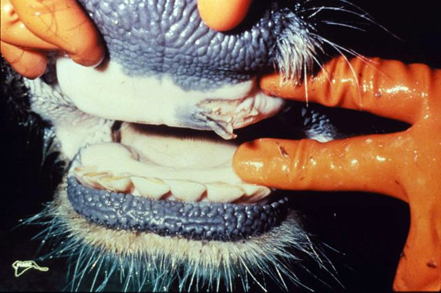
FMD produces vesicles, which are fluid-filled blisters, on the muzzle, tongue, gums, teats, and feet of cloven-hoofed animals. These vesicles rupture and leave painful erosions. Affected animals drool heavily, smack their lips, stamp their feet, and become lame. They go off feed, lose weight, and drop in milk production. Fever is common in the early phase.
The clinical picture varies by species. Cattle and buffaloes are highly susceptible and typically show severe oral and foot lesions [5]. Pigs tend to show more severe foot lesions and lameness. Sheep and goats often show milder signs, which is dangerous because mild cases can go unnoticed and spread the virus. The table below summarizes the species, typical lesion sites, and severity.
Species, Lesion Sites, and Severity
| Species | Typical lesion sites | Typical severity | Notes |
|---|---|---|---|
| Cattle | Tongue, gums, muzzle, interdigital space, teats | Severe | Heavy drooling, marked lameness, sharp milk drop |
| Buffalo | Mouth, feet | Severe | Highly susceptible, comparable to cattle [5] |
| Pigs | Feet, snout, oral cavity | Moderate to severe | Lameness often dominates the picture |
| Sheep | Mouth, feet | Mild to moderate | Mild signs are easily missed |
| Goats | Mouth, feet | Mild to moderate | Similar to sheep |
The severity of disease in a herd also depends on the virus serotype and the immune status of the animals. Endemic circulation of multiple serotypes in the same region is well documented. In Bangladesh, buffalo herds showed co-circulation of serotype O and Asia-1, with an individual-level prevalence of 41.0 percent in sampled animals and 88.1 percent of herds testing positive [5]. In Sudan, serotypes A and O circulated together between 2019 and 2022, with molecular testing detecting viral RNA in 94.3 percent of suitable samples compared with only 25.5 percent by antigen ELISA [6]. The practical lesson is that laboratory confirmation and serotyping are essential, because the clinical signs alone do not tell you which serotype you are dealing with.
Why Laboratory Confirmation Matters
Diagnosis cannot rest on clinical signs alone. Molecular methods such as real-time reverse transcription PCR are far more sensitive than antigen detection ELISA, and they are the preferred tools for confirming an outbreak [6]. Serological testing is used to assess immune status after vaccination and to detect infection. A competitive chemiluminescent enzyme immunoassay based on recombinant VP2 protein can detect antibodies against serotypes O and A simultaneously with high specificity and no cross-reactivity against classical swine fever virus, porcine reproductive and respiratory syndrome virus, or Senecavirus A [7]. A lateral flow strip based on recombinant non-structural protein can differentiate infected from vaccinated animals, which is critical when vaccination is part of the control strategy [1].
Distinguishing FMD from Look-Alike Diseases
Several diseases produce vesicles and lameness that resemble FMD. Getting the diagnosis wrong has serious consequences because the regulatory response differs.
Vesicular Stomatitis
Vesicular stomatitis affects cattle, horses, and pigs and produces vesicles on the mouth, teats, and feet that look almost identical to FMD. The key difference is that it also affects horses, which are not susceptible to FMD. It is caused by a different virus and is not typically subject to the same eradication measures.
Swine Vesicular Disease
Swine vesicular disease affects only pigs and produces vesicles on the snout, mouth, and feet. It is clinically indistinguishable from FMD in pigs without laboratory testing. It is caused by an enterovirus and is generally milder, but it is also notifiable in many countries.
Bluetongue
Bluetongue is a viral disease of ruminants spread by biting midges. It causes fever, oral ulcers, lameness, and swelling of the face and tongue, so it can be confused with FMD. It does not affect pigs. The oral lesions are typically erosions rather than true vesicles, and the epidemiology is tied to midge activity.
Other Differential Diagnoses
Foot rot, bovine viral diarrhea, and photosensitization can all cause oral or foot lesions that resemble FMD. None of them produce the combination of vesicles, drooling, and lameness across multiple species that FMD does. When in doubt, treat every vesicular lesion in a cloven-hoofed animal as a suspected FMD case until laboratory testing says otherwise.
The Principle of Outbreak Response
The core principle is simple. Stop the virus from moving. FMD spreads through direct contact between animals, through contaminated equipment, vehicles, clothing, and footwear, and through aerosol transmission over short distances. The virus is also present in meat, milk, and animal products, which is why trade restrictions follow every outbreak.
Control relies on four pillars, applied according to national policy.
- Quarantine and isolation. Separate affected and exposed animals from the rest of the herd immediately.
- Movement restriction. Stop all animal movements, vehicle traffic, and people movement on and off the premises.
- Culling or vaccination. Some countries cull infected and contact animals. Others use vaccination to create a buffer. Some use a combination.
- Surveillance and diagnostics. Test animals to confirm the serotype and monitor the spread.
The choice between culling and vaccination is a policy decision made by the national veterinary authority. It depends on the serotype, the availability of a matching vaccine, the size of the outbreak, and trade considerations. In countries that vaccinate, the vaccine must match the circulating serotype because there is no cross-protection between serotypes [2][3].
Materials and Supplies for Supportive Care
The table below lists the supplies needed to provide supportive care to affected animals. It does not include drug doses, which must be determined by the attending veterinarian.
| Item | Purpose | Notes |
|---|---|---|
| Clean water | Hydration | Offer frequently, especially to animals with oral lesions |
| Soft feed or gruel | Nutrition | Easier to eat than dry forage when the mouth is painful |
| Wound cleaning solution | Lesion hygiene | Use as directed by the veterinarian |
| Fly control | Prevent myiasis | Flies lay eggs in open lesions |
| Clean bedding | Comfort | Reduces pressure on sore feet |
| Disinfectant footbath | Biosecurity | For personnel and equipment, not for treating animals |
| Personal protective equipment | Biosecurity | Gloves, boots, coveralls, changed between areas |
| Dedicated tools | Biosecurity | Do not share between affected and clean areas |
Step-by-Step Supportive Care Procedure
The following steps describe how to care for an individual affected animal while maintaining biosecurity. Each step includes the reason it matters.
- Report the suspicion before doing anything else. Contact your state or federal animal health authority or your veterinarian immediately. FMD is notifiable in most jurisdictions and reporting is mandatory. Delay allows the virus to spread.
- Isolate the animal. Move it to a separate pen or paddock away from healthy animals. Use a barrier that prevents nose-to-nose contact. The virus spreads readily through direct contact.
- Establish a biosecurity line. Designate a clean zone and a dirty zone. Put on dedicated boots and coveralls before entering the dirty zone. Change or disinfect before returning to the clean zone. This prevents you from carrying the virus on your clothing and footwear.
- Provide soft, palatable feed. Oral lesions make eating painful. Offer soft feed, soaked hay, or gruel. The goal is to keep the animal eating so it does not lose condition.
- Ensure free access to clean water. Dehydration is a major risk when animals refuse to drink because of mouth pain. Offer water frequently and consider multiple water points.
- Clean oral lesions gently. Follow your veterinarian's instructions for cleaning and treating oral erosions. Gentle cleaning removes debris and reduces the risk of secondary bacterial infection. Do not use harsh chemicals.
- Clean and protect foot lesions. Wash the feet with a mild solution as directed, then apply any dressing or protective wrap your veterinarian recommends. Keep the animal on clean, dry bedding. Mud and manure worsen foot lesions and invite infection.
- Control flies. Flies lay eggs in open lesions and can cause myiasis, which is fly strike. Use approved fly control measures around the pen.
- Monitor temperature, appetite, and lameness daily. Record what you observe. Worsening lameness, refusal to eat, or a rising temperature may indicate secondary infection that needs veterinary attention.
- Disinfect equipment and vehicles. Use an approved disinfectant on boots, tools, and vehicle tires. FMD virus is susceptible to many common disinfectants, but they must be used at the correct concentration and contact time.
- Do not move animals off the premises. Movement restrictions are the single most effective tool for stopping spread. Follow the instructions of the animal health authority exactly.
- Keep records. Document which animals are affected, when signs started, and what care was given. This information helps the authorities trace the outbreak and plan the response.
Expected Results and How to Read Them
With supportive care, most affected animals survive the acute phase. The vesicles rupture, the erosions gradually heal, and the animal returns to eating and walking. Recovery takes weeks, not days. Foot lesions often take longer to heal than oral lesions because the animal keeps putting weight on them.
The outcome you are looking for is a gradual return of appetite, a reduction in drooling, and improved willingness to stand and walk. If the animal is still not eating after several days, or if the foot lesions are getting worse rather than better, that is a signal to call the veterinarian.
In severe outbreaks, mortality can occur, especially in young animals and in cases complicated by secondary infection. The prognosis depends on the serotype, the species, the age of the animal, and the quality of supportive care.
Workflow for a Suspected Outbreak
The diagram below shows the decision path from first suspicion to resolution.
flowchart TD
    A[Suspect FMD] --> B[Isolate animal]
    B --> C[Report to authority]
    C --> D[Laboratory testing]
    D --> E{Confirmed FMD}
    E -->|No| F[Investigate other causes]
    E -->|Yes| G[Apply national control policy]
    G --> H[Cull or vaccinate]
    G --> I[Supportive care]
    H --> J[Movement restrictions]
    I --> J
    J --> K[Surveillance and monitoring]
    K --> L[Declare free]
Troubleshooting Table
| Symptom | Likely cause | Fix |
|---|---|---|
| Animal refuses to eat for more than 2 days | Severe oral pain or secondary infection | Call the veterinarian, offer softer feed, check for infection |
| Foot lesions worsening | Mud, manure, or fly strike | Move to clean dry bedding, clean lesions, control flies |
| Fever persists beyond the expected window | Secondary bacterial infection | Veterinary examination and treatment |
| New animals showing signs | Virus spread before isolation | Review biosecurity, report to authority |
| Lameness in a previously unaffected animal | Early FMD lesion or foot rot | Isolate and have it examined |
| Sudden death in a young animal | Severe disease or secondary complication | Report immediately, do not move the carcass |
Variations in Control Policy
Control policy varies by country and sometimes by region within a country.
Culling policy. Infected and in-contact animals are slaughtered and disposed of. This is the fastest route to eradication but has a heavy economic and welfare cost.
Vaccination policy. Animals are vaccinated with a matching serotype vaccine to create a buffer of immunity. This reduces spread and allows animals to survive, but it complicates trade because vaccinated animals may test positive on some serological assays. Non-structural protein tests are used to differentiate infected from vaccinated animals [1].
Combination policy. Some countries vaccinate to slow the outbreak and then cull the vaccinated animals later. This is sometimes called vaccinate-to-live or vaccinate-to-die, depending on the end goal.
Whichever policy applies, the owner's role is the same. Report, isolate, restrict movement, and provide supportive care until the authorities direct otherwise.
Storage and Stability Notes
FMD virus is stable in cool, moist conditions and can survive in meat, milk, and animal products. It is inactivated by heat and by many common disinfectants. Store disinfectants according to the label, and do not dilute them beyond the recommended concentration. Vaccine storage requirements depend on the specific product and must follow the manufacturer's instructions. A monovalent inactivated vaccine formulated as a double oil emulsion has been shown to induce protective immunity in cattle, buffaloes, and sheep [2], but it must be stored and handled correctly to remain effective.
Biosecurity in Practice
Biosecurity is not a single action. It is a set of habits that must be maintained throughout the outbreak.
- Change boots and coveralls between the affected area and the clean area.
- Disinfect vehicles entering and leaving the premises.
- Do not share tools, feed buckets, or equipment between affected and healthy animals.
- Limit visitors and keep a log of everyone who enters.
- Wash hands thoroughly after handling affected animals.
- Do not take animal products off the premises.
These measures are tedious, but they are the difference between a contained outbreak and a regional disaster.
Limitations and When to Contact a Veterinarian
This article provides general guidance for a serious notifiable disease. Individual cases require a veterinarian, and suspected FMD must be reported to the animal health authority.
Contact a veterinarian or the animal health authority immediately if:
- You see vesicles, blisters, or erosions on the mouth, feet, or teats of any cloven-hoofed animal.
- Multiple animals in a group develop lameness or drooling at the same time.
- An animal stops eating and drinking for more than 24 hours.
- Foot lesions worsen, smell bad, or are infested with maggots.
- A young animal dies suddenly.
- You have moved animals on or off the premises in the past two weeks and now see signs of disease.
- You are unsure whether a lesion is FMD or another disease.
Do not wait for laboratory confirmation to isolate animals and report the suspicion. Early reporting is the most important action an owner can take.
Clinical Reasoning Behind Supportive Care in Foot and Mouth Disease
Understanding why supportive care works requires understanding what the virus does to the animal. FMD virus replicates in the epithelial cells of the mouth, pharynx, and feet, causing the cells to balloon with fluid and form vesicles. When those vesicles rupture, they leave raw, painful erosions. The animal's refusal to eat and drink is not a behavioral choice; it is a pain response. Every swallow and every step hurts. The clinical reasoning behind supportive care is therefore straightforward: reduce pain, maintain intake, and prevent the open lesions from becoming a portal for bacteria.
The immune response to FMD is serotype-specific. Antibodies produced against one serotype do not neutralize another, which is why an animal that recovers from serotype O can still be infected by serotype A or Asia-1 [2][3]. This has two practical implications. First, natural infection does not confer broad protection, so recovered animals remain susceptible to other serotypes circulating in the region. Second, any vaccination used during an outbreak must be matched to the serotype identified by laboratory testing. A mismatched vaccine wastes resources and creates a false sense of security.
The duration of protective immunity after natural infection or vaccination is finite. A monovalent inactivated vaccine against an emerging serotype induced protective antibody levels in cattle and buffaloes by 14 days after vaccination and in sheep by 21 days, with immunity lasting up to 32 weeks in cattle and buffaloes and 28 weeks in sheep [2]. This window shapes how control programs are timed. Vaccination campaigns must be repeated to maintain herd immunity, and the interval between campaigns depends on the duration of protection and the ongoing risk of exposure.
Secondary bacterial infection is the most common complication of FMD and the most common reason an animal that should recover instead deteriorates. Open erosions in the mouth and on the feet are ideal environments for bacteria, especially when the animal is standing in mud or manure. The feet are particularly vulnerable because they are in constant contact with the ground. Fly strike is another complication; flies lay eggs in open lesions, and the resulting maggots cause additional tissue damage and distress. These complications are preventable with diligent hygiene and fly control, which is why the supportive care protocol emphasizes clean bedding and lesion cleaning.
Diagnostic Workflow from Suspicion to Confirmation
The diagnostic workflow for suspected FMD follows a defined sequence, and each step has a purpose. Understanding the sequence helps owners and farm workers cooperate efficiently with the investigating authorities.
The first step is clinical suspicion. This occurs when an animal or group of animals shows vesicles, erosions, drooling, or lameness. At this point, no laboratory test has been performed, but the clinical picture is consistent with FMD. The correct action is to isolate and report, not to wait for confirmation. The reason is that the virus can spread from an infected animal before laboratory results are available, and every hour of delay increases the risk to neighboring farms.
The second step is sample collection by a veterinarian or trained animal health professional. Suitable samples include vesicular fluid, epithelial tissue from ruptured vesicles, and swabs from the mouth or feet. The quality of the sample directly affects the sensitivity of the test. In a Sudanese study, molecular testing detected viral RNA in 94.3 percent of suitable samples, compared with only 25.5 percent by antigen ELISA [6]. The difference is partly due to sample quality and partly due to the inherent sensitivity of the two methods. This is why molecular testing is preferred for confirmation.
The third step is laboratory testing. Real-time reverse transcription PCR is the most sensitive method for detecting viral RNA and is the preferred tool for confirming an outbreak [6]. Antigen ELISA is faster and cheaper but less sensitive, so a negative ELISA result does not rule out FMD. Serotyping follows confirmation, because the serotype determines which vaccine, if any, will be used.
The fourth step is serological testing, which serves a different purpose. Serology detects antibodies rather than the virus itself, so it is used to assess immune status after vaccination and to detect past infection. A competitive chemiluminescent enzyme immunoassay based on recombinant VP2 protein can detect antibodies against serotypes O and A simultaneously with high specificity and no cross-reactivity against classical swine fever virus, porcine reproductive and respiratory syndrome virus, or Senecavirus A [7]. This matters because false positives from cross-reactive antibodies would trigger unnecessary control measures.
The fifth step is differentiation of infected from vaccinated animals. When vaccination is part of the control strategy, serological tests that detect antibodies against non-structural proteins are used to identify animals that have been infected rather than merely vaccinated. A lateral flow strip based on recombinant non-structural protein can perform this differentiation [1]. This is critical for trade and for deciding when an outbreak can be declared over.
The diagnostic workflow is not a single test but a sequence of tests, each answering a different question. Clinical suspicion answers whether FMD is possible. PCR answers whether the virus is present. Serotyping answers which strain is circulating. Serology answers whether animals have been exposed or vaccinated. Non-structural protein testing answers whether a vaccinated animal has also been infected. Each answer informs the next decision in the control program.
Evidence Limitations in FMD Treatment and Control
The evidence base for FMD treatment and control has important limitations that owners and veterinarians should understand. Recognizing these limitations prevents overconfidence in any single intervention.
The first limitation is that there is no randomized controlled trial evidence for supportive care protocols in FMD. The recommendations for soft feed, clean water, lesion cleaning, and fly control are based on general principles of wound care and supportive medicine, not on FMD-specific trials. This does not mean the recommendations are wrong; it means they are extrapolated from similar conditions. The absence of trial evidence is a consequence of the disease's notifiable status, which makes controlled studies ethically and logistically difficult.
The second limitation is that vaccine efficacy data are serotype-specific and cannot be generalized. A vaccine that induces protective immunity against one serotype may not protect against another [2][3]. Even within a serotype, vaccine efficacy depends on the match between the vaccine strain and the circulating field strain, the adjuvant used, the dose, and the animal's immune status. The finding that a monovalent inactivated vaccine induced protective antibody levels in cattle and buffaloes by 14 days and in sheep by 21 days applies to that specific vaccine and that specific serotype [2]. It does not mean that all vaccines against all serotypes will perform the same way.
The third limitation is that diagnostic test performance varies with sample quality, timing, and the test method used. The Sudanese study found that molecular testing detected viral RNA in 94.3 percent of suitable samples compared with 25.5 percent by antigen ELISA [6]. This is a large difference, but it does not mean PCR is perfect. A poorly collected sample, a sample taken too late in the disease course, or a sample from an animal with low viral load can all produce a false negative. A negative test result in a clinically suspect animal should not override the clinical picture.
The fourth limitation is that prevalence data from one region cannot be applied to another. The finding that buffalo herds in Bangladesh showed co-circulation of serotype O and Asia-1, with an individual-level prevalence of 41.0 percent and 88.1 percent of herds testing positive [5], describes that specific population at that specific time. It does not predict what will happen in a different country or a different production system. Prevalence depends on vaccination coverage, animal movement patterns, trade, and the circulating serotypes.
The fifth limitation is that the economic and welfare costs of different control policies are not fully captured by efficacy data alone. Culling stops an outbreak quickly but has a heavy welfare and economic cost. Vaccination allows animals to survive but complicates trade and requires repeated campaigns. The choice between policies is a value judgment as much as a scientific one, and the evidence does not make the choice for the decision-maker.
What Owners Should Observe and Record Before the Veterinary Visit
When FMD is suspected, the quality of the information the owner provides to the veterinarian and the animal health authority directly affects the speed and accuracy of the response. Owners should observe and record specific details before the veterinary visit.
The first detail is the timeline. When did the first animal show signs? When did the next animal show signs? How quickly did the signs spread through the group? A rapid spread over one to three days is more consistent with FMD than a slow spread over weeks. The timeline helps the veterinarian assess the stage of the outbreak and the likely source of introduction.
The second detail is the specific signs observed in each animal. Which animals are drooling? Which are lame? Which have visible lesions, and where are the lesions located? Are the lesions on the mouth, the feet, the teats, or multiple sites? Are the lesions vesicles, erosions, or ulcers? The pattern of lesions helps distinguish FMD from look-alike diseases.
The third detail is the number of animals affected and the number of animals at risk. If five of fifty animals are affected, the attack rate is ten percent. If forty of fifty are affected, the attack rate is eighty percent. The attack rate helps the veterinarian assess the severity of the outbreak and the likely control measures required.
The fourth detail is the species affected. If only pigs are affected, swine vesicular disease is a possibility. If horses are affected alongside cattle, vesicular stomatitis is more likely because horses are not susceptible to FMD. If only ruminants are affected and midges are active, bluetongue should be considered.
The fifth detail is any recent animal movement. Have animals been introduced to the premises in the past two weeks? Have animals been moved off the premises? Have vehicles, equipment, or people visited from other farms? Movement history is critical for tracing the source of the outbreak and identifying premises at risk.
The sixth detail is the vaccination history. Have the animals been vaccinated against FMD? With which vaccine, and when? The vaccination history affects the interpretation of serological tests and the choice of control measures.
The seventh detail is the feed and water history. Have there been any changes in feed or water that could explain the signs? Have animals had access to potentially contaminated feed or water? FMD virus can survive in meat, milk, and animal products, so feed containing such products is a potential source.
Owners should record these details in writing and provide them to the veterinarian at the first contact. Photographs of the lesions are also valuable, especially if the lesions change over time. The more precise the information, the faster the diagnostic and control response.
Preparing the Premises for a Veterinary Visit
Preparing the premises before the veterinarian arrives reduces the risk of spreading the virus and makes the visit more efficient. The following steps should be taken as soon as FMD is suspected.
Designate a clean zone and a dirty zone. The dirty zone is the area where affected animals are housed. The clean zone is everywhere else. The boundary between the two zones should be clearly marked, and movement between them should be controlled.
Set up a biosecurity station at the boundary. The station should have dedicated boots, coveralls, gloves, and disinfectant. Anyone entering the dirty zone must put on clean protective equipment. Anyone leaving the dirty zone must remove and disinfect or discard the equipment before entering the clean zone.
Park vehicles outside the dirty zone. Vehicles that enter the dirty zone can carry the virus on their tires and undercarriage. The veterinarian's vehicle should be parked in the clean zone, and the veterinarian should change into dedicated boots and coveralls before entering the dirty zone.
Provide a safe route for the veterinarian to examine the animals. The route should avoid unnecessary contact with unaffected animals. If possible, the affected animals should be moved to a separate pen or paddock before the veterinarian arrives.
Have records ready. The timeline, the list of affected animals, the lesion descriptions, the movement history, and the vaccination history should be written down and available for the veterinarian.
Limit the number of people present. Only essential personnel should be in the dirty zone. Each additional person is an additional biosecurity risk.
Prepare for sample collection. The veterinarian may need to collect vesicular fluid, epithelial tissue, or swabs. Having the animals restrained and accessible makes sample collection faster and less stressful for the animals.
Preparing the premises is not a substitute for reporting the suspicion. Reporting should happen first, and preparation should happen while waiting for the veterinary visit.
Prevention at the Herd Level
Prevention of FMD is a herd-level activity, not an individual-animal activity. The virus spreads through populations, so the measures that prevent introduction and spread operate at the population level.
The first preventive measure is vaccination. In countries where FMD is endemic, routine vaccination with a vaccine matched to the circulating serotype is the primary tool for reducing disease incidence. The vaccine must match the circulating serotype because serotypes do not cross-protect [2][3]. Vaccination does not prevent all infections, but it reduces the severity of disease and the amount of virus shed, which reduces transmission.
The second preventive measure is movement control. Animals, animal products, vehicles, equipment, and people can all carry the virus. Controlling their movement is the most effective way to prevent introduction. Quarantine new animals before introducing them to the herd. Disinfect vehicles entering and leaving the premises. Limit visitors and keep a log of everyone who enters.
The third preventive measure is biosecurity. Biosecurity is a set of habits that reduce the risk of introducing and spreading the virus. Change boots and coveralls between areas. Disinfect equipment. Do not share tools, feed buckets, or equipment between affected and healthy animals. Wash hands thoroughly after handling animals.
The fourth preventive measure is surveillance. Early detection of FMD depends on owners and veterinarians recognizing the signs and reporting them promptly. Surveillance systems that combine clinical reporting with laboratory testing can detect outbreaks before they spread widely.
The fifth preventive measure is zoning and compartmentalization. Some countries establish zones or compartments with different FMD status, which allows trade to continue from free zones while control measures are applied in affected zones. This requires strict separation and surveillance to maintain the status of the free zones.
Prevention is more cost-effective than control. An outbreak imposes costs on the affected farm, on neighboring farms, on the regional economy, and on international trade. The cost of prevention is small compared with the cost of an outbreak.
Prognosis and Factors That Influence Recovery
The prognosis for an individual animal with FMD depends on several factors, and understanding these factors helps owners set realistic expectations.
The first factor is the species. Cattle and buffaloes are highly susceptible and typically show severe disease [5]. Pigs tend to show severe foot lesions. Sheep and goats often show milder signs. The species affects the severity of disease and the speed of recovery.
The second factor is the age of the animal. Young animals are more vulnerable to severe disease and mortality. Their immune systems are less mature, and they are more susceptible to dehydration and secondary infection. Sudden death in a young animal is a recognized complication of FMD.
The third factor is the serotype. Different serotypes vary in virulence. Some cause mild disease, while others cause severe disease with high mortality. The serotype also determines which vaccine, if any, will be effective.
The fourth factor is the immune status of the animal. Animals that have been vaccinated or previously infected with the same serotype may have partial immunity, which reduces the severity of disease. Animals with no prior exposure are fully susceptible.
The fifth factor is the quality of supportive care. Animals that receive soft feed, clean water, lesion cleaning, and fly control recover faster and with fewer complications than animals that do not. The quality of care is one of the few factors that owners can control.
The sixth factor is the presence of secondary infection. Animals with secondary bacterial infection or fly strike take longer to recover and may not recover at all without veterinary treatment. Preventing secondary infection is a key goal of supportive care.
The seventh factor is the control policy applied. In a culling policy, affected animals are slaughtered, so the prognosis for the individual animal is poor even if the animal would have recovered with supportive care. In a vaccination policy, affected animals may be allowed to recover. The control policy is determined by the national veterinary authority, not by the owner or the veterinarian.
The prognosis for the herd depends on the same factors plus the speed of detection and the effectiveness of movement control. An outbreak detected early and contained quickly has a better herd prognosis than an outbreak detected late and spread widely.
Special Population Considerations
Certain populations of animals require special consideration in FMD treatment and control.
Young animals are more vulnerable to severe disease, dehydration, and mortality. They need more frequent monitoring and more aggressive supportive care. Soft feed and easy access to water are especially important. Sudden death in a young animal should be reported immediately.
Pregnant animals are at risk of abortion and other reproductive complications. The stress of disease and the pain of lesions can trigger abortion. Pregnant animals need careful monitoring and veterinary attention.
Lactating animals are at risk of mastitis because the teats may have lesions that allow bacteria to enter the udder. Teat lesions should be cleaned and protected, and milk production should be monitored for signs of mastitis.
High-producing dairy cattle are at risk of metabolic complications because they may stop eating and drinking. The drop in feed intake can trigger ketosis and other metabolic disorders. These animals need aggressive nutritional support.
Sheep and goats often show milder signs, which means they may not be identified as affected and may continue to spread the virus. Surveillance in sheep and goat flocks must be more sensitive than in cattle herds because the clinical signs are less obvious.
Buffaloes are highly susceptible and can maintain endemic circulation alongside cattle [5]. In regions where buffaloes and cattle are raised together, both species must be included in surveillance and vaccination programs.
Pigs tend to show severe foot lesions, which can make movement and feeding difficult. Pigs with severe foot lesions need special attention to bedding and flooring to reduce pain and prevent secondary infection.
Animals in mixed-species farms present a particular challenge because different species may show different signs and may be affected at different times. Surveillance must cover all species on the farm, and control measures must be applied to all species.
Animals in transit or at markets are at high risk of exposure and spread. Movement restrictions during an outbreak are especially important for these populations because they can carry the virus over long distances.
Conclusion
Foot and mouth disease has no curative treatment, but it is not untreatable. The treatment is supportive care, and the control is biosecurity, movement restriction, and the national control policy. Owners who understand the clinical reasoning behind supportive care, the diagnostic workflow, the evidence limitations, and the factors that influence prognosis are better prepared to act quickly and effectively when FMD is suspected. The most important actions are to isolate, report, and restrict movement. Everything else follows from those three steps.
This article is educational and is not a substitute for veterinary diagnosis or treatment. Suspected FMD must be reported to the animal health authority immediately.
Frequently Asked Questions
Can foot and mouth disease be cured with medication?
No. There is no specific antiviral treatment for FMD. Care is supportive and focuses on comfort, nutrition, hydration, and preventing secondary infection.
Is foot and mouth disease the same as hand, foot, and mouth disease in people?
No. They are different diseases caused by different viruses. Animal FMD affects cloven-hoofed livestock, while human hand, foot, and mouth disease is caused by enteroviruses such as coxsackievirus and EV-A71 [4][8].
Do I have to report suspected foot and mouth disease?
Yes. FMD is a notifiable disease in most jurisdictions, and reporting suspected cases is mandatory. Contact your animal health authority or veterinarian immediately.
Which animals can get foot and mouth disease?
Cattle, buffalo, sheep, goats, pigs, and other cloven-hoofed animals are susceptible. Buffaloes are highly susceptible and can maintain endemic circulation alongside cattle [5].
How is foot and mouth disease confirmed in a laboratory?
Molecular tests such as real-time reverse transcription PCR are the most sensitive method and can detect viral RNA in a high proportion of suitable samples, outperforming antigen detection ELISA [6].
Can vaccination stop an outbreak?
Vaccination can reduce spread when the vaccine matches the circulating serotype, because serotypes do not cross-protect [2][3]. It is used as part of a national control policy, not as a standalone cure.
How long does it take for an animal to recover?
Recovery takes weeks with supportive care. Oral lesions often heal faster than foot lesions, and full recovery depends on the severity of disease and the quality of care.
What should I do first if I suspect an outbreak?
Isolate the affected animal, report the suspicion to the animal health authority, and restrict all movement on and off the premises. Do these three things before anything else.
Related Articles
- How To Treat Periodontal Disease In Cats
- How To Treat Lyme Disease In Dogs
- How To Treat Dental Disease In Cats
- How To Treat Gum Disease In Dogs
- What To Do If Your Dog Is Stung By A Bee In The Mouth
- Dog Stung By Bee In Mouth
- Foot and Mouth Disease Symptoms in Animals
- Foot and Mouth Disease Treatment and Control
Sources
- Development of a mu3ABC-Based Lateral Flow Immunochromatographic Strip for Rapid DIVA-Compatible Detection of Antibodies Specific to Foot-and-Mouth Disease Virus.
- Preparation and evaluation of a monovalent foot and mouth disease vaccine as a rapid response to the emerging serotype SAT1 in Egypt.
- Phylogenetic Analysis of Foot-and-Mouth Disease Virus from Cattle in Nigeria, 2017-2020.
- Hand-foot-and-mouth disease: a new look at a classic viral rash.
- Endemic Circulation and Genetic Characterization of Foot-and-Mouth Disease Virus in Buffalo Populations of Bangladesh.
- Molecular epidemiology of foot-and-mouth disease viruses collected from Khartoum, Blue Nile, and Northern States of Sudan (2019-2022).
- Development of a Competitive Chemiluminescent Enzyme Immunoassay Based on Recombinant FMDV VP2 Protein for Rapid and Simultaneous Detection of Antibodies Against Foot-and-Mouth Disease Virus Serotypes O and A.
- Coxsackievirus A6-induced hand-foot-mouth disease.