Foot and Mouth Disease Symptoms in Animals
By Dr. Zubair Khalid, DVM, MS, PhD ·
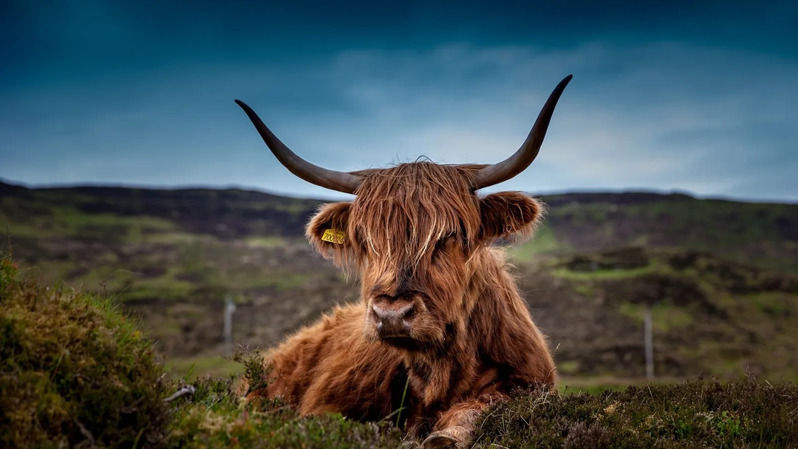
Foot and mouth disease (FMD) in animals produces fever followed by blisters, called vesicles, on the tongue, dental pad, gums, teats and hooves, along with heavy drooling, lameness and a sudden drop in milk yield. Cattle and pigs usually show obvious signs, while sheep and goats often show mild or inapparent signs that are easy to miss.
Foot and mouth disease is a highly contagious viral disease of cloven-hoofed livestock caused by foot and mouth disease virus (FMDV), a member of the genus Aphthovirus in the family Picornaviridae. It is not a human disease. It spreads rapidly within and between herds, and it is notifiable in the United States and most countries worldwide, meaning suspected cases must be reported to animal health authorities immediately. This article covers the clinical signs across cattle, pigs, sheep and goats, the biology behind those signs, the diseases that look similar, and what the signs mean for detection and control.
This article is educational and is not a substitute for veterinary diagnosis or treatment.
The Cardinal Signs of Foot and Mouth Disease
The clinical picture of FMD follows a predictable sequence. After an incubation period of roughly 2 to 14 days, an animal develops a fever, then vesicles appear, then the vesicles rupture and leave erosions that heal slowly. The most commonly reported signs in outbreaks are profuse salivation, vesiculation, lameness or ataxia, and ulceration, and these appear across species [1].
Fever and the Incubation Period
The incubation period for FMD is generally 2 to 14 days, depending on the virus dose, the route of exposure, and the species. Fever is often the first sign and can precede visible lesions by 24 hours or more. In experimentally infected bison, fever, lameness, inappetence and ptyalism (excessive salivation) developed alongside oral and foot lesions [2]. In cattle, FMDV can be transmitted at least 24 hours before clinical signs appear, which means an animal can infect pen mates while still looking normal [3]. This preclinical transmission window is one reason outbreaks spread faster than expected. In one modeling study, accounting for transmission during the incubation phase increased the number of affected farms in a between-farm simulation by 33.7 percent [3].
Vesicles on the Tongue, Dental Pad, Teats and Hooves
Vesicles are fluid-filled blisters that form on mucous membranes and skin. In cattle, the classic sites are the tongue, the dental pad (the tough pad on the upper jaw in front of the molars), the gums, the inner lips, the muzzle, the coronary band (the junction where the hoof meets the skin), and the interdigital skin between the claws. A review of the 2010/2011 FMD epidemic in the Republic of Korea found that about 54 percent of infected dairy farms reported vesicles on the teats as the primary clinical sign, while vesiculation on the nose, including the snout and muzzle, was the major lesion in infected beef cattle and pig farms [1]. In pigs, the teat and the feet were the second most frequently vesiculated body parts [1].
Vesicles rupture within a day or two, leaving raw erosions. The erosions on the tongue and dental pad make eating painful, which is why affected animals go off feed and drool. The erosions on the feet make walking painful, which is why they become lame.
Drooling and Salivation
Profuse salivation, also called ptyalism or drooling, is one of the most visible signs. It happens because vesicles and erosions on the tongue, gums and dental pad cause pain and interfere with normal swallowing. Saliva drips from the mouth, sometimes in long strings, and the animal may smack its lips or grind its teeth. In the Korean outbreak review, profuse salivation was among the most commonly observed clinical signs across all infected species [1]. In a study of cattle in Egypt, profuse salivation was recorded alongside fever, panting, lameness and reduced milk production in cows confirmed to have FMD [4].
Lameness and Foot Lesions
Lameness is a hallmark of FMD and often the sign that prompts a farmer to call a veterinarian. Vesicles on the coronary band and interdigital skin cause pain with every step. Animals may stand with an arched back, shift weight from foot to foot, or lie down more than usual. In severe cases, the hoof capsule can slough. In experimentally infected bison, lameness accompanied oral and foot lesions, and physical examination revealed vesicles and erosions on the coronary bands and interdigital skin [2]. In the Korean outbreak, lameness or ataxia was among the most frequently reported signs regardless of species [1].
Drop in Milk Yield
A sudden drop in milk production is a common early sign in dairy cattle, sometimes appearing before visible lesions. In Egyptian Holstein cows with confirmed FMD, reduced milk production was recorded along with fever, panting, profuse salivation and lameness [4]. Milk yield can fall sharply during the acute phase and recover slowly as lesions heal. Vaccination itself can cause transient reductions in milk production, which is one reason newer vaccination routes are being studied for their effect on productivity [5].
Species Comparison: Cattle, Pigs, Sheep and Goats


The same virus produces different clinical pictures in different species. Cattle and pigs tend to show severe signs. Sheep and goats often show mild or inapparent signs, which makes them dangerous silent spreaders.
| Feature | Cattle | Pigs | Sheep | Goats |
|---|---|---|---|---|
| Fever | Common, often first sign | Common | Variable, often mild | Variable, often mild |
| Oral vesicles | Tongue, dental pad, gums, muzzle | Snout, tongue | Tongue, often small | Tongue, often small |
| Teat vesicles | Common in dairy cows (about 54% of affected dairy farms) [1] | Teat and feet are second most affected sites [1] | Uncommon | Uncommon |
| Foot lesions | Coronary band, interdigital skin, lameness | Severe foot lesions, often the dominant sign | Mild, sometimes unnoticed | Mild, sometimes unnoticed |
| Drooling | Profuse | Profuse | Mild to moderate | Mild to moderate |
| Lameness | Common | Common and often severe | Mild | Mild |
| Milk drop | Marked in dairy cattle | Not applicable | Marked in dairy ewes | Marked in dairy does |
| Overall severity | Moderate to severe | Severe foot lesions | Mild or inapparent | Mild or inapparent |
Cattle
Cattle are the species most often used to describe the textbook picture of FMD. They develop fever, then vesicles on the tongue, dental pad, gums, muzzle, teats and feet. Drooling is profuse. Lameness is common. Milk yield falls in lactating cows. In the Korean outbreak, dairy cattle and pigs showed clearer clinical signs than beef cattle, deer and goats on the same infected farms [1]. Dairy cattle were especially likely to show teat vesicles, which is a useful early clue in milking herds [1].
Pigs
Pigs tend to show more severe foot lesions than cattle. Vesicles on the coronary band and interdigital skin can be extensive, and lameness can be the dominant sign. In the Korean outbreak, vesiculation on the snout and muzzle was the major lesion in pig farms, and the teat and feet were the second most frequently vesiculated body parts [1]. Pigs also drool heavily. Because pigs are often housed on hard floors, foot lesions can worsen quickly and lead to secondary bacterial infection.
Sheep
Sheep are the species most likely to be missed. Clinical signs are often mild or inapparent, and the lesions are small and easy to overlook. A retrospective analysis of submissions to the World Reference Laboratory for Foot-and-Mouth Disease found that only 30.0 percent of small ruminant samples tested positive for FMDV, compared with 72.2 percent of large ruminant samples and 73.4 percent of pig samples [6]. This lower detection rate reflects both milder disease and lower sampling effort. When sheep do show signs, they include fever, lack of appetite, salivation, vesiculation and erosions of the buccal mucosa, nose and feet, with signs ranging from mild to severe [7]. Sheep may also show bloody diarrhea and rapid respiration in some outbreaks [7]. Because sheep shed virus while showing few signs, they can spread FMD through a region before anyone realizes the disease is present.
Goats
Goats resemble sheep in showing mild or inapparent signs. In the Korean outbreak, goats on infected farms showed less clear clinical signs than dairy cattle and pigs [1]. Goats are susceptible and can transmit the virus, but they are often overlooked in surveillance because they do not look sick. In one vaccination and challenge study, goats vaccinated intradermally showed no clinical signs after challenge with a field isolate, while unvaccinated controls did [5]. This confirms that goats can be protected, but it also confirms that unvaccinated goats can carry and spread the virus.
Why the Signs Appear: The Biology in Plain Language
FMDV enters the body through the mouth or respiratory tract, then replicates in the epithelial cells that line the pharynx and the mucous membranes. From there it spreads through the bloodstream to other epithelial tissues, including the tongue, dental pad, teats, coronary band and interdigital skin. The virus destroys these cells, which causes fluid to accumulate under the surface and form vesicles.
The vesicles are the body's attempt to contain the damage. When they rupture, they leave erosions that are painful and slow to heal. The pain explains most of the visible signs. Pain in the mouth causes drooling and loss of appetite. Pain in the feet causes lameness. Pain in the teats causes cows to resist milking, which contributes to the drop in milk yield.
After the acute phase, about half of infected cattle develop a persistent infection in the upper respiratory tract that can last for weeks or months [8]. During this persistent phase, the virus continues to replicate and mutate, and the animals show no clinical signs [9]. This is why some cattle are called carriers. The carrier state is important for epidemiology but does not change the acute signs described in this article.
Differential Diagnosis: What Else Looks Like FMD
Several diseases produce vesicles, drooling and lameness in livestock. Because FMD is notifiable and has enormous economic consequences, any suspected case must be reported and tested. The main look-alikes are vesicular stomatitis, swine vesicular disease and bluetongue.
Vesicular Stomatitis
Vesicular stomatitis (VS) is caused by a vesiculovirus in the family Rhabdoviridae. It affects cattle, horses, pigs and sometimes humans. The vesicles look identical to FMD vesicles, which is why laboratory testing is required to tell them apart. VS is reportable in the United States. Unlike FMD, VS affects horses, which is a useful clinical clue because horses are not susceptible to FMD.
Swine Vesicular Disease
Swine vesicular disease (SVD) is caused by a virus in the genus Enterovirus. It affects pigs only and produces vesicles on the snout, feet and mouth that are indistinguishable from FMD. SVD is notifiable in many countries. It is generally milder than FMD, but laboratory testing is the only way to confirm the diagnosis.
Bluetongue
Bluetongue is caused by a virus in the genus Orbivirus and affects sheep, cattle and other ruminants. It does not produce true vesicles, but it can cause erosions and ulcers in the mouth, drooling, lameness from coronitis, and fever. The key difference is that bluetongue is spread by biting midges, not by direct contact, and it does not affect pigs. Bluetongue is also notifiable.
Other Conditions to Consider
Other conditions that can resemble FMD include bovine viral diarrhea (BVD), which can cause oral erosions, and photosensitization, which can cause skin lesions on unpigmented areas. Foot rot and digital dermatitis cause lameness but not oral vesicles. The presence of vesicles on the tongue, dental pad, teats or hooves in a cloven-hoofed animal should always raise suspicion of FMD until proven otherwise.
Clinical Relevance, Limitations and Common Mistakes
The clinical relevance of FMD signs is enormous. A single missed case in a sheep flock or goat herd can seed an outbreak that spreads across a region. In countries that are free of FMD, the appearance of vesicles in livestock triggers immediate regulatory action, including quarantine, movement controls and diagnostic testing. In countries where FMD is endemic, the signs are used for passive surveillance, but recognition is often poor. A study in Brazil, a country recognized as FMD-free without vaccination, found that only 23.74 percent of cattle producers correctly identified all clinical signs of FMD, and only 37.69 percent correctly recognized all susceptible species [10]. This knowledge gap is a real risk for early detection.
Common mistakes in recognizing FMD include:
- Assuming sheep and goats are not susceptible because they look healthy. They are susceptible and can spread the virus while showing few or no signs [6].
- Waiting for vesicles before reporting. Fever, drooling and a sudden drop in milk yield can precede visible lesions by 24 hours or more, and transmission can occur before clinical signs appear [3].
- Confusing FMD with vesicular stomatitis or swine vesicular disease. These diseases look identical, and only laboratory testing can tell them apart.
- Ignoring teat lesions in dairy cows. Teat vesicles were the primary sign on about 54 percent of infected dairy farms in one outbreak review [1].
- Underestimating foot lesions in pigs. Pigs can show severe foot lesions that dominate the clinical picture [1].
Limitations of clinical diagnosis are real. Individual animals can show atypical signs, and the severity depends on the virus strain, the dose, the route of exposure and the animal's immune status. A veterinarian should evaluate any animal with suspected FMD, and laboratory confirmation is required for a definitive diagnosis.
Frequently Asked Questions
What are the first signs of foot and mouth disease?
Fever and a drop in milk yield are often the first signs, followed within 24 hours by vesicles on the tongue, dental pad, teats or feet. Drooling and lameness appear as the vesicles rupture.
Can sheep and goats get foot and mouth disease?
Yes. Sheep and goats are susceptible to FMD, but they often show mild or inapparent signs. This makes them difficult to detect and allows them to spread the virus silently.
Is foot and mouth disease the same as hand, foot and mouth disease in humans?
No. Foot and mouth disease in animals is caused by FMDV and does not infect humans. Hand, foot and mouth disease in humans is caused by unrelated enteroviruses.
How long after exposure do symptoms appear?
The incubation period is generally 2 to 14 days. The exact timing depends on the virus dose, the route of exposure and the species.
Why do animals with FMD drool so much?
Vesicles and erosions on the tongue, gums and dental pad cause pain and interfere with swallowing. Saliva accumulates and drips from the mouth.
Do pigs or cattle get worse foot lesions?
Pigs tend to get more severe foot lesions than cattle. In pigs, vesicles on the coronary band and interdigital skin can be extensive and are often the dominant sign.
Can FMD be confused with other diseases?
Yes. Vesicular stomatitis, swine vesicular disease and bluetongue can all produce similar signs. Laboratory testing is required to tell them apart.
What should I do if I suspect FMD in my animals?
Contact your veterinarian and animal health authorities immediately. FMD is notifiable, and suspected cases must be reported. Do not move animals or animal products off the premises until authorities advise you.
Related Articles
- What To Do If Your Dog Is Stung By A Bee In The Mouth
- Dog Stung By Bee In Mouth
- Liver Disease Symptoms In Dogs
- Lyme Disease Symptoms In Dogs
- Symptoms Of Kidney Disease In My Cat
- Symptoms Of Pancreatitis Disease In Dogs
- How to Treat Foot and Mouth Disease in Animals
- Foot and Mouth Disease Treatment and Control
Sources
- Clinical manifestations of foot-and-mouth disease during the 2010/2011 epidemic in the Republic of Korea.
- Foot-and-mouth disease in North American bison (Bison bison) and elk (Cervus elaphus nelsoni): susceptibility, intra- and interspecies transmission, clinical signs, and lesions.
- Incubation phase transmission of foot-and-mouth disease virus in cattle: experimental evidence and simulated impacts.
- Clinical, haematological and biochemical alterations in heat intolerance (panting) syndrome in Egyptian cattle following natural foot-and-mouth disease (FMD).
- Intradermal Vaccination Induces Protective Immunity Against Foot-and-Mouth Disease While Mitigating Milk Yield Reduction in Ruminants.
- Retrospective analysis of submissions to the World Reference Laboratory for foot-and-mouth disease: What can these data tell us about the role of small ruminants in disease epidemiology?
- Outbreak of foot and mouth disease and peste des petits ruminants in sheep flock imported for immediate slaughter in Riyadh.
- Distinct mutations emerge in the genome of serotype O foot-and-mouth disease virus during persistence in cattle.
- Persistent foot-and-mouth disease virus infection of the bovine nasopharynx is associated with suppression of innate and cellular immunity.
- Assessment of knowledge on foot and mouth disease in free zones where vaccination is not practiced in Brazil.