Hydrocephalus in Dogs: Symptoms, Causes, and Diagnosis
By Dr. Zubair Khalid, DVM, MS, PhD ·
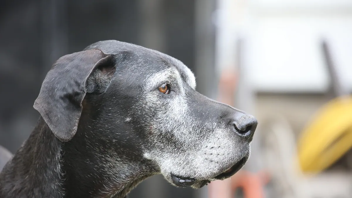
Hydrocephalus in dogs symptoms start with a change in behavior or movement long before the skull changes shape. The word means "water on the brain," and it describes a buildup of cerebrospinal fluid (CSF) that presses on brain tissue. Recognizing the early signs helps your dog get evaluated sooner, when treatment options are widest.
This article is educational and is not a substitute for veterinary diagnosis or treatment.
Quick Answer
- A domed or enlarged skull in a puppy, plus circling, head pressing, or seizures, is the classic picture of hydrocephalus in dogs.
- The condition can be congenital (present at birth) or acquired later from infection, trauma, or a mass.
- MRI is the imaging test that confirms the diagnosis and shows how much brain tissue is affected.
- Treatment ranges from medication to surgery, and a shunt is the standard surgical option for the congenital internal form [1].
- Any sudden worsening, collapse, or seizure that will not stop is an emergency, not a wait-and-see problem.
What Hydrocephalus in Dogs Symptoms Actually Look Like
The meaning of hydrocephalus is simple. CSF, the fluid that normally cushions the brain, builds up in the ventricles (the fluid-filled spaces inside the brain). When that fluid cannot drain properly, pressure rises and brain tissue gets compressed [2].
Signs in puppies
Congenital hydrocephalus often shows up in the first months of life. Puppies may have a rounded, dome-shaped head with an open soft spot. They may walk with a wide, unsteady gait, bump into walls, or seem slow to learn house training compared with littermates. Some cry out for no clear reason or press their head against furniture.
Signs in adult dogs
Adult-onset cases tend to show up as behavior change first. You might notice circling in one direction, staring at walls, or a head tilt. Seizures are common, and some dogs develop weakness in the limbs. In a study of toy-breed dogs with hydrocephalus, presenting signs included seizures, head turning, ataxia (wobbliness), and tetraparesis (weakness in all four legs) [3].
What is normal
It helps to know what is not hydrocephalus. A puppy who is clumsy because of fast growth, or an older dog who is stiff from joint pain, is a different picture. If your dog is limping or slow to rise, that points toward a joint problem, and you can read more in Arthritis In Dogs Symptoms And Diagnosis. Head shaking that comes and goes has its own list of causes, covered in Comprehensive Guide to Head Shaking in Dogs: Causes, Diagnostics, and Treatment.
Hydrocephalus in Dogs Causes
Hydrocephalus has several causes, and the cause shapes the treatment plan. Some dogs are born with it. Others develop it after an injury or illness.
| Cause | Typical clues | How urgent |
|---|---|---|
| Congenital (hereditary) | Young puppy, domed skull, unsteady walk, possible family history | Urgent, see a vet within days |
| Infection or inflammation | Fever, neck pain, recent illness, sudden behavior change | Emergency if severe |
| Trauma | Known head injury, worsening neurologic signs | Emergency now |
| Mass or blockage | Adult dog, progressive signs, seizures | Urgent, imaging needed |
| Fluid buildup without clear cause | Ventriculomegaly found on routine imaging | Routine follow-up |
Hereditary and congenital causes
Hydrocephalus is one of the most common malformations in dogs [4]. A case report of a Beagle pedigree found suspected hereditary lateral ventricular enlargement confirmed by MRI, with a sire showing hydrocephalus and a second cousin also affected [4]. That pattern supports a genetic basis in some lines. If a breeder tells you a relative had hydrocephalus, that is worth mentioning to your veterinarian.
Ventriculomegaly versus true hydrocephalus
Not every enlarged ventricle is a problem. Ventriculomegaly means the ventricles are bigger than usual, and it is often found by accident during routine imaging [5]. A study of 95 dogs with normal neurologic exams found that dogs with ventriculomegaly had measurable changes in brain regions including the cingulate cortex, caudate nucleus, thalamus, hippocampus, and corpus callosum [5]. In other words, the brain adapts to extra fluid, and the line between a harmless variant and true disease depends on clinical signs plus imaging.
Acquired causes
Infections, trauma, and masses can block CSF flow or cause pressure to rise. One report described a 6-month-old Bernese Mountain Dog with a subdural mass, midline shift, compression of the left ventricle, and herniation, traced back to an oral cavity source [6]. That case shows how a problem far from the brain can still raise intracranial pressure. If your dog has a swollen face, a bad tooth, or a recent mouth injury plus neurologic signs, tell your veterinarian.
Red Flags That Make This an Emergency
Call an emergency clinic right away if you see any of these:
- A seizure that lasts more than a few minutes or repeats without recovery
- Sudden collapse, inability to stand, or loss of consciousness
- Rapidly worsening confusion, circling, or head pressing
- Breathing that becomes irregular or labored
- A puppy with a bulging soft spot who becomes limp or unresponsive
These signs suggest rising pressure inside the skull, which can damage brain tissue quickly. Do not wait for a regular appointment.
What You Can Safely Do at Home While You Wait
There is no home treatment for hydrocephalus. Your job is to keep your dog safe and gather information for the veterinarian.
- Keep your dog in a quiet, padded area away from stairs and sharp corners.
- Block access to pools, decks, and furniture your dog could fall from.
- Do not give any human medication, including pain relievers, without veterinary direction. The dose depends on your dog's weight and health, and your veterinarian will calculate it.
- Write down what you see: when signs started, how often they happen, what triggers them, and any video of an episode.
- Note your dog's age, breed, and any family history of neurologic problems.
If your dog has a seizure, time it and keep your hands away from the mouth. Dogs do not swallow their tongues, and you can be bitten during the episode.
What Your Veterinarian Will Check and Test
Your veterinarian starts with a full neurologic exam. They watch how your dog walks, check the pupils and reflexes, and look for a domed skull or open fontanelle in puppies. They also ask about the timeline, since a slow onset points one way and a sudden onset points another.
Bloodwork and baseline tests
Routine bloodwork helps rule out metabolic causes of neurologic signs, such as liver or kidney problems. It also confirms your dog is stable enough for anesthesia if imaging is needed.
MRI and what it shows
MRI is the imaging test of choice for hydrocephalus. It shows the ventricles, the brain tissue around them, and any mass, fluid pocket, or blockage. In the Beagle case, MRI was used to calculate the ventricle-to-brain index (VBI), a number that quantifies how much of the brain space is fluid [4]. A VBI of 0.72 in that dog reflected severe hydrocephalus, while the dam's VBI of 0.52 reflected minimal enlargement [4]. Your veterinarian may use a similar measurement to track your dog over time.
MRI also helps distinguish hydrocephalus from other problems. In the Bernese Mountain Dog case, MRI showed a mass, midline shift, and herniation that pointed to an infection instead of simple fluid buildup [6]. That distinction changes treatment completely.
Other tests
Depending on findings, your veterinarian may recommend CSF analysis, which is collected during the same anesthetic event as the MRI. They may also check for underlying infections or inflammatory disease. If surgery is planned, imaging helps map the safest path for a shunt.
Treatment and Monitoring After Diagnosis
Treatment depends on the cause, the severity, and your dog's age.
Medical management
Mild cases may be managed with medication that reduces CSF production. Your veterinarian will choose the drug and calculate the dose from your dog's weight and health. Recheck exams and repeat imaging track whether the medication is holding pressure steady.
Surgical shunting
For congenital internal hydrocephalus, ventriculoperitoneal shunting (VPS) is the standard treatment [1]. A tube drains fluid from the ventricle into the abdomen. Shunt obstruction is a frequent and serious complication, and a study of 100 dogs treated with VPS looked at risk factors including age, sex, breed, weight, ventricular size, catheter position, and preoperative medications [1]. Shunt design itself is an active area of research, with flow velocity, pressure difference, and shear stress identified as key performance factors [2].
Newer techniques aim to place the catheter more accurately. A study of 11 toy-breed dogs used electromagnetic neuronavigation to guide catheter placement during VP shunt surgery, with accurate tip position checked by postoperative CT [3]. This is a specialized option available at referral centers.
Medication cautions
Dogs with hydrocephalus may react differently to common drugs. One report described two dogs with hydrocephalus who developed myoclonus (sudden muscle jerks) after premedication with butorphanol and remifentanil, which resolved after midazolam was given [7]. The authors advised caution with opioids in dogs who have pre-existing central nervous system disorders [7]. Always tell any veterinarian treating your dog that hydrocephalus is part of the history.
Monitoring at home
Keep a simple log of seizures, circling episodes, and changes in appetite or energy. Bring it to every recheck. Report any new weakness, vomiting, or fever right away, since shunt complications can appear suddenly.
Prevention and Long-Term Outlook
You cannot prevent every case, but you can lower risk in a few practical ways.
- Ask breeders about neurologic history in the parents and littermates. Hereditary patterns have been documented in at least one pedigree [4].
- Keep puppies and adult dogs away from head trauma, and use a harness instead of a neck collar if your dog has balance problems.
- Treat dental disease and mouth injuries promptly, since an oral source was linked to intracranial infection in one report [6].
- Keep routine wellness visits, especially in the first year of life when congenital signs appear.
- If your dog has a shunt, follow the recheck schedule closely. Obstruction is common enough that monitoring matters [1].
Some dogs with mild ventriculomegaly live normal lives with no treatment. Others need lifelong management. Your veterinarian's plan will be based on your dog's specific imaging and exam findings.
Limitations and When to Contact a Veterinarian
This article cannot tell you whether your dog has hydrocephalus. Only a veterinarian can examine your dog, review imaging, and make that call. Individual cases vary widely, and two dogs with the same MRI findings can need very different care.
Contact your veterinarian the same day if you notice new circling, head pressing, a head tilt, or a change in behavior that lasts more than a few hours. Go to an emergency clinic now if your dog has a seizure that will not stop, collapses, cannot stand, or becomes unresponsive. Schedule a routine visit if you have a puppy with a domed head but no other signs yet, or if you are considering breeding a dog with a family history of neurologic disease.
If you are worried about other neurologic or gastrointestinal signs, Dog Obstruction Symptoms covers a different set of emergencies that can look similar at home.
Frequently Asked Questions
What is the life expectancy of a dog with hydrocephalus?
It varies a lot based on the cause and how well treatment controls pressure. Some dogs with mild ventriculomegaly live a normal lifespan without surgery, while dogs with severe congenital hydrocephalus may need lifelong management. Your veterinarian can give you a realistic range after seeing the MRI.
Can hydrocephalus in dogs be cured?
There is no cure that reverses brain tissue changes, but treatment can manage pressure and improve quality of life. Surgery with a shunt is the standard option for congenital internal hydrocephalus, though shunt obstruction remains a common complication. Medical management helps some milder cases.
Is hydrocephalus painful for dogs?
The pressure itself is not typically described as pain, but the resulting headaches and neurologic signs can make a dog uncomfortable and distressed. Dogs with rising pressure may pace, press their head, or become withdrawn. Treating the underlying pressure is the way to relieve that distress.
Do all dogs with big ventricles need treatment?
No. Ventriculomegaly is often found by accident in dogs with normal neurologic exams, and many of those dogs do fine without treatment. The decision depends on whether your dog has clinical signs and how the brain tissue looks on imaging.
Can hydrocephalus be prevented in puppies?
You cannot prevent a congenital case once a puppy is conceived, but you can reduce risk through careful breeding choices. Ask about neurologic history in the parents and relatives, since hereditary patterns have been documented. Early veterinary visits help catch signs in the first months of life.
Related Articles
- Comprehensive Guide to Head Shaking in Dogs: Causes, Diagnostics, and Treatment
- What Are The Signs And Symptoms Of A Dog With Worms
- Dog Obstruction Symptoms
- Arthritis In Dogs Symptoms And Diagnosis
- Signs Of Worms In A Dog
- Dog Dehydration Symptoms: How to Tell and What to Do
- Hydrocephalus in Cats: Symptoms, Causes, and Risk
- Head Pressing in Dogs: Causes, Symptoms, and What to Do
- Stroke in Dogs: Symptoms, Causes, and What to Do
References
- Evaluation of risk factors for obstruction of the intraventricular catheter after ventriculoperitoneal shunting in dogs with congenital internal hydrocephalus. Frontiers in veterinary science, 2025.
- Mitigating failures and enhancing reliability of a canine ventricular shunt through robust multi-objective design method. Medical engineering & physics, 2026.
- Novel application of electromagnetic neuronavigation for guided ventriculoperitoneal shunt placement in toy-breed dogs with hydrocephalus: a preliminary descriptive study. American journal of veterinary research, 2026.
- Case Report: Suspected hereditary spontaneous hydrocephalus in a Beagle pedigree. Frontiers in veterinary science, 2026.
- Subcortical and white matter alterations in canine ventriculomegaly: an observational volumetric and voxel-based-morphometry study. Fluids and barriers of the CNS, 2025.
- Subdural Empyema in a Dog Originating From the Oral Cavity. Veterinary radiology & ultrasound : the official journal of the American College of Veterinary Radiology and the International Veterinary Radiology Association, 2026.
- Opioid-Induced Myoclonus During Premedication in Two Dogs With Hydrocephalus Proactively Managed With Antiepileptic Medications. Veterinary medicine and science, 2025.