Erythema Migrans Rash: Lyme Disease Explained
By Dr. Zubair Khalid, DVM, MS, PhD ·
Erythema migrans is the expanding skin rash that marks the early localized stage of Lyme borreliosis, the tick-borne infection caused by Borrelia burgdorferi sensu lato. It usually appears 3 to 30 days after the bite of an infected Ixodes tick, grows outward over days, and often shows central clearing that gives it a bullseye look, although a uniform red patch is just as common [1][2].
The rash is the single most useful clinical clue in early Lyme disease. It is also the stage at which treatment works best, which is why recognizing it matters more than any laboratory result. This guide explains what erythema migrans looks like, how it changes over time, how it differs from look-alike skin problems, and how the same infection behaves in dogs, horses, and other animals.
This article is educational and is not a substitute for veterinary diagnosis or treatment.
What Erythema Migrans Is and Why It Happens
Erythema migrans is not an allergic reaction to a tick bite. It is the visible result of Borrelia spirochetes multiplying in the skin and spreading outward from the bite site. The rash is the first clinical manifestation of Lyme borreliosis and defines what clinicians call early localized disease [1][3].
The name has a long history. Older literature used terms such as erythema chronicum migrans, erythema chronica migrans, erythema migrans chronicum, and erythema chronicum migrans rash. Those older labels described the same lesion and are still used interchangeably in some texts and search queries, but the modern term is erythema migrans. The word "migrans" refers to the way the border creeps outward across the skin, not to the rash moving to other parts of the body.
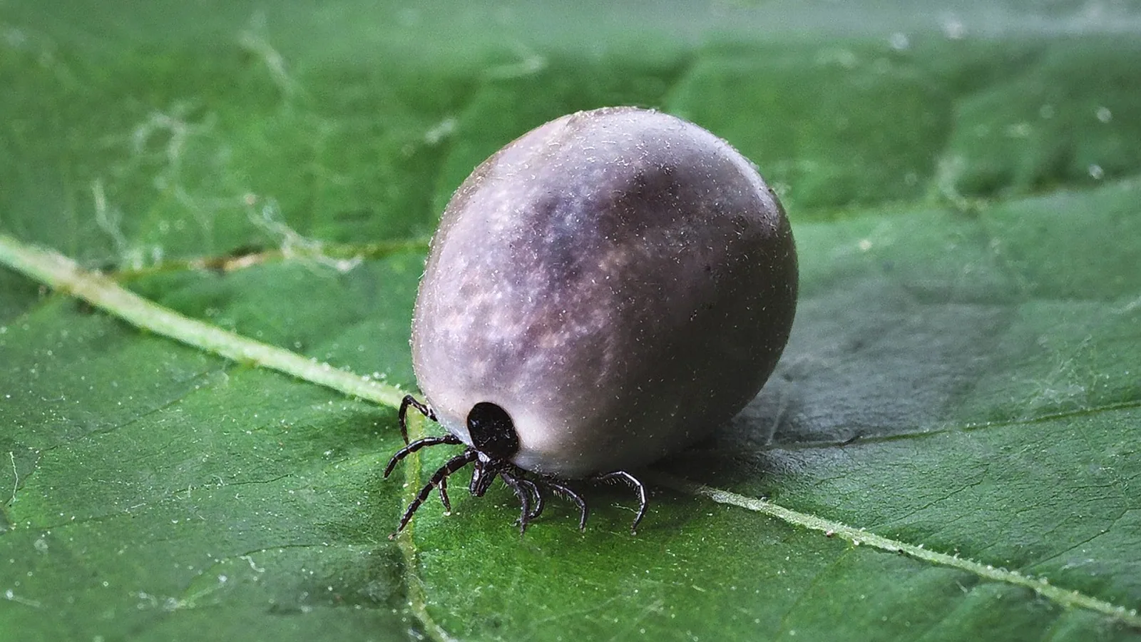
The infection is transmitted by ticks of the genus Ixodes. In North America the main vector is Ixodes scapularis in the Northeast and Upper Midwest and Ixodes pacificus on the West Coast. In Europe and parts of Asia, Ixodes ricinus carries the infection [4][5]. The bacteria involved are a group of related species called Borrelia burgdorferi sensu lato. In Europe, Borrelia afzelii and Borrelia garinii are the leading causes of Lyme borreliosis, while Borrelia burgdorferi sensu stricto dominates in North America [6].
Different Borrelia species tend to produce different clinical patterns. B. afzelii is strongly associated with skin findings, which is one reason erythema migrans is such a common presentation in European case series [4][6]. The BOLD study, a prospective European study of 797 Lyme borreliosis cases across six countries, found that 615 cases (77.2%) had erythema migrans and 182 (22.8%) had disseminated disease, most of which was Lyme arthritis [5].
The bullseye myth and the uniform red reality
Most people picture a perfect target with a red ring, a pale center, and a red dot in the middle. That image is real but incomplete. Central clearing produces the classic bullseye, and it is common enough to be the textbook description [1]. However, many erythema migrans lesions are uniformly red or dusky with no clearing at all. A solid expanding red patch is not a milder or atypical form. It is simply another normal presentation of the same lesion.
This matters because a person or an owner waiting for a bullseye before taking action can miss the diagnosis entirely. The key feature is expansion over days, not the presence of a ring.
The Timeline: Onset and Expansion
Erythema migrans typically appears 3 to 30 days after the tick bite [1]. In the case report literature, the interval can stretch longer. One patient who developed Lyme carditis with third-degree atrioventricular block described a tick bite and migrating erythema at the bite site one month before arriving at the hospital [7]. Another critically ill patient in northern China recalled an expanding erythematous lesion that followed a tick bite roughly 15 months earlier [8].
The lesion usually starts as a small red macule or papule at the bite site. Over several days to a couple of weeks it enlarges, sometimes reaching several centimeters or more across. The advancing edge is often the most intensely colored part, and the center may fade as the border moves outward. The lesion is usually not painful and often not intensely itchy, which is one reason it can be overlooked.
Lesions can occur at more than one site. A European badger with multiple erythematous lesions on its ventral body surface, heavily infested with Ixodes ricinus ticks, is a documented example of a multifocal presentation in an animal [4]. In people, multiple lesions generally indicate that the bacteria have already spread through the bloodstream rather than remaining at the original bite site.
Table: Stages of Lyme Borreliosis and Their Typical Features
| Stage | Timing after bite | Typical features | Notes |
|---|---|---|---|
| Early localized | 3 to 30 days | Erythema migrans at the bite site, expanding over days, often with central clearing but frequently uniform red | The most common clinical manifestation of early Lyme disease [3]. Treatment at this stage is most effective. |
| Early disseminated | Weeks to a few months | Multiple erythema migrans lesions, Lyme carditis, Lyme neuroborreliosis, early arthritis | In the BOLD study, 22.8% of European cases had disseminated disease, most commonly Lyme arthritis [5]. Cardiac involvement such as third-degree AV block is a recognized complication [7]. |
| Late | Months to years | Lyme arthritis, late neurologic findings, chronic skin changes | In the BOLD study, Lyme arthritis accounted for the large majority of disseminated cases [5]. Diagnosis often relies on serology because the rash is long gone. |
The table reflects general patterns. Individual patients do not always move neatly from one stage to the next, and some skip the rash entirely.
Recognizing Erythema Migrans in Photos and in Person


A photo-based approach helps because the lesion is defined by shape and change over time rather than by a single measurement.
Look for these features:
- An expanding red or dusky patch that grows over several days.
- A border that is often more intensely colored than the center.
- Central clearing that may or may not be present.
- A shape that is roughly circular or oval, sometimes irregular.
- A location that matches a site where a tick could attach, including the groin, armpit, waistband, behind the knee, or the scalp.
- Absence of severe pain, which distinguishes it from many other skin infections.
Serial photographs taken a day or two apart are more useful than a single image, because the defining feature of erythema migrans is expansion. A lesion that looks the same size on two occasions is less concerning for erythema migrans and more consistent with a fixed reaction.
One important limitation concerns skin tone. The characteristic erythema migrans rash is less visible on darker skin, and qualitative research with people of color diagnosed with Lyme disease identified this reduced visibility as a factor in diagnostic delays [9]. Clinicians and patients should not rely on redness alone. A dusky, violaceous, or subtly darker expanding patch deserves the same attention as a bright red one.
What Erythema Migrans Is Often Confused With
Several skin conditions look like erythema migrans at first glance. Distinguishing them changes management.
Tick-bite hypersensitivity
A local allergic reaction to a tick bite can produce redness, swelling, and itching at the attachment site. This reaction typically appears within hours to a day or two of the bite and does not continue to expand over days. It also tends to be itchy in a way that erythema migrans usually is not. The timing is the key discriminator. Hypersensitivity is immediate. Erythema migrans is delayed by 3 to 30 days [1].
Cellulitis
Cellulitis is a bacterial skin infection that produces a warm, tender, spreading redness. It is usually painful, may be accompanied by fever, and often has a less defined border than erythema migrans. Cellulitis does not typically show central clearing. It also responds to antibiotics that target skin flora rather than Borrelia, so mistaking one for the other leads to the wrong treatment.
Southern tick-associated rash illness
Southern tick-associated rash illness, often abbreviated STARI, produces an erythema migrans-like rash after the bite of the lone star tick, Amblyomma americanum. The rash can look nearly identical to Lyme disease, but the illness is not caused by Borrelia burgdorferi and is not Lyme disease. Geography and tick identification help separate the two, and the distinction matters because the diagnostic tests for Lyme disease will not confirm STARI.
Other considerations
A fixed drug eruption, a fungal skin infection, and a healing insect bite can all resemble erythema migrans. The expanding border and the delayed onset after a tick bite remain the most reliable clues.
Diagnosis: Clinical Recognition First, Testing Second
In early Lyme disease with a clear erythema migrans lesion, diagnosis is clinical. Guidelines from the Public Health Agency of Canada, the Association of Medical Microbiology and Infectious Disease Canada, the Infectious Diseases Society of America, and the European Society of Clinical Microbiology and Infectious Diseases all support early recognition based primarily on the characteristic rash and exposure to Lyme-endemic areas [10].
Laboratory testing is most useful when the rash is absent or atypical. Standard two-tier serologic testing has high specificity but low sensitivity in early disease, which means a negative test during the first weeks of infection does not rule out Lyme disease [3][11]. Several newer approaches aim to close that gap. A single-tier Hybrid Lyme ELISA that requires simultaneous binding to VlsE and the C6 peptide showed greater sensitivity than both standard and modified two-tier testing while maintaining equivalent specificity [3]. A multiplexed single-tier immunoassay combining ten Borrelia antigens with machine-learning classification identified all 30 early Lyme disease cases in one cohort and achieved an AUC of 0.98 in two independent cohorts [12]. The InBios Lyme Detect Multiplex ELISA detected 21 of 79 clinically diagnosed samples that standard two-tier testing missed [11]. Single-step immunoblot tests using recombinant protein antigens have also shown greater sensitivity than cleared two-tier tests for early-stage samples [13].
These advances matter because false negatives in early Lyme disease lead to delayed treatment and increase the likelihood of severe symptoms [11]. Even so, no serologic test replaces clinical judgment when a classic erythema migrans lesion is present.
Distinguishing Lyme disease from other febrile illnesses with similar symptoms is a separate challenge. Broad profiling of antibody binding has identified Borrelia protein biomarkers that differentiate Lyme disease from look-alike diseases, including in patients who tested negative on standard tests [14]. This line of work is still developing.
Treatment Principles and Follow-Up
Antibiotic treatment is the standard of care for Lyme borreliosis at every stage. The choice of agent, dose, and duration depends on the stage, the organs involved, and the individual patient. This article does not provide human antibiotic doses, because those decisions belong to a treating clinician.
What is well established is that earlier treatment produces better outcomes. The BOLD study documented that doxycycline was prescribed in every participating European country, although the overall pattern of antibiotic use varied [5]. In a case of Lyme carditis with third-degree AV block, ceftriaxone was started, and the patient ultimately required a permanent two-chamber pacemaker despite therapy [7]. At follow-up one year later he was symptom-free with no AV block on ECG [7]. That case illustrates why cardiac involvement is treated as a serious complication rather than a routine rash.
Follow-up timing varies with the clinical picture. A patient treated for early localized disease is typically reassessed to confirm that the rash is resolving and that no new symptoms have appeared. A patient with cardiac or neurologic involvement needs closer monitoring. Persistent or recurrent symptoms after treatment raise the question of post-treatment Lyme disease, which is a recognized outcome in a minority of cases [11].
Veterinary Relevance: Dogs, Horses, and Other Animals
Lyme borreliosis affects animals as well as people, but the clinical picture differs in important ways.
Dogs are the most commonly affected domestic species. They become infected through the same Ixodes ticks that transmit the disease to people, and infection can lead to arthritis, kidney disease, and other problems. What dogs rarely develop is erythema migrans. The expanding bullseye rash that defines early human Lyme disease is not a reliable clinical sign in dogs, which means veterinarians rely on history, physical examination, and serology rather than a visible rash.
Horses can also develop borreliosis, and again erythema migrans is not a typical finding. Diagnosis in horses rests on clinical signs and laboratory testing rather than on skin lesions.
The reason for this species difference is not fully understood. It may relate to differences in skin structure, immune response, or the Borrelia species involved. What is clear is that a dog or horse with lameness, joint swelling, fever, or unexplained illness in a Lyme-endemic area deserves veterinary evaluation even without a rash.
Wildlife can show the lesion. A European badger in Romania developed multiple erythematous lesions on its ventral body surface, and molecular analysis confirmed Borrelia afzelii DNA in the skin lesions. Histology showed a mild chronic inflammatory reaction consistent with erythema migrans-like lesions as seen in humans [4]. That report is important because it shows the lesion is not exclusive to people, and it highlights the role of wildlife as reservoir hosts in the disease cycle [4].
For pet owners, the practical takeaway is straightforward. Tick prevention and prompt tick removal reduce risk for dogs, cats, horses, and the people who live with them. The Companion Animal Parasite Council publishes year-round tick control guidelines for dogs and cats, and the American Veterinary Medical Association offers owner-facing resources on tick-borne disease prevention. A veterinarian can recommend a product appropriate for the individual animal.
Geographic Spread and Rising Risk
Lyme disease is the most common vector-borne illness in the United States, with an estimated 476,000 cases diagnosed and treated annually [9]. Incidence is increasing in Canada as well [10]. Warming temperatures and changing ecosystems have expanded tick populations and altered seasonal activity, raising the likelihood of exposure in areas previously considered low risk [15].
This has two consequences. First, clinicians must maintain a high index of suspicion even in patients without traditional geographic exposure [15]. Second, a history of travel matters. The first reported case of Lyme disease in a returning traveler to the Kingdom of Saudi Arabia involved a healthy woman who presented with erythema migrans after traveling to Scotland, where the disease is endemic [1]. Lyme disease has also been reported in Japan, with most domestic cases from Hokkaido and a median delay from onset to diagnosis of 28.5 days [16].
For anyone assessing a rash, the relevant question is not only where you live but where you have been.
Common Myths and Misunderstandings
Several beliefs about erythema migrans cause people to miss or misread the rash.
The first myth is that the rash always looks like a bullseye. Central clearing is common but not required [1]. A uniformly red expanding patch is a normal presentation.
The second myth is that you will always remember a tick bite. Many people never see the tick. The patient in one case report recalled a bite and a migrating rash a month before hospitalization [7]. The patient in another case had a tick bite roughly 15 months before the illness that brought him to intensive care [8]. In many cases there is no recalled bite at all, and the rash may be the only clue.
The third myth is that a negative blood test rules out Lyme disease. Early serology has limited sensitivity, and a negative result in the first weeks does not exclude the diagnosis [3][11].
The fourth myth is that the rash is the whole illness. Erythema migrans marks the early localized stage. Without treatment, the infection can disseminate to the heart, joints, and nervous system [7][5].
The fifth myth is that the rash looks the same on every skin tone. Erythema migrans is less visible on darker skin, and this reduced visibility contributes to diagnostic delays [9].
Limitations and When to Contact a Veterinarian
This article covers the biology and recognition of erythema migrans in people and explains why the same rash is uncommon in animals. It does not replace an examination. Individual cases need a veterinarian, and a person with a suspicious rash needs a physician.
Contact a veterinarian promptly if a dog or horse in a Lyme-endemic area develops lameness, joint swelling, fever, lethargy, loss of appetite, or unexplained illness, especially if a tick was found recently. Seek urgent veterinary care for collapse, difficulty breathing, a distended abdomen, or sudden weakness, since these can signal serious complications such as kidney involvement in dogs.
For people, seek medical care if an expanding rash appears within 3 to 30 days of a tick bite, if a rash is accompanied by fever, headache, neck stiffness, palpitations, fainting, or joint swelling, or if symptoms persist or worsen after treatment. Cardiac symptoms such as a slow or irregular heartbeat warrant emergency evaluation, because Lyme carditis can cause high-grade heart block [7].
Frequently Asked Questions
What does an erythema migrans rash look like?
It is an expanding red or dusky patch that grows over days, often with a paler center that creates a bullseye, but a uniform red patch is equally common [1][2].
How soon after a tick bite does erythema migrans appear?
It typically appears 3 to 30 days after the bite, though documented cases have shown longer intervals [7][1][8].
Can you have Lyme disease without the rash?
Yes. Not everyone recalls a tick bite or notices the rash, and some people present later with joint, heart, or neurologic findings [7][5].
Is erythema migrans always a bullseye?
No. Central clearing is common but not universal, and many lesions are solid red without a ring [1].
How is erythema migrans different from a tick-bite reaction?
A tick-bite hypersensitivity reaction appears within hours to a day or two and is often itchy, while erythema migrans is delayed by days to weeks and expands steadily [1].
Can dogs get erythema migrans?
Dogs can develop Lyme borreliosis, but erythema migrans is rarely seen in them, so diagnosis relies on clinical signs and testing rather than a rash.
Does a negative Lyme test mean you do not have Lyme disease?
No. Standard two-tier testing has low sensitivity in early disease, and newer single-tier assays were developed specifically to improve early detection [3][11].
Why is early recognition of erythema migrans so important?
Treatment is most effective in early localized disease, and delayed treatment increases the likelihood of severe complications [7][11].
Related Articles
- Lyme Disease In Dogs
- How To Treat Lyme Disease In Dogs
- Lyme Disease Symptoms In Dogs
- Lymes Disease In Cats: Comprehensive Veterinary Reference Guide
- Lyme Disease in Dogs: Borrelia Diagnosis and Doxycycline Treatment
- Lyme Disease Symptoms In Dogs: Comprehensive Veterinary Reference Guide
- What Is Anthrax Disease? Causes and Symptoms
- Sand Fly: Biology, Diseases, and Veterinary Importance
- Tularemia (Rabbit Fever): Symptoms and Transmission
Sources
- Lyme disease presenting with erythema migrans: First reported case in a returning traveler to the Kingdom of Saudi Arabia.
- Pathology Outlines - Erythema migrans
- A novel single-tier serologic test to diagnose all stages of Lyme disease.
- Erythema migrans-like lesions associated with Borrelia afzelii infection in a European badger, Meles meles.
- Clinical Manifestations of Lyme Borreliosis in Europe: Burden of Lyme Disease Study (BOLD), 2021-2022.
- Genomic characterization and antibiotic susceptibility of biofilm-forming Borrelia afzelii and Borrelia garinii from patients with erythema migrans.
- Association of Lyme Disease and Erythema Migrans with Atrioventricular Block.
- A rare presentation of clinically diagnosed lyme disease with probable neuroborreliosis, septic shock, and bone marrow suppression: a case report.
- Perspectives on Lyme disease among persons of color in the United States: Qualitative research findings.
- Update on Lyme disease diagnostic approaches and protocols in Canada: Overview of existing testing strategies and limitations.
- The multiplexed single-tier InBios Lyme Detect Multiplex ELISA is more sensitive than standard two-tier tests in the early stages of Lyme disease.
- Multiplexed single-tier immunoassay for improved Lyme disease diagnosis across all disease stages.
- Single-step immunoblot tests with recombinant protein antigens for detecting IgG and IgM antibodies in Lyme disease.
- Serologic biomarker discovery for differentiating Lyme disease from diseases with similar clinical symptoms using broad profiling of antibody binding.
- Lyme disease in a warming world: Rising cases, shifting patterns, and clinical implications.
- Epidemiology of Lyme disease, a growing tick-borne disease of concern, in Japan from May 2013 to March 2024: a descriptive study.