Dropped Jaw in Dogs: Causes and What to Do
By Dr. Zubair Khalid, DVM, MS, PhD ·
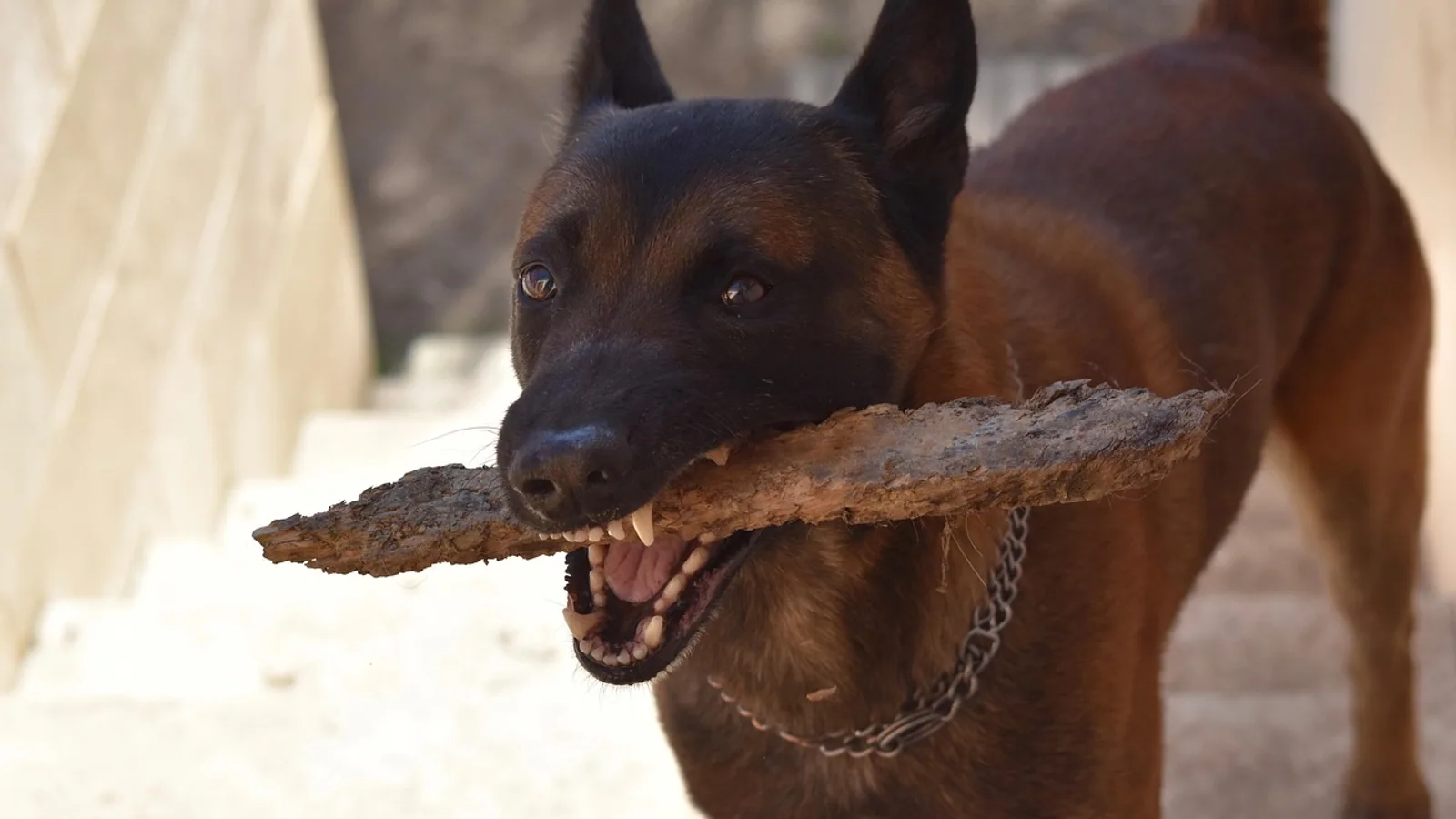
A dropped jaw is a sudden inability to hold the mouth closed. The lower jaw hangs open, the dog cannot bring the teeth together, and the mouth may stay open even when the dog is resting. In most dogs this is a nerve problem, not a broken bone. The three conditions veterinarians separate first are trigeminal neuropathy, masticatory muscle myositis, and jaw trauma or temporomandibular joint luxation. Each has a different cause, a different level of pain, and a different treatment plan.
This article is educational and is not a substitute for veterinary diagnosis or treatment.
Owner Triage: What to Do Right Now
A dropped jaw is a veterinary problem, not a wait-and-see problem. The jaw is the gateway to eating, drinking, breathing, and panting. A dog that cannot close its mouth cannot pick up food or lap water normally, and the tongue can become dry, swollen, or trapped.
Do these things first:
- Keep the dog calm and cool. A dog that cannot close its mouth cannot pant efficiently, so heat and stress make things worse.
- Do not force the mouth closed. If the jaw is fractured or the joint is dislocated, forcing it can cause more damage and severe pain.
- Do not try to feed hard food. Soft food or a slurry placed on the tongue may be safer, but only if the dog can swallow.
- Check for breathing trouble. Noisy breathing, blue gums, or a tongue that looks swollen and dark needs emergency care.
- Call your veterinarian or an emergency clinic and describe exactly what you see.
The single most useful thing you can tell the vet is whether the jaw hangs loosely or feels locked, and whether the dog seems painful when the mouth is touched. Those two observations point in very different directions.
At a Glance: Comparing the Main Causes
The table below summarizes how the three main causes differ. It is a guide to the pattern, not a diagnosis.
| Feature | Trigeminal neuropathy | Masticatory myositis | Jaw trauma or TMJ luxation |
|---|---|---|---|
| Typical onset | Sudden, over hours to days | Acute phase over days, or chronic | Immediately after injury |
| Pain | Usually none | Often painful in the acute phase | Painful |
| Jaw position | Hangs open, flaccid | May be open, or locked closed in chronic cases | Open, deviated, or asymmetric |
| Ability to open mouth | Jaw drops open easily, the problem is closing it | Often cannot open the mouth, trismus | Limited or abnormal movement, often with a click or shift |
| Muscle changes | Normal early, atrophy of chewing muscles later | Swollen and firm early, shrunken later | Swelling from trauma, not immune |
| Key test | Neurologic exam, MRI for the cause | 2M antibody test, EMG, muscle biopsy | Skull radiographs or CT |
| Sensation on the face | Usually spared | Normal | May be altered by swelling or nerve injury |
The Anatomy Behind a Dropped Jaw
To understand why the jaw drops, it helps to know which structures hold it up. Closing the mouth is an active job. The muscles of mastication, mainly the temporalis, masseter, and pterygoid muscles, pull the mandible up against the upper jaw. These muscles are supplied by the mandibular branch of the trigeminal nerve, the fifth cranial nerve. When that nerve stops working, the muscles go quiet and the jaw falls open under its own weight.
The trigeminal nerve does more than move muscles. It carries sensation from the face, the mouth, and the teeth, and it supplies the muscle that opens the pharyngeal opening of the auditory tube, the tensor veli palatini [1]. That last detail explains a finding veterinarians look for on imaging. When the trigeminal nerve is damaged, the auditory tube can stop opening properly, fluid builds up in the middle ear, and effusion appears in the tympanic cavity on the same side as the nerve problem [1][2].
The chewing muscles themselves are unusual. They are built from a distinctive fiber type composition that sets them apart from the muscles of the leg or back [3]. That specialization may be one reason these muscles are vulnerable to a specific immune attack, which is the basis of masticatory myositis.
Trigeminal Neuropathy: The Classic Dropped Jaw
Trigeminal neuropathy is the most common reason a dog suddenly develops a dropped jaw with no history of injury. The nerve stops conducting signals, the chewing muscles lose their tone, and the mandible hangs. Owners often describe it as the dog looking like it has forgotten how to close its mouth.
What Trigeminal Neuropathy Looks Like
The jaw hangs loosely and symmetrically in many cases. The dog can still feel the face in most instances. The palpebral reflex, the blink that happens when the inner corner of the eye is touched, is typically spared because that reflex runs through the facial nerve. This is a key point for owners and for veterinarians. A dog with a dropped jaw, normal facial sensation, and a normal blink is more likely to have a trigeminal problem than a widespread cranial nerve disease.
A less obvious sign is saliva. Dogs with trigeminal dysfunction can develop thick, foamy saliva that collects in the back of the mouth on the affected side [4]. The reason is that the salivary glands lose part of their nerve supply, and the saliva they produce changes in volume and consistency [4]. On MRI, the salivary glands on the affected side can look smaller and brighter than normal [4]. Owners may notice bad breath, drooling, or a messy mouth before they notice the jaw itself.
As the condition continues, the chewing muscles shrink. Atrophy of the temporalis and masseter muscles is a consistent finding in dogs with trigeminal nerve disease, including nerve sheath tumors and neuritis [5][6]. The change can be subtle at first and obvious after a few weeks. One side of the head may look hollow above the eye.
Why Trigeminal Neuropathy Happens
Veterinarians group the causes of trigeminal neuropathy into three buckets based on imaging: neoplasia, neuritis, and idiopathic trigeminal neuropathy [7].
- Neoplasia means a tumor, usually a nerve sheath tumor, growing on or near the nerve. These are typically unilateral. In one review of 49 dogs with trigeminal neuropathy, about 31 percent had suspected neoplasia, and all of those were unilateral [7]. Dogs with tumors were also older than dogs with neuritis or idiopathic disease [7].
- Neuritis means inflammation of the nerve. It can be unilateral or bilateral. In the same review, about 16 percent of dogs had neuritis [7]. MRI in dogs with trigeminal neuritis shows diffuse enlargement of the nerve without a distinct mass [5].
- Idiopathic trigeminal neuropathy means no cause is found. This was the largest group in that review at about 53 percent, and it was strongly associated with bilateral clinical signs [7]. A dog with both sides of the jaw affected is most likely to fall into this category.
Idiopathic trigeminal neuropathy is the condition many veterinarians call dropped jaw syndrome. It often follows a vaccination or an infectious illness in the weeks before the signs appear. The nerve inflammation is presumed to be immune mediated, but the exact trigger is not always identified. The good news is that idiopathic cases often improve over weeks to months with supportive care.
The Post-Vaccinal Pattern
Owners frequently ask whether a vaccine caused the dropped jaw. The honest answer is that a temporal association is sometimes seen, and the condition is often described as post-vaccinal, but proving cause and effect in an individual dog is difficult. What matters clinically is the pattern. A young dog with a sudden bilateral dropped jaw, normal facial sensation, a normal blink, and no pain fits idiopathic trigeminal neuropathy well. A veterinarian will still recommend imaging to rule out a tumor or an inflammatory lesion that needs different treatment.
Masticatory Myositis: Painful, Swollen, Then Shrunken
Masticatory muscle myositis is an immune-mediated disease that targets the chewing muscles specifically. It is the most common inflammatory myopathy in dogs [8]. The immune system attacks a protein found in masticatory muscle fibers, and the resulting inflammation damages the muscles.
The Two Phases of Masticatory Myositis
Masticatory myositis has an acute phase and a chronic phase, and they look almost opposite.
In the acute phase, the muscles are swollen, firm, and painful. The dog may resist having the mouth opened. The jaw may be held slightly open, or the dog may be unable to open it fully. Owners often notice swelling over the top of the head and along the cheeks. The dog may be reluctant to eat, may drop food, and may cry when the mouth is handled.
In the chronic phase, the muscles shrink and scar. The head takes on a sunken, bony appearance. The jaw may become stiff and difficult to open, a condition called trismus. In severe chronic cases, the mouth can be locked nearly shut. This is the opposite problem from a dropped jaw, and it is a common reason owners seek help months after the acute episode was missed.
Diagnosing Masticatory Myositis
The diagnosis rests on three tools.
The 2M antibody test is the most specific blood test. Dogs with masticatory myositis develop antibodies against a muscle protein called masticatory myosin binding protein-C, often shortened to 2M [9]. A positive titer supports the diagnosis. One reported case had a positive 2M titer at 1:500 [10]. The test is not perfect, and some dogs with the disease do not have circulating antibodies at the time of testing [8], so a negative result does not fully rule it out.
Electromyography, or EMG, measures the electrical activity of muscle. Dogs with masticatory myositis show abnormal spontaneous activity in the chewing muscles. EMG is also useful in trigeminal neuropathy, where spontaneous activity in the temporal and masseter muscles can reflect nerve damage [11].
Muscle biopsy provides the definitive tissue diagnosis. Biopsies are best taken from areas that look most abnormal on imaging [12]. The histology shows inflammation, muscle fiber necrosis, and atrophy. Immunohistochemistry can show MHC class I and class II antigens on the muscle fibers, which supports an immune-mediated process [13]. The cellular infiltrate includes CD8-positive T cells, which are thought to drive the muscle damage [8].
Imaging in Masticatory Myositis
CT and MRI can both show changes. On CT, the chewing muscles may be swollen or atrophied, and they often enhance with contrast in an inhomogeneous pattern [12]. The head and neck lymph nodes can be enlarged as well [12]. These findings help guide where to biopsy.
Why the Chewing Muscles Are Targeted
The masticatory muscles have a unique fiber type composition that is not found in most other muscles [3]. They also express immune markers on their surface that may help start and sustain the inflammation [13]. The result is a disease that attacks the muscles a dog uses to chew, which is why the jaw is the first thing affected.
Masticatory myositis is usually limited to the chewing muscles. When a dog has weakness or atrophy in the legs as well, veterinarians consider polymyositis or an underlying disease such as leishmaniasis, which can cause myositis in both masticatory and skeletal muscles [14]. Myasthenia gravis can also occur alongside masticatory myositis in the same dog, which complicates the picture [15].
Jaw Trauma and Temporomandibular Joint Luxation
Trauma is the third major cause of a dropped jaw, and it is the one with the clearest history. A dog that was hit by a car, fell from a height, fought with another animal, or ran into a hard object can fracture the mandible, fracture the temporomandibular joint, or dislocate the joint.
What Trauma Looks Like
The jaw may hang open, but it often hangs unevenly. One side may be higher than the other. The dog may have swelling, bruising, bleeding from the mouth, or a wound on the face. The mouth may not close at all, or it may close with a click or a shift. The dog is usually painful, though some dogs with severe injuries are surprisingly stoic.
Temporomandibular joint luxation means the condyle of the mandible has popped out of the joint socket. It can happen with relatively minor trauma in some dogs. The jaw deviates to one side, and the dog cannot close the mouth normally. This is a mechanical problem that needs to be reduced, meaning put back into place, by a veterinarian.
Diagnosing Trauma
Skull radiographs are the first imaging step. They can show fractures of the mandible, the condylar process, or the temporal bone. CT is more sensitive and is often used when the injury is complex or when surgery is being planned. A neurologic exam helps determine whether a nerve injury happened at the same time as the trauma.
How Veterinarians Tell These Apart
The history and physical exam narrow the list quickly. A dog with a sudden dropped jaw and no trauma, no pain, and normal facial sensation is most likely to have trigeminal neuropathy. A dog with a painful, swollen head and resistance to opening the mouth is more likely to have acute masticatory myositis. A dog with a known injury and an asymmetric jaw is more likely to have trauma.
The neurologic exam is the next step. Veterinarians test facial sensation, the palpebral reflex, the corneal reflex, and the ability to move the jaw. They look for atrophy of the temporalis and masseter muscles. They check for Horner's syndrome, which can occur with trigeminal disease and was present in about 12 percent of dogs in one case series [7].
Imaging follows. MRI is the best test for the trigeminal nerve itself. It can show a mass, diffuse nerve enlargement from neuritis, or normal findings in idiopathic cases [5][7]. MRI can also show middle ear effusion, which is common in dogs with trigeminal nerve disease and is associated with more severe muscle loss [1][2]. CT is useful for bone detail in trauma and for assessing the chewing muscles in myositis [12].
Blood work and specialized tests complete the picture. The 2M antibody test supports masticatory myositis [9][10]. EMG helps confirm muscle or nerve dysfunction [11]. Muscle biopsy gives a tissue diagnosis when the other tests are inconclusive [12].
The flowchart below shows the main decision path a veterinarian follows when a dog arrives with a dropped jaw.
flowchart TD
    A[Dog arrives with dropped jaw] --> B{History of trauma}
    B -->|Yes| C[Image the skull and joint]
    B -->|No| D{Is the mouth painful}
    D -->|Yes| E[Test for masticatory myositis]
    D -->|No| F{Can the mouth be opened}
    F -->|Yes jaw hangs open| G[Evaluate the trigeminal nerve]
    F -->|No jaw is locked| E
    C --> H[Fracture or luxation confirmed]
    E --> I[2M antibody and EMG and biopsy]
    G --> J[MRI of the trigeminal nerve]
    J --> K{Tumor or neuritis or idiopathic}
    I --> L[Immune suppression and supportive care]
    H --> M[Reduction or surgical repair]
    K --> N[Treat cause and support eating]
Diagnostic Steps in Detail
Neurologic Examination
The neurologic exam is the foundation. The veterinarian watches the dog eat and drink if it is safe. They assess jaw tone by gently opening and closing the mouth. They test the gag reflex and the ability to swallow. They check the facial nerves by watching for symmetry of the ears, lips, and eyelids. They test sensation around the eyes, muzzle, and inside the mouth.
A key distinction is between the ability to close the mouth and the ability to open it. Trigeminal neuropathy causes a failure to close. Masticatory myositis in the chronic phase causes a failure to open. Trauma can cause either, depending on the injury.
Blood Tests
Routine blood work looks for underlying disease. A complete blood count and chemistry panel can show inflammation, muscle enzyme elevation, or organ dysfunction that changes the treatment plan. Creatine kinase and lactate dehydrogenase can be elevated in myositis, but they are not always abnormal [14].
The 2M antibody test is specific for masticatory myositis. A positive result is strong evidence. A negative result does not exclude the disease, because not all affected dogs have circulating antibodies at the time of testing [8].
Electrodiagnostics
EMG and nerve conduction studies assess the electrical health of muscles and nerves. In masticatory myositis, EMG shows spontaneous activity in the chewing muscles. In trigeminal neuropathy, EMG can show denervation changes in the same muscles [11]. EMG is especially useful when the physical exam is ambiguous.
Imaging
MRI is the imaging test of choice for the trigeminal nerve. It can distinguish a nerve sheath tumor from neuritis from a normal-appearing nerve in idiopathic disease [5][7]. It also shows middle ear effusion, which is a common secondary finding [1][2].
CT is the test of choice for bone trauma and is also useful in myositis. In one series of seven dogs with masticatory myositis, CT showed muscle swelling or atrophy, contrast enhancement, and enlarged head and neck lymph nodes [12].
Muscle Biopsy
Muscle biopsy is the definitive test for masticatory myositis. The biopsy is taken from the most abnormal area seen on imaging [12]. The tissue is examined for inflammation, necrosis, atrophy, and immune markers [8][13]. Biopsy is also used to rule out other causes of muscle disease, including infectious myositis.
Evidence-Based Management
Treatment depends on the cause. There is no single drug or protocol that fits all three conditions.
Managing Trigeminal Neuropathy
When the cause is idiopathic, the mainstay is supportive care. The nerve often recovers on its own over weeks to months. The veterinarian's job is to keep the dog nourished, hydrated, and comfortable while that happens.
Feeding is the biggest challenge. Soft food, meatballs, or a slurry may be easier to swallow than kibble. Some dogs need a feeding tube for a period. Water may need to be offered in a raised bowl or by syringe if the dog cannot lap.
If the cause is neuritis, anti-inflammatory or immunosuppressive treatment may be used. If the cause is a tumor, treatment depends on the tumor type, location, and whether surgery is possible. Some nerve sheath tumors can be removed, and one dog in a case series survived 27 months after surgery [6]. Untreated tumors tend to progress, with survival times ranging from five to 21 months in one report [6].
Managing Masticatory Myositis
Masticatory myositis is treated with immune suppression, usually corticosteroids. The goal is to reduce inflammation, preserve muscle, and restore jaw movement. Treatment is often long, and relapses can happen if the drug is stopped too quickly.
One case report described a dog with masticatory myositis that developed a life-threatening complication during anesthesia. The dog had trismus, and when the tongue slipped out during anesthesia, it could not be put back because the mouth would not open. The tongue swelled from venous congestion, and a mandibular symphysiotomy was needed to relieve the pressure [16]. After treatment with corticosteroids, jaw range of motion improved to about 70 percent of normal at six months [16]. This case shows why anesthesia in dogs with masticatory myositis needs careful planning.
Dogs with masticatory myositis should not have their mouths forced open. Physical therapy and gentle range-of-motion exercises may help in the chronic phase, but they should be guided by a veterinarian. Monitoring the tongue during anesthesia is important, and pulse oximeter probes should not be placed on the tongue in these dogs [16].
Managing Trauma and Luxation
Trauma and temporomandibular joint luxation are mechanical problems. Treatment may involve manual reduction of the joint, splinting, wiring, or surgery. The specifics depend on the injury. Pain control and soft food are part of recovery. Some dogs need a temporary feeding tube while the jaw heals.
What Not to Do at Home
Several home remedies can make a dropped jaw worse.
Do not force the mouth closed. If the jaw is fractured or dislocated, force can cause more damage. If the dog has masticatory myositis, force can tear muscle and cause severe pain.
Do not give human pain medications. Many common human drugs are toxic to dogs, and dosing is not something an owner can safely guess.
Do not try to splint or tape the jaw at home. Improper immobilization can lead to wounds, infection, and permanent joint damage.
Do not wait to see if it gets better. A dropped jaw can progress to dehydration, weight loss, and aspiration pneumonia. Early evaluation gives the best chance of a good outcome.
Prevention and Risk Reduction
Not all causes of a dropped jaw can be prevented. Trauma is the most preventable. Keep dogs on a leash near roads, supervise them around larger animals, and avoid situations where they can fall from height.
For immune-mediated conditions, there is no proven prevention. Owners should report any swelling or pain around the head to their veterinarian early, because early treatment of masticatory myositis may limit muscle loss.
For trigeminal neuropathy, there is no reliable way to prevent the idiopathic form. If a dog has had a previous episode, the veterinarian may consider adjusting the vaccination schedule, but this is an individual decision.
Prognosis
The prognosis varies widely by cause.
Idiopathic trigeminal neuropathy often improves over weeks to months with supportive care. Some dogs recover fully. Others have residual weakness or muscle atrophy.
Masticatory myositis responds to immune suppression, but recovery can be partial. One dog regained about 70 percent of normal jaw range of motion at six months [16]. Chronic cases with muscle scarring may have permanent stiffness.
Trauma and luxation have a good prognosis when treated promptly. Delayed treatment can lead to permanent joint changes.
Nerve sheath tumors have a guarded prognosis. Surgery can extend survival in some cases, but recurrence is possible [6].
Limitations and When to Contact a Veterinarian
This article is educational and is not a substitute for veterinary diagnosis or treatment. Individual dogs need individual evaluation.
Contact a veterinarian immediately if your dog has any of these signs:
- A jaw that suddenly hangs open and does not close
- Difficulty breathing, noisy breathing, or a swollen tongue
- Inability to eat or drink for more than a few hours
- Pain when the mouth or face is touched
- Swelling over the head, cheeks, or around the eye
- A known or suspected injury to the head or jaw
- Bleeding from the mouth that does not stop
- Weakness, collapse, or difficulty walking
- A jaw that is locked shut and cannot be opened
If your dog has a dropped jaw and you are not sure what caused it, call your veterinarian or an emergency clinic. Do not wait for it to resolve on its own.
Frequently Asked Questions
What is the most common cause of a dropped jaw in dogs?
Idiopathic trigeminal neuropathy is the most common cause of a sudden dropped jaw without trauma. It was the largest diagnostic group in a review of dogs with trigeminal neuropathy, and it was strongly associated with bilateral signs [7].
Is a dropped jaw in dogs painful?
It depends on the cause. Trigeminal neuropathy is usually not painful. Masticatory myositis is often painful in the acute phase. Jaw trauma and temporomandibular joint luxation are typically painful.
Can a dog recover from a dropped jaw?
Many dogs recover, especially those with idiopathic trigeminal neuropathy. Recovery can take weeks to months. Dogs with masticatory myositis often improve with immune suppression but may have lasting stiffness.
How do veterinarians test for masticatory myositis?
The main tests are the 2M antibody blood test, EMG, and muscle biopsy. The 2M test detects antibodies against a masticatory muscle protein [9]. Biopsy provides a tissue diagnosis and is often taken from the most abnormal area on imaging [12].
What is the difference between trigeminal neuropathy and masticatory myositis?
Trigeminal neuropathy is a nerve problem that causes the jaw to hang open with little or no pain. Masticatory myositis is a muscle problem that causes painful swelling early and stiffness or shrinkage later. The 2M antibody test and muscle biopsy help separate them.
Can a dog with a dropped jaw still eat?
Some dogs can eat soft food or a slurry, but many cannot manage normal kibble. A veterinarian may recommend a feeding tube if the dog cannot take in enough calories or water.
Is a dropped jaw an emergency?
Yes, it should be evaluated promptly. A dog that cannot close its mouth is at risk of dehydration, weight loss, and aspiration. Breathing difficulty or a swollen tongue is a true emergency.
Can a dropped jaw be caused by a vaccine?
A temporal association with vaccination has been described for idiopathic trigeminal neuropathy, but proving cause and effect in an individual dog is difficult. The pattern of a sudden bilateral dropped jaw in a young dog with no pain fits idiopathic disease.
Related Articles
- Differential Diagnosis of Egg Drop in Laying Hens: Infectious and Management Causes
- Actinomycosis (Lumpy Jaw) in Cattle: Actinomyces bovis Infection and Wooden Tongue
- Zymox Ear Drops For Dogs
- Dog Eye Goop Drops
- Leopard Gecko Tail Drop Care
- Dog Anti Itch Ear Drops
- Jaw Bone Anatomy: Mandible Structure and Landmarks
- Keratosis Pilaris Causes: Follicular Plugging Explained
- Dog Digestive Issues: Causes, Symptoms, and When to Worry
Sources
- Prevalence of effusion in the tympanic cavity in dogs with dysfunction of the trigeminal nerve: 18 cases (2004-2013).
- The association of middle ear effusion with trigeminal nerve mass lesions in dogs.
- Canine muscle fiber types and susceptibility of masticatory muscles to myositis.
- A Salivation Abnormality with Trigeminal Nerve Dysfunction in Dogs.
- Magnetic resonance imaging of acquired trigeminal nerve disorders in six dogs.
- Clinical features of trigeminal nerve-sheath tumor in 10 dogs.
- Diagnostic Values of Clinical and Magnetic Resonance Findings in Presumptive Trigeminal Neuropathy: 49 Dogs.
- Evidence for MHC I-restricted CD8+ T-cell-mediated immunopathology in canine masticatory muscle myositis and polymyositis.
- Autoantibodies in canine masticatory muscle myositis recognize a novel myosin binding protein-C family member.
- Dynamic exophthalmos and lateral strabismus in a dog caused by masticatory muscle myositis.
- Acute bilateral trigeminal neuropathy associated with nervous system lymphosarcoma in a dog.
- Computed tomographic appearance of masticatory myositis in dogs: 7 cases (1999-2006).
- Expression of major histocompatibility complex class I and class II antigens in canine masticatory muscle myositis.
- Masticatory and skeletal muscle myositis in canine leishmaniasis (Leishmania infantum).
- Myasthenia gravis and masticatory muscle myositis in a dog.
- Life threatening complication associated with anesthesia in a dog with masticatory muscle myositis.