Do Dogs Get Zits? Causes of Pimples and Skin Bumps
By Dr. Zubair Khalid, DVM, MS, PhD ·
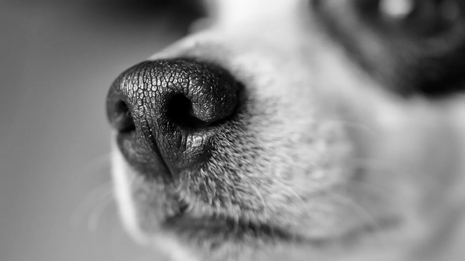
Yes, dogs can get zits. Owners frequently notice small red bumps, whiteheads, or crusty spots on a dog's muzzle, chin, and lips, and these can look almost identical to teenage acne in people. The important caveat is that true acne is uncommon in dogs. Most bumps that owners call zits turn out to be bacterial folliculitis, a surface or hair-follicle infection, and that infection is usually secondary to something else such as trauma, allergies, or skin mites.
This article explains what those bumps actually are, how veterinarians tell them apart, and when a bump on your dog deserves a professional look rather than watchful waiting.
This article is educational and is not a substitute for veterinary diagnosis or treatment.
The Short Answer: Dogs Get Bumps, Not Usually Acne
When people ask "can dogs get zits," the honest answer is that dogs develop acne-like lesions far more often than they develop true acne. The canine version of a pimple is usually a papule (a small raised red bump) or a pustule (a small pus-filled bump), and both are hallmarks of infection or inflammation around the hair follicle.
Superficial bacterial folliculitis is the single most common skin infection in dogs, and it is more common in dogs than in most other mammals [1]. The bacteria involved are overwhelmingly staphylococci, with Staphylococcus pseudintermedius identified as the major pathogen in superficial canine pyoderma [2][3]. So when you see a scattering of small pimples on your dog's chin, the most likely explanation is a staphylococcal follicle infection, not clogged pores in the human sense.
True acne, meaning a disorder of the sebaceous (oil) glands and hair follicles with blackheads and whiteheads, is rare in dogs. What veterinarians do see regularly is a group of conditions that mimic acne. Distinguishing them matters because the treatments are completely different. An antibacterial plan will not fix a mite infestation, and a steroid-responsive inflammatory disease will get worse if it is treated as a simple infection.
Why the Muzzle, Chin, and Lips Are Common Sites
Short-coated breeds and dogs with prominent muzzles tend to show these bumps most often on the chin, lips, and the top of the muzzle. Several factors converge in that area.
The hair follicles on the muzzle are relatively large and densely packed. The skin there is thin and constantly exposed to moisture from the nose, mouth, food, and water bowls. Dogs also rub their faces on the ground, on toys, and on furniture. That friction creates micro-trauma to the follicles, and micro-trauma is a well-recognized trigger for folliculitis. Once the follicle barrier is disrupted, resident staphylococci that normally live harmlessly on the skin can overgrow and cause infection.
This is why the classic picture of "dog acne" is a young dog with a bumpy chin. The bumps are real. The label is often wrong.
What Is Actually Causing the Bumps
Bacterial Folliculitis
Folliculitis means inflammation of the hair follicle. When bacteria are the cause, it is called bacterial folliculitis, and it is the most common reason a dog develops pimple-like bumps. In a study of dogs with superficial pyoderma, the bacterial communities in the lesions were far less diverse than on healthy skin, and Staphylococcus species, particularly S. pseudintermedius, dominated the infected sites [2]. Genomic work has confirmed S. pseudintermedius as the major causative agent of superficial pyoderma in dogs, including the two common variants, superficial bacterial folliculitis and epidermal collarettes [3].
Folliculitis is almost always secondary. It develops because something else has already compromised the skin. Common triggers include allergies (especially atopic dermatitis and food allergy), flea bites, friction or rubbing, excessive licking, and skin mites such as Demodex. When a dog has an underlying allergy, the skin barrier and the normal bacterial balance are disrupted, which opens the door to infection [2].
Impetigo and Epidermal Collarettes
Impetigo is a superficial, non-follicular pustular eruption. It tends to appear as small pus-filled bumps that rupture and leave thin crusts, often on the belly or groin of young dogs. It sits higher in the skin than folliculitis and does not center on the hair follicle.
Epidermal collarettes are a related pattern. They look like circular, expanding rings of peeling skin with a red or crusted edge, and they are a classic sign of exfoliative superficial pyoderma. A detailed study of these lesions found that the advancing edge consists of splits within the outer skin layer containing neutrophils, bacterial cocci, and fluid, and that all bacterial isolates from the lesions were S. pseudintermedius [4]. The same study found that the bacteria were located within the superficial skin layers, which helps explain why topical therapy can be effective for these lesions.
Demodicosis
Demodex mites live in hair follicles and sebaceous glands. When they overgrow, especially in young dogs or dogs with a suppressed immune system, they cause folliculitis that looks very much like bacterial acne. Demodicosis is a critical diagnosis to rule out, because treating it as a simple bacterial infection will not work. Demodex is one of the conditions that veterinarians specifically screen for when a dog has recurrent or non-healing folliculitis [5].
Eosinophilic Folliculitis and Furunculosis
This is an uncommon inflammatory condition that can be mistaken for infection. In three reported cases, dogs developed rapidly appearing itchy papules, pustules, and ulcers on the top of the muzzle, and in two dogs the lesions stayed confined to the face [6]. The condition is centered on the hair follicle and sebaceous gland unit, with heavy eosinophil infiltration, and it did not respond to antibacterial therapy alone. The dogs improved quickly once glucocorticoids were added, which suggests this is an inflammatory disease rather than a primary infection [6]. This is a good example of why a bump that fails to respond to antibiotics needs a different diagnostic approach.
Deep Pyoderma and Unusual Bacteria
Sometimes the infection goes deeper than the follicle. Deep pyoderma involves the deeper skin layers and can produce nodules, draining tracts, and ulcers. These cases are more serious and often involve underlying immune suppression or unusual bacteria. One report described deep skin infections caused by Burkholderia cepacia complex in six dogs, all of which were receiving the immunosuppressive drug ciclosporin at the time [7]. Another series described four dogs with Burkholderia dermatitis, two of which were on ciclosporin and one on oclacitinib, with severe neutrophilic folliculitis and furunculosis on biopsy [8]. Pseudomonas aeruginosa has also been reported as a cause of pyoderma, including a form that presents with sudden pain along the back and lesions such as red papules, hemorrhagic blisters, and ulcers [9].
The practical takeaway is that most bumps are ordinary staphylococcal folliculitis, but a minority reflect deeper or unusual infections that need culture and tailored treatment.
Mast Cell Tumors and Other Bumps That Are Not Zits
Not every bump is infectious. Mast cell tumors are a common canine skin cancer that can initially look like a small raised bump, sometimes red and sometimes hairless. A bump that persists, changes size, or does not respond to standard skin treatment should be evaluated. Inflammatory skin disease can also contain mast cells, and one study found a specific linear pattern of mast cells in the skin in a small number of inflammatory conditions, but this was far more common in Malassezia (yeast) dermatitis than in other diseases [10]. The point for owners is simple: a bump that behaves unusually deserves a veterinarian's eyes and often a sample.
Comparison Table: Telling the Bumps Apart
| Condition | Typical appearance | Common location | Underlying cause |
|---|---|---|---|
| Bacterial folliculitis | Red papules and pustules centered on hair follicles, may crust | Muzzle, chin, trunk, anywhere | Staphylococcus pseudintermedius, usually secondary to allergy, trauma, or mites [2][3] |
| Impetigo | Small superficial pustules that rupture and crust | Belly, groin, sometimes chin | Surface bacterial infection, often in young dogs |
| Epidermal collarettes | Expanding circular rings of peeling skin with red or crusted edges | Trunk, sometimes generalized | Exfoliative superficial pyoderma, S. pseudintermedius [4] |
| Demodicosis | Patchy hair loss, redness, follicular bumps, sometimes blackheads | Face, paws, trunk | Demodex mite overgrowth [5] |
| Eosinophilic folliculitis | Rapidly appearing itchy papules, pustules, ulcers | Top of the muzzle, face | Inflammatory, eosinophil-rich, steroid-responsive [6] |
| Deep pyoderma | Nodules, draining tracts, ulcers, pain | Trunk, surgical sites | Deeper bacterial infection, sometimes unusual organisms or immune suppression [7][8] |
| Mast cell tumor | Firm raised bump, may be red or hairless, changes over time | Anywhere | Cancer of mast cells |
How Veterinarians Sort Out a Bumpy Dog
Diagnosis starts with the history. Your veterinarian will want to know how long the bumps have been present, whether they itch, whether they come and go, and whether the dog has a history of allergies, ear infections, or prior antibiotic use. Recurrent or chronic skin infection is a common reason owners seek help, and recurrence often points to an underlying trigger.
The physical exam focuses on the distribution and character of the lesions. Folliculitis centered on the muzzle in a young short-coated dog suggests one path. Generalized collarettes suggest another. Patchy hair loss with blackheads raises the possibility of demodicosis.
Skin cytology is a quick and valuable step. A sample from a pustule or the edge of a lesion is examined under the microscope for bacteria and inflammatory cells. In epidermal collarettes, cytology from the lesion margins shows neutrophils with bacteria inside them, which supports a bacterial cause [4]. Cytology is not perfect on its own. In one study of acral lick dermatitis, surface culture and cytology did not reliably predict what was found on deep tissue culture, and 48 percent of deep cultures grew multidrug-resistant organisms [11]. That finding is specific to a different condition, but it illustrates a general principle: surface sampling can miss deeper infection.
Skin scrapings are used to look for Demodex mites. Bacterial culture and susceptibility testing become important when infection is recurrent, deep, or not responding to initial treatment, because resistance is a growing problem. Methicillin-resistant S. pseudintermedius and methicillin-resistant S. aureus are increasingly found in canine superficial bacterial folliculitis, which complicates antibiotic selection [1]. In one study of severe or recurrent pyoderma, most dogs responded well to an appropriate antibiotic, but resistance did develop in a treated case, and relapses occurred in a portion of dogs over follow-up [12].
Skin biopsy is reserved for cases where the diagnosis is unclear, where deep infection is suspected, or where an inflammatory or neoplastic condition is on the list. Biopsy was what identified the eosinophilic folliculitis cases that failed antibacterial therapy [6], and it is how deep infections such as Burkholderia dermatitis are characterized [8].
Treatment Principles, Not Home Remedies
The treatment of bacterial folliculitis is antibacterial therapy, and the choice depends on severity, depth, and prior history. In a blinded comparison of two dose rates of oral co-amoxiclav for canine folliculitis, furunculosis, and cellulitis, folliculitis was cured in 91.5 percent of cases in a mean of about 25 days, and doubling the dose gave no added benefit [13]. That study is old, and resistance patterns have shifted since then, but it established that folliculitis generally responds to appropriate antibiotics given for several weeks.
Other antibiotics have been studied in this space. Marbofloxacin produced an excellent response in most dogs with severe or recurrent pyoderma in one report, with relapses in a portion of dogs during follow-up [12]. Cefovecin given as a single injection was as effective as oral cefadroxil for secondary superficial pyoderma, abscesses, and infected wounds in a large trial [14]. The recurring theme is that the drug must match the infection, and that is why culture and susceptibility testing matter in complicated cases.
Topical therapy has become increasingly important. Because resistance is rising, topical antimicrobials are now a necessary part of treatment for some cases of superficial bacterial folliculitis, and options include chlorhexidine, benzoyl peroxide, ethyl lactate, and diluted sodium hypochlorite [1]. Topical fusidic acid penetrates canine skin and reaches the hair follicle, which supports its use for staphylococcal skin infection [15]. A commercial product containing natural antimicrobial peptides and plant extracts was compared with chlorhexidine-miconazole shampoo and oral cephalexin for superficial pyoderma, and all groups improved by day 28, with the natural product group showing lower itch scores early in treatment [16]. A propolis-based gel has also been developed and tested for antibacterial activity against staphylococcal skin infection, reflecting ongoing interest in alternatives to systemic antibiotics [17]. Fluorescent light energy has been studied as a non-antibiotic management option for superficial bacterial folliculitis, and in one trial it reduced the time to clinical resolution compared with oral antibiotics [18].
What all of this means for an owner is straightforward. Do not reach for human acne products. Benzoyl peroxide, salicylic acid, and retinoids formulated for people can irritate or damage a dog's skin, and some are toxic if licked off. Do not squeeze or pop the bumps, because that drives infection deeper. Do not use leftover antibiotics from a previous prescription. The right treatment depends on what is actually causing the bump, and that determination belongs to a veterinarian.
Practical Care at Home
Home care has a real but limited role. It supports veterinary treatment and reduces the factors that trigger new lesions.
Keep the muzzle and chin clean and dry. Wipe food and moisture away after meals, and consider whether your dog's food or water bowl is a source of friction or bacterial buildup. Stainless steel or ceramic bowls that are washed daily are easier to keep clean than scratched plastic.
Address the underlying trigger. If allergies are driving the infection, the infection will keep returning until the allergy is managed. This is the single most important long-term step, and it is why recurrent "zits" in an allergic dog are a sign that the allergy plan needs review.
Do not share ointments, creams, or shampoos intended for people or for other pets. Some topical products are safe for dogs and some are not, and the difference is not obvious from the label.
Keep a simple log. Note when bumps appear, where they are, whether they itch, and what changed in the dog's routine beforehand. This history helps your veterinarian find the trigger faster.
Limitations and When to Contact a Veterinarian
Individual dogs vary, and a bump that looks minor in one dog can be the first sign of a serious problem in another. Contact a veterinarian if any of the following apply.
- The bumps are painful, bleeding, ulcerated, or draining pus.
- The lesions are spreading or increasing in number.
- The bumps keep coming back after treatment.
- Your dog is licking, scratching, or rubbing the area persistently.
- There is hair loss, thickening, or darkening of the skin around the bumps.
- Your dog seems unwell, is off food, or has a fever.
- A single bump is growing, changing color, or has been present for more than a few weeks.
- Your dog is on an immunosuppressive medication and develops new skin lesions.
Recurrent or non-healing skin infection is not something to manage indefinitely at home. It usually means there is an underlying disease to find, and the sooner that is identified, the better the outcome.
Frequently Asked Questions
Can dogs get zits?
Yes, dogs can develop pimple-like bumps, but true acne is rare. Most bumps that look like zits are bacterial folliculitis or another skin condition that mimics acne.
Do dogs get pimples on their chin?
Yes, the chin, lips, and muzzle are common sites for folliculitis in dogs, especially short-coated breeds. Friction, moisture, and hair follicle anatomy make that area prone to infection.
Can dogs have zits that are not infected?
Some bumps are inflammatory rather than infectious. Eosinophilic folliculitis, for example, is an inflammatory condition of the muzzle that does not respond to antibiotics alone and improves with glucocorticoids [6].
Are dog zits contagious to people?
The staphylococci that cause canine folliculitis are generally adapted to dogs, and ordinary contact is not a major concern for healthy people. People with weakened immune systems should take standard hygiene precautions and discuss any concern with their physician.
Can dogs get spots from allergies?
Yes. Allergic skin disease disrupts the skin barrier and predisposes dogs to bacterial folliculitis, which is why recurrent bumps are common in allergic dogs [2].
Should I pop my dog's pimples?
No. Squeezing or popping a bump can push infection deeper and cause pain, bleeding, and scarring. Leave the lesions alone and let a veterinarian assess them.
Can I use human acne cream on my dog?
No. Human acne products are not formulated for canine skin and some ingredients are harmful if the dog licks them off. Ask your veterinarian before applying anything to your dog's skin.
How do I know if a bump is a mast cell tumor?
You cannot tell by looking. A bump that is growing, changing, or not responding to treatment should be sampled by a veterinarian to rule out cancer and other serious causes.
Related Articles
- Cat Skin Rash with Bumps: Common Causes and Next Steps
- Dog Skin Rash with Bumps: What Could It Be?
- Dog Skin Bumps and Lumps: When to See the Vet
- Dog Skin Bumps: Comprehensive Veterinary Reference Guide
- Cat Skin Bumps and Lumps: When to See a Vet and When to Monitor
- Cat Skin Bumps and Lumps: What the Vet Will Do and How Much It Costs
- Do Dogs Get Hiccups? Causes and When to Worry
- Dark Patches on Dog Skin: Causes and When to Worry
Sources
- Canine superficial bacterial folliculitis: current understanding of its etiology, diagnosis and treatment.
- Characterization of Cutaneous Bacterial Microbiota from Superficial Pyoderma Forms in Atopic Dogs.
- Complete Genome Sequences of Six Staphylococcus pseudintermedius Strains from Dogs with Superficial Pyoderma in Georgia, USA.
- Clinical, microscopic and microbial characterization of exfoliative superficial pyoderma-associated epidermal collarettes in dogs.
- Use of cystine, lactose, electrolyte-deficient medium for isolation and identification of bacteria and yeasts from the skin and ears of dogs.
- Canine eosinophilic folliculitis and furunculosis in three cases.
- Deep pyoderma caused by Burkholderia cepacia complex associated with ciclosporin administration in dogs: a case series.
- Clinical and histopathological features of Burkholderia cepacia complex dermatitis in dogs: a series of four cases.
- Pyoderma caused by Pseudomonas aeruginosa infection in dogs: 20 cases.
- Subepidermal linear alignment of mast cells in inflammatory dermatoses of the dog.
- Microbiological and histopathological features of canine acral lick dermatitis.
- Therapy of difficult cases of canine pyoderma with marbofloxacin: a report of 39 dogs.
- Treatment of canine pyoderma with co-amoxyclav: a comparison of two dose rates.
- Efficacy and safety of cefovecin in treating bacterial folliculitis, abscesses, or infected wounds in dogs.
- Opportunities for topical antimicrobial therapy: permeation of canine skin by fusidic acid.
- Comparative study of a commercial formula containing natural antimicrobials versus oral cephalexin or topical chlorhexidine-miconazole therapies for treating superficial pyoderma in dogs.
- Efficacy Study of Propolis Eutectic Extract in Gel Formulations for the Treatment of Bacterial Skin Diseases in Dogs.
- Exploring fluorescent light energy as management option for canine superficial bacterial folliculitis.