Discoid Lupus Erythematosus in Dogs Explained
By Dr. Zubair Khalid, DVM, MS, PhD ·
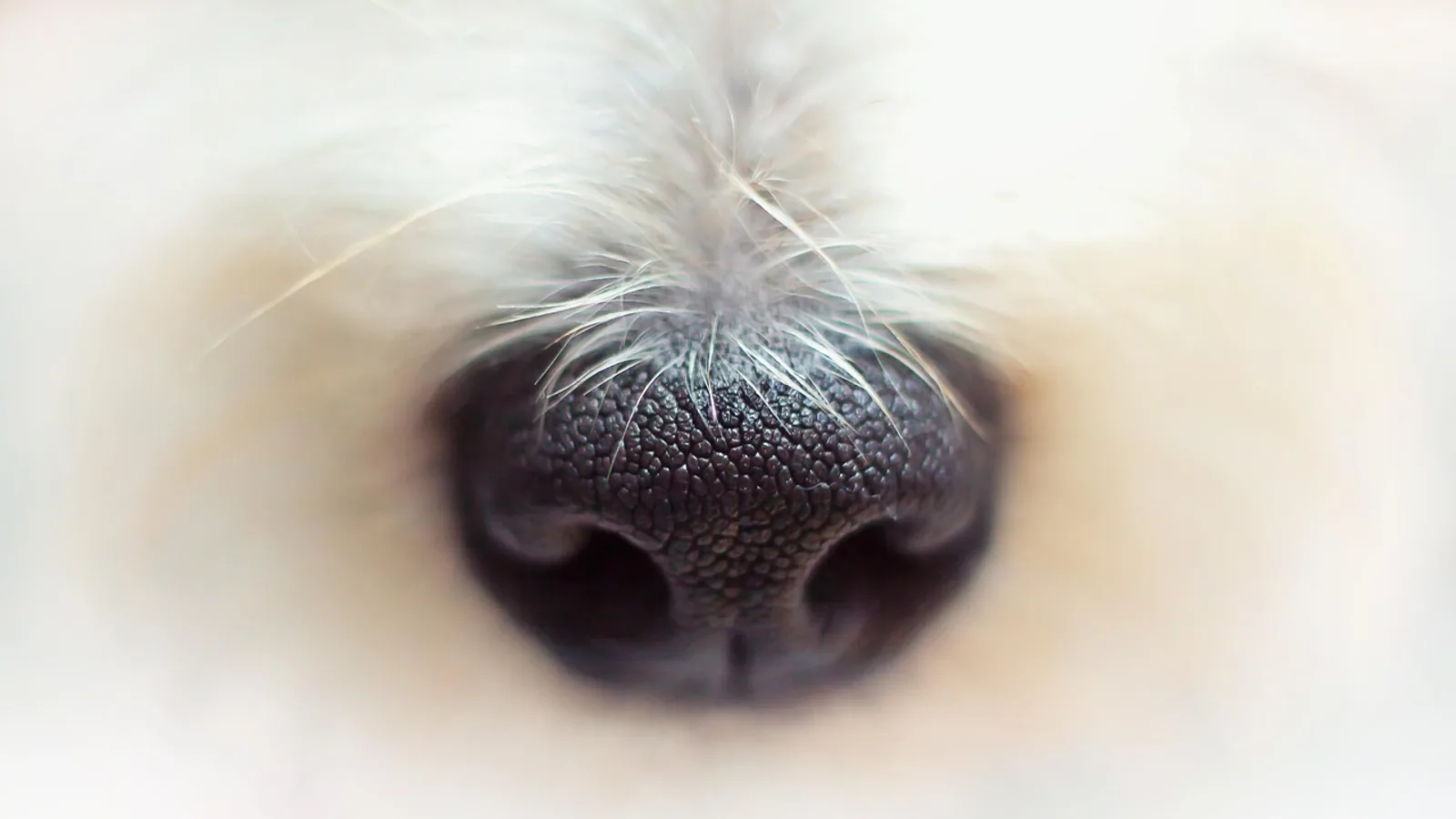
Discoid lupus erythematosus (DLE) is a benign, immune-mediated, photosensitive skin disease that most often affects a dog's nose. It is a form of chronic cutaneous lupus erythematosus, meaning the immune reaction stays in the skin and does not spread to internal organs the way systemic lupus erythematosus (SLE) can [1].
That distinction is the single most important fact for an owner to hold onto. A dog with discoid LE is not a dog with systemic lupus. The disease is confined to the skin, it is treatable, and most dogs live a normal lifespan with management. The trade-off is that DLE is chronic. It tends to wax and wane with sun exposure, and it needs ongoing attention rather than a short course of pills.
This guide walks through what discoid LE is, how the nasal lesions progress, how veterinarians confirm the diagnosis, and how sun avoidance, topical therapy, and systemic medication fit together. It also covers the conditions that look almost identical to DLE, because several of them carry a very different prognosis.
This article is educational and is not a substitute for veterinary diagnosis or treatment.
What Discoid Lupus Erythematosus Actually Is


DLE is an autoimmune skin disorder. The immune system mounts an inflammatory attack at the boundary between the outer layer of skin (the epidermis) and the tissue beneath it (the dermis). That boundary is called the basement membrane zone. When immune cells cluster there, the result is a pattern pathologists call interface dermatitis, with damage to the basal cells that anchor the epidermis to the dermis [2][1].
The disease was first described in dogs in 1979, in two dogs with erythema and depigmentation of the nasal planum [3]. Since then, the spectrum of canine cutaneous lupus erythematosus has expanded considerably, and veterinarians now recognize several variants, including facial discoid LE, generalized discoid LE, vesicular CLE, exfoliative CLE, and mucocutaneous lupus erythematosus [1][4].
Facial DLE, the classic nasal form, is the most common presentation and the focus of this article. Generalized DLE is a distinct variant in which annular, scaly plaques appear on the trunk, neck, and limbs rather than being limited to the face [5][6].
Why the Nose Is the Target
The nasal planum is hairless, thinly pigmented or unpigmented in many dogs, and directly exposed to ultraviolet light. That combination makes it a prime site for a photosensitive immune reaction. Ultraviolet radiation is thought to trigger or worsen the inflammatory cascade in genetically susceptible dogs [7].
A 2021 comparative study using gene expression profiling found that skin from dogs with DLE shares strong interferon signatures with human DLE and with a mouse model of cutaneous lupus. The genes CXCL10, ISG15, and an S100 family member were among the most significantly upregulated across all three species, and the affected skin showed marked infiltration by T cells and B cells [7]. In plain terms, dog DLE is not a look-alike of the human disease. It runs on many of the same molecular pathways, which is why canine DLE is studied as a model for the human condition.
DLE Is Not Systemic Lupus
Owners frequently search for "lupus in dogs" and land on material about SLE, which is a much more serious multisystem disease. The two are not the same.
In a study of 10 dogs with generalized DLE, none progressed to systemic lupus erythematosus over a median follow-up of 2.5 years [5]. In the case that first described generalized DLE treated with hydroxychloroquine, the dog had no systemic signs, and laboratory testing was unremarkable, which is what excluded concurrent SLE [6]. When a dog is diagnosed with DLE, veterinarians typically look for evidence of systemic involvement precisely to rule it out. A normal physical exam, normal blood work, and a negative antinuclear antibody test support a purely cutaneous diagnosis [8][9].
How DLE Progresses on the Nose
DLE lesions evolve in a fairly predictable sequence. Recognizing the early stages helps owners seek care before scarring sets in.
Stage 1: Depigmentation. The first visible change is usually a loss of pigment on the nasal planum. Black noses turn gray, pink, or mottled. At this point the surface may still look smooth.
Stage 2: Erythema and scaling. The depigmented areas become red and develop fine, adherent scale. The normal cobblestone texture of the nasal planum begins to blur.
Stage 3: Crusting and erosion. Crusts form over the surface, and shallow erosions appear. These can be painful. The cobblestone architecture is progressively lost.
Stage 4: Ulceration and scarring. Deeper ulceration and, eventually, scarring and distortion of the nasal planum can occur if the disease is not controlled.
A key clinical feature is that DLE usually spares the nasal mucosa, the pink lining inside the nostril. The lesion sits on the planum itself and the adjacent haired skin of the muzzle. In one reported case, however, the majority of the nasal mucosa was affected alongside pigment atrophy and heavy peeling of the rostral planum, which shows that mucosal involvement is possible and does not automatically rule DLE in or out [10].
Lesions can also extend beyond the nose. The bridge of the muzzle is commonly involved, and less often the periocular region, ear pinnae, distal limbs, and mucocutaneous junctions such as the lips, oral cavity, and genital region [11]. In generalized DLE, the pattern shifts entirely to well-demarcated annular or polycyclic plaques with pigment change, a red margin, adherent scaling, follicular plugging, and central hair loss, sometimes with central scarring [5].
Sun Exposure Drives Flares
DLE is photosensitive. Lesions worsen in summer, in dogs who spend time outdoors, and in dogs with light pigmentation. Two German Shepherd Dogs that developed squamous cell carcinoma within chronic, untreated, unprotected DLE nasal lesions had gone 4 to 6 years without treatment or photoprotection [12]. That outcome is uncommon, but it is the reason long-standing, uncontrolled nasal DLE deserves respect rather than watchful waiting.
Which Dogs Get DLE
DLE can affect a wide range of breeds. Reported cases include German Shepherd Dogs, Shetland Sheepdogs, Spitz-type dogs, Golden Retrievers, Shih Tzu, Chinese Crested, American Staffordshire Terriers, and mixed-breed dogs [13][5][6][14][9][10][15].
Age at diagnosis clusters in adulthood. In the 10-dog generalized DLE case series, dogs were affected in their mid-to-late adulthood [5]. Individual reports include a 3-year-old Spitz, a 6-year-old crossbred dog, a 9-year-old Shetland Sheepdog, and an 11-year-old Golden Retriever and Shih Tzu [8][14][9][10][15]. Nasal DLE itself can appear in younger adult dogs, so age alone does not exclude it.
There is no strong sex predilection across the literature. Both male and female dogs appear in reported cases [8][5][14][10].
Diagnosis: How Veterinarians Confirm DLE
Diagnosis rests on three pillars: history, clinical signs, and skin biopsy with histopathology [1]. There is no single blood test that confirms DLE.
Step 1: History and Physical Examination
Your veterinarian will ask about the timeline of pigment loss, sun exposure, seasonal worsening, and whether the dog is itchy. DLE is often minimally pruritic, though some dogs are uncomfortable. The exam focuses on the distribution of lesions and whether the nasal mucosa, eyelids, lips, or other mucocutaneous junctions are involved.
Step 2: Ruling Out Infection and Other Causes
Because nasal planum dermatitis has many causes, baseline testing usually includes skin cytology and fungal culture to exclude bacterial and fungal infection. In regions where leishmaniosis is endemic, it becomes a major differential for erosive nasal dermatitis, and testing for it is warranted [2].
Step 3: Blood Work and Antinuclear Antibody Testing
A complete blood count, serum chemistry panel, and urinalysis help confirm that the dog is systemically well, which supports a cutaneous-only diagnosis [8]. An antinuclear antibody (ANA) test is often run to help exclude SLE. In DLE, the ANA is typically negative [9]. A negative ANA does not by itself diagnose DLE, and a positive result does not by itself diagnose SLE.
Step 4: Skin Biopsy and Histopathology
Biopsy is the definitive step. The characteristic finding is lymphocyte-rich interface dermatitis, often with thickening of the basement membrane zone, suprabasal apoptosis, and dermal fibrosis [5]. Hydropic degeneration of basal cells, pigmentary incontinence, and a lichenoid infiltrate are also typical [14][11]. In one case, the infiltrate was predominantly plasmacytic, which shows that the cellular makeup can vary [14].
Biopsy technique matters. Samples should be taken from the edge of an active lesion, not from the center of a scar, and the tissue should be handled so a pathologist can assess the interface. Discuss with your veterinarian whether a single punch or multiple samples are needed.
Step 5: Direct Immunofluorescence
Direct immunofluorescence (DIF) looks for immunoglobulin and complement deposited along the basement membrane zone. It is supportive when positive, but it is inconsistent. In the generalized DLE case series, DIF and ANA serology were pursued "whenever possible," which reflects how variable the yield is in practice [5]. A negative DIF does not rule out DLE, and diagnosis should not hinge on it.
Table: DLE and Its Main Look-Alikes
| Condition | Typical location | Key clinical clues | How it is distinguished |
|---|---|---|---|
| Discoid lupus erythematosus | Nasal planum, muzzle bridge, less often periocular, pinnae, limbs, mucocutaneous junctions | Depigmentation, erythema, scaling, crusting, erosions, loss of cobblestone texture, usually spares nasal mucosa | History, clinical signs, biopsy showing lymphocyte-rich interface dermatitis [1][11] |
| Pemphigus foliaceus | Face and nasal planum, often generalized | Pustules and crusts, frequently more widespread and more inflammatory | Cytology and biopsy showing pustular acantholysis. Can coexist with generalized DLE as polyautoimmunity [13] |
| Nasal solar dermatitis | Nonpigmented nasal planum and adjacent skin | Sun-dependent, seasonal, confined to sun-exposed unpigmented skin | Distribution and history. Overlaps clinically with DLE and can be difficult to separate without biopsy |
| Zinc-responsive dermatosis | Muzzle, periocular, pressure points, footpads | Crusting and scaling around the face and paws, often in specific breeds | Signalment, dietary history, response to zinc supplementation |
| Lymphoma (cutaneous or nasal) | Nasal planum, muzzle, or nasal cavity | Rapidly progressive, often ulcerated or nodular, may involve mucosa or cause nasal discharge | Biopsy with immunohistochemistry |
Treatment: Sun Avoidance Comes First
Every treatment plan for DLE starts with photoprotection. Medication alone, without reducing ultraviolet exposure, tends to produce partial and temporary improvement.
Sun Avoidance and Photoprotection
Keep the dog indoors during peak ultraviolet hours, roughly mid-morning to late afternoon. Walk in early morning, evening, or after dark. Consider protective cover for the nose if your dog tolerates it. Discuss pet-safe sunscreen with your veterinarian, since human products are not always appropriate for dogs that lick their noses. The hydroxychloroquine case report explicitly included restriction of sun exposure as part of the treatment regimen, alongside medication [6].
Topical Therapy
Topical corticosteroids are a mainstay for localized nasal lesions. They reduce inflammation at the site with far less systemic exposure than oral steroids.
Topical 0.1% tacrolimus is an alternative or adjunct. In a study of 10 dogs with DLE and 2 with pemphigus erythematosus, 8 of 10 DLE dogs improved after 8 weeks of topical tacrolimus, and in 6 of the 8 responders, other medications were discontinued. No adverse clinical or laboratory effects were noted [16]. Tacrolimus is particularly useful for lesions around the eyes and mucocutaneous junctions where steroid-related skin thinning is a concern.
In the generalized DLE case, 0.1% tacrolimus ointment was alternated with 0.1% prednicarbate cream once daily for 5 to 10 days to control flares [6].
Systemic Immunosuppression and Immunomodulation
When topical therapy is not enough, or when lesions are generalized, systemic options come into play. The choice depends on severity, the dog's overall health, and monitoring capacity.
Tetracycline and niacinamide. This combination is an immunomodulating regimen rather than a classic immunosuppressant. An 11-year-old Shih Tzu with generalized DLE achieved complete remission on oral tetracycline and niacinamide, the first documented case of generalized DLE responding to this combination [15]. A separate study evaluated long-term tetracycline and niacinamide in dogs, including 9 with DLE, and found no significant difference in post-vaccination antibody titers against parvovirus and distemper virus compared with healthy controls. All dogs had protective distemper titers, and 8 of 10 dogs in each group had protective parvovirus titers [17]. That is reassuring for owners worried about vaccine response during long-term therapy.
Hydroxychloroquine. This antimalarial immunomodulator was used successfully in a 9-year-old hairless Chinese Crested with generalized DLE. The dog received 5 mg/kg once daily along with topical tacrolimus and sun restriction, and complete remission was maintained over the following year, with three relapses that were controlled by adjusting topical therapy or dosing frequency [6].
Ciclosporin. A 6-year-old crossbred dog with generalized DLE that had not responded to doxycycline and niacinamide was treated with oral dexamethasone and ciclosporin, with dexamethasone discontinued after one month and ketoconazole added. The dog improved [8].
Oclacitinib. A 2023 report described seven dogs with chronic cutaneous lupus erythematosus variants treated with oral oclacitinib. Complete remission occurred in the dog with exfoliative cutaneous lupus, both dogs with mucocutaneous lupus, and three of four dogs with facial discoid lupus. The remaining facial DLE dog had a good response, defined as at least 50 percent lesion reduction. First visible improvement appeared within 2 to 3 weeks, and time to complete remission was around 2 months. No clinical adverse effects were seen, and blood counts remained stable [18]. This is an off-label use in dogs and requires veterinary supervision.
Glucocorticoids. Systemic corticosteroids were the original reported treatment for canine DLE and produced favorable responses in the first two documented cases [3]. They remain an option for severe or refractory disease, but they carry the well-known risks of long-term steroid use. In the cannabinoid case report, elevated liver enzymes indicated corticosteroid-induced side effects, which is exactly the scenario where steroid-sparing options become attractive [19].
Cannabinoids. A case report described a 2-year-old mixed-breed dog with DLE that had worsening lesions and elevated liver enzymes on conventional treatment. A CBD-rich full-spectrum cannabis oil was started, the dose was gradually adjusted to the minimum effective level, and the dog improved within weeks, with stable disease at one year on a low dose [19]. This is a single case. It is not a standard recommendation, and cannabis products are regulated differently from veterinary medications. Any use should be discussed with your veterinarian.
Vitamin E
Vitamin E is sometimes used as an antioxidant adjunct in immune-mediated skin disease. It is not a substitute for photoprotection or anti-inflammatory therapy, and it should be part of a plan your veterinarian approves rather than a self-prescribed add-on.
How Treatment Is Monitored
Expect rechecks. In the oclacitinib report, complete blood counts were performed at variable intervals during treatment [18]. In the hydroxychloroquine case, relapses were detected and managed during the first year of therapy [6]. A reasonable approach is to reassess at 4 to 8 weeks after starting or changing treatment, then extend the interval once the dog is stable. Photographs taken under consistent lighting are genuinely useful for tracking pigment return and lesion size over time.
The flowchart below shows the general decision path from nasal depigmentation to diagnosis and treatment.
flowchart TD
    A[Nasal depigmentation] --> B[Veterinary examination]
    B --> C[Rule out infection and leishmaniosis]
    C --> D[Blood work and antinuclear antibody test]
    D --> E[Skin biopsy for histopathology]
    E --> F{Interface dermatitis present}
    F -->|Yes| G[Diagnosis of discoid lupus]
    F -->|No| H[Reconsider differentials]
    G --> I[Sun avoidance and photoprotection]
    I --> J[Topical steroid or tacrolimus]
    J --> K{Response adequate}
    K -->|Yes| L[Maintain and monitor]
    K -->|No| M[Systemic immunomodulation]
Living With a Dog That Has DLE
Most dogs with DLE do well. The prognosis for canine cutaneous lupus erythematosus variants is generally good once the diagnosis is made and treatment is started [1]. The disease is managed, not cured, and owners should expect a long-term relationship with their veterinarian.
Practical habits that help:
- Track sun exposure and note whether lesions flare in summer.
- Photograph the nose monthly under the same lighting.
- Keep recheck appointments even when the nose looks good.
- Do not stop topical or systemic therapy abruptly without veterinary guidance, since relapse is common when treatment is reduced too quickly [6].
- Watch for new lesions on the eyelids, lips, ears, or paws, and report them.
Common Myths and Confusions
"DLE is a mild form of systemic lupus." This is the most persistent misunderstanding. DLE is a separate cutaneous condition. In a 10-dog generalized DLE series, no dog progressed to SLE over a median 2.5 years of follow-up [5].
"A negative biopsy means it is not DLE." Biopsy is the strongest diagnostic tool, but sampling site and timing affect results. A biopsy taken from scarred, inactive tissue may not show the interface pattern. Direct immunofluorescence is supportive but inconsistent [5].
"Once the nose repigments, the dog is cured." Repigmentation is a good sign, but DLE is chronic and photosensitive. Sun exposure can trigger a new flare.
"Steroids are the only option." Tetracycline and niacinamide, hydroxychloroquine, ciclosporin, oclacitinib, and topical tacrolimus have all been reported as effective in specific cases [8][6][18][15][16]. The right choice depends on the individual dog.
"Any nasal crust is DLE." Pemphigus foliaceus, nasal solar dermatitis, zinc-responsive dermatosis, and lymphoma can all look similar. Biopsy is often what separates them [13][1].
What Is Still Uncertain
Several questions remain open. The exact genetic and environmental triggers that start DLE in a given dog are not fully mapped, though comparative gene expression work has identified shared interferon-driven pathways across dogs, humans, and mice [7]. The role of direct immunofluorescence in routine diagnosis is still debated, since results are inconsistent [5]. Optimal long-term monitoring protocols for newer agents like oclacitinib are not standardized [18]. Cannabinoid therapy has one published case report and needs far more evidence before it can be considered a mainstream option [19]. And the true rate of progression from chronic DLE to squamous cell carcinoma is unknown, though it has been documented in at least two dogs with long-standing untreated lesions [12].
Limitations and When to Contact a Veterinarian
This article describes general patterns. Every dog is an individual, and only a veterinarian who has examined your dog can diagnose and treat DLE.
Contact a veterinarian promptly if:
- Your dog develops new depigmentation, redness, scaling, or crusting on the nose.
- Existing nasal lesions worsen, bleed, or develop ulcers.
- Your dog shows pain, reluctance to have the muzzle touched, or reduced appetite.
- Lesions spread to the eyelids, lips, ears, paws, or other body areas.
- Your dog is on treatment and new lesions appear or old ones return.
- Your dog is receiving systemic medication and develops vomiting, diarrhea, lethargy, or jaundice.
- A nasal lesion has been present and untreated for years, since chronic DLE has been linked to squamous cell carcinoma in at least two reported cases [12].
Frequently Asked Questions
What is discoid lupus erythematosus in dogs?
DLE is a benign, immune-mediated, photosensitive skin disease that most commonly affects the nasal planum. It stays in the skin and does not involve internal organs the way systemic lupus erythematosus can [1].
Is discoid lupus the same as systemic lupus in dogs?
No. DLE is a chronic cutaneous form of lupus confined to the skin. In a study of 10 dogs with generalized DLE, none progressed to systemic lupus over a median follow-up of 2.5 years [5].
What does DLE look like on a dog's nose?
It typically starts with loss of pigment, then progresses to redness, scaling, crusting, and shallow erosions, with loss of the normal cobblestone texture. The nasal mucosa is usually spared, though mucosal involvement has been reported [10].
How is discoid lupus diagnosed?
Diagnosis is based on history, clinical signs, and skin biopsy showing lymphocyte-rich interface dermatitis. Blood work and an antinuclear antibody test help exclude systemic disease, and direct immunofluorescence is supportive but inconsistent [5][1].
Does my dog with DLE need to stay out of the sun?
Yes. Sun avoidance is the cornerstone of management. Ultraviolet exposure triggers flares, and photoprotection should accompany any topical or systemic treatment [6].
What treatments are available for DLE?
Options include topical corticosteroids, topical tacrolimus, tetracycline and niacinamide, hydroxychloroquine, ciclosporin, oclacitinib, and systemic glucocorticoids. The choice depends on lesion severity and the individual dog [8][18][15][16].
Can DLE be cured?
DLE is managed rather than cured. Most dogs respond well to treatment and maintain good quality of life, but relapses can occur when therapy is reduced or sun exposure increases [6][1].
Can DLE turn into skin cancer?
It is uncommon, but squamous cell carcinoma has been reported arising within chronic, untreated, unprotected DLE nasal lesions in two German Shepherd Dogs after 4 to 6 years [12]. Controlling the disease and limiting sun exposure reduces this risk.
Related Articles
- Ferret Adrenal Disease Signs
- Dental Disease in Pet Rabbits
- Cushing Disease Dog Behavior Problems
- Hamster Diseases: Common Conditions and Treatment
- Chinchilla Dental Disease: Diagnosis and Treatment
- Cushing Disease Dog Behavior Problems At Night
- What Is Anthrax Disease? Causes and Symptoms
- Sand Fly: Biology, Diseases, and Veterinary Importance
- Tularemia (Rabbit Fever): Symptoms and Transmission
Sources
- Cutaneous lupus erythematosus in dogs: a comprehensive review.
- A retrospective study comparing histopathological and immunopathological features of nasal planum dermatitis in 20 dogs with discoid lupus erythematosus or leishmaniosis.
- Canine discoid lupus erythematosus.
- Canine Cutaneous Lupus Erythematosus: Newly Discovered Variants.
- Clinical and microscopic features of generalized discoid lupus erythematosus in dogs (10 cases).
- Successful treatment of a novel generalized variant of canine discoid lupus erythematosus with oral hydroxychloroquine.
- Shared inflammatory and skin-specific gene signatures reveal common drivers of discoid lupus erythematosus in canines, humans and mice.
- Ciclosporin therapy for canine generalized discoid lupus erythematosus refractory to doxycycline and niacinamide.
- A canine case of discoid lupus erythematosus with circulating autoantibody.
- Case report of canine discoid lupus erythematosus in Guatemala.
- A case of interface perianal dermatitis in a dog: is this an unusual manifestation of lupus erythematosus?
- Squamous Cell Carcinoma Arising in Chronic Discoid Lupus Erythematosus Nasal Lesions in Two German Shepherd Dogs.
- Cutaneous polyautoimmunity in two unrelated dogs: pemphigus foliaceus and generalized discoid lupus erythematosus.
- Discoid lupus erythematosus (DLE) in a Spitz dog.
- Generalized canine discoid lupus erythematosus responsive to tetracycline and niacinamide therapy.
- Topical 0.1% tacrolimus for the treatment of discoid lupus erythematosus and pemphigus erythematosus in dogs.
- Influence of long-term treatment with tetracycline and niacinamide on antibody production in dogs with discoid lupus erythematosus.
- Effective treatment of canine chronic cutaneous lupus erythematosus variants with oclacitinib: Seven cases.
- Case report: Cannabinoid therapy for discoid lupus erythematosus in a dog.