Cat Ear Pinna: Anatomy, Problems, and When to See a Vet
By Dr. Zubair Khalid, DVM, MS, PhD ·
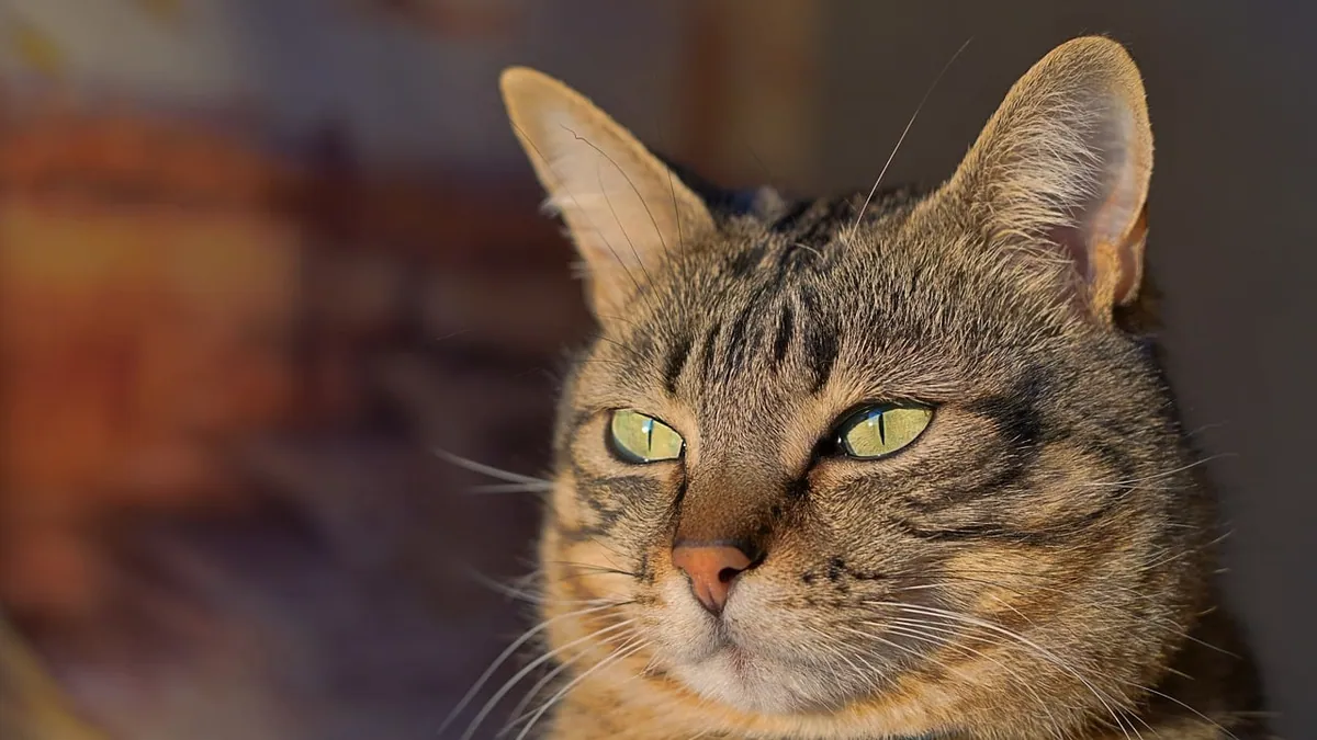
The cat ear pinna is the visible flap of cartilage covered in skin and fur on each side of your cat's head. It collects sound and funnels it into the ear canal, and because it sits on the outside of the body, it is easy to injure and easy to examine at home. Problems with the pinna range from sun damage and crusted skin disease to tears and tumors, and some of them need veterinary care quickly.
This article is educational and is not a substitute for veterinary diagnosis or treatment.
Quick Answer
- The pinna is the outer ear flap. It is made of cartilage covered by skin and fur, and it moves independently on each side to help your cat locate sounds [1].
- Common pinna problems include infections, crusted immune-mediated disease, sun-related damage, trauma, and growths [2][3][4].
- Warning signs include crusts, redness, swelling, discharge, a bad smell, head shaking, scratching, and any wound or lump on the flap.
- A torn or bleeding pinna, sudden swelling, or a cat that cannot hold the ear normally needs same-day veterinary care.
- Many pinna diseases look alike, so diagnosis often requires an exam, ear cytology, and sometimes a skin biopsy [2].
What the Cat Ear Pinna Is and What It Does
The ear has three parts: outer, middle, and inner. The pinna and the ear canal make up the outer ear [1]. The pinna is the flap you see. It is cartilage covered by skin and fur, and its job is to gather sound and send it down the canal to the eardrum [1].
Cats have excellent hearing. They detect a wider range of frequencies than people and most dogs, and the left and right pinnae can move independently, which helps them pinpoint where a sound came from [1]. A cat's ear canal is also deeper than a human's. That shape funnels sound well, but it can trap dirt and wax, which can lead to inflammation and infection [1].
Because the pinna is thin, poorly padded, and fully exposed, it takes the first hit from scratches, bites, sun, and cold. That is why so many ear problems in cats start on the flap itself.
Signs Owners Notice on the Pinna
You do not need special equipment to spot many pinna problems. Look at both ears in good light once a week. Signs that something is wrong include:
- Redness, swelling, or warmth on the flap
- Dark red, brown, or black crusts that stick to the skin [4]
- Scabs, sores, or areas of hair loss
- Discharge, wetness, or a bad smell from the ear
- Head shaking, ear scratching, or rubbing the ear on furniture
- Holding one ear lower or flatter than the other
- A notch, split, or wound along the edge
- A lump, bump, or thickened patch
- Folded or drooping ear tips that stay that way [2]
Some cats with ear disease show almost nothing at home. In one study of owned cats, ear mites were found in 6% of the cats, and about half of those cats were exclusively indoor and completely asymptomatic [5]. That is a good reason to check the ears yourself and let your vet look inside at every exam.
Common Conditions That Affect the Cat Ear Pinna
Infections and Mites
Otitis externa means inflammation of the outer ear, including the canal and often the pinna. In a study of owned cats, the prevalence of otitis externa was 17% [5]. Ear cytology was abnormal in 19% of cats, and mites were found in 6% [5]. When bacteria or yeast were present in abnormal numbers, about 29% of those cats had only Malassezia yeast, 65% had only cocci bacteria, and 6% had a mix [5].
Cats with itchy, smelly, or discharge-filled ears often have otitis externa. If you notice a bad odor, see Cat Ear Infection Smell: Causes and When to See a Vet. Green or yellow discharge is another clue that deserves a closer look, covered in Cat Ear Infection with Green Discharge: When to See a Vet.
Proliferative and Necrotizing Otitis Externa (PNOE)
PNOE is a crusting disease that most often affects the concave (inner) surface of the pinna and the vertical ear canal, usually on both sides [4]. The crusts are thick and adhere to the skin, and they can be dark red, brown, or black [4]. Lesions may also appear on the face, and a generalized form is rare [4]. The cause is unknown, but it responds to drugs that calm the immune system, so it is thought to be immune-mediated [4]. Only about 21 cases have been described in the veterinary literature [4].
Folded Ear Tips
Folded ear tips in cats are most often linked to long-term glucocorticoid (steroid) therapy, such as daily eye or ear medications [2]. Sun damage can also cause them [2]. The folding may not be reversible [2]. If your cat is on long-term steroid drops for any reason, ask your vet whether the ears should be checked at follow-up visits.
Sun Damage and Skin Tumors
Ear tumors are common in light-coated, unpigmented cats with long-term sun exposure [3]. Constant tissue trauma from intense itching in chronic otitis is another factor [3]. Any tissue in the pinna can develop abnormal cell growth, including blood vessel tumors such as hemangiosarcomas, which are less common [3]. White cats and cats with white ears are at higher risk, so keep them out of strong midday sun and check the ear edges often.
Trauma, Tears, and Avulsions
Ear canal tears and avulsions are usually caused by acute trauma [6]. In a review of 24 dogs and cats, 92% had a visible break in the ear canal on otoscopy, including complete avulsion, partial avulsion, and perforating tears [6]. These injuries are serious and often need surgery [6]. Fight wounds, dog bites, and car accidents are typical causes.
Polyps and Other Growths
Masses in the ear canal are not rare in cats. In one study comparing CT and video-otoscopy, 13 of 32 examined ears had a mass, and on histopathology 9 of 13 were polyps, 1 was a carcinoma, and 1 was granulomatous tissue from otitis externa [7]. A separate case report described a tympanic papillary adenoma, a benign glandular tumor, in a cat with discharge and a polyp-like mass [8]. Growths can be benign or malignant, so any lump needs a diagnosis.
Other Skin Disease on the Pinna
Many skin conditions that affect the body also affect the ears. If your cat has crusts, scabs, or hair loss on the pinna along with problems elsewhere, see Cat Skin Problems: When to See the Vet and How to Help. Patchy hair loss on the ears and head is also a common reason owners seek help, covered in Cat Hair Loss: Causes and When to See a Vet.
Causes and Risk Factors
| Risk factor | What it means for the pinna |
|---|---|
| White or light coat, no pigment | Higher risk of sun-related tumors [3] |
| Long-term steroid eye or ear drops | Can cause folded ear tips [2] |
| Chronic ear inflammation or itching | Tissue trauma can contribute to tumors [3] |
| Outdoor access or contact with other cats | Fight wounds, bites, and trauma [6] |
| Living indoors only | Does not rule out mites, since asymptomatic indoor cats can carry them [5] |
| Short hair and a history of skin disease | Linked to higher odds of otitis externa [5] |
| Multiple cats in the home | More opportunity for mites and wounds to spread |
How Veterinarians Diagnose Pinna Problems
Your vet starts with a full history, including any medications your cat takes now or took recently, because drugs can cause pinna changes [2]. Then comes a physical exam of the ear, face, and skin.
Otoscopy lets the vet look down the canal. Video-otoscopy gives better magnification than standard otoscopy, which helps identify fine structures and the eardrum [9]. In cats, CT and video-otoscopy agree very well for detecting ear canal masses, with perfect agreement in one study [7]. CT is also useful for seeing the middle ear and planning surgery [8].
Cytology (a sample of ear wax and debris viewed under a microscope) checks for mites, bacteria, and yeast [2][5]. If the pinna itself has crusts or lumps, the vet may take a skin biopsy, which is often needed for a definitive diagnosis [2]. Blood work may be recommended before anesthesia or long-term immune-suppressing treatment.
Treatment Options
Treatment depends on the diagnosis, not just the appearance of the ear.
- Ear infections: cleaning plus medication chosen for the specific organism found on cytology. Your vet calculates any dose from your cat's weight and health.
- Ear mites: prescription parasite treatment for your cat and, when needed, the other pets in the home.
- PNOE: topical and systemic immune-modulating drugs [4]. One report describes successful use of oclacitinib in refractory cases of proliferative and necrotizing otitis externa [2].
- Folded ear tips: often no treatment, and the fold may be permanent [2]. If a steroid medication caused it, your vet may adjust the drug.
- Sun damage and tumors: surgery is common. In one cat with a recurrent ear tumor, total ear canal ablation plus cryotherapy of the surgical bed was used to improve the chance of long-term control [3].
- Tears and avulsions: surgical repair. In a case series, treatments included total ear canal ablation with lateral bulla osteotomy, vertical ear canal ablation, and primary repair [6].
- Polyps and other masses: removal, sometimes through the ear canal and sometimes with more extensive surgery [8][7].
Never put human ear drops or leftover pet medication into your cat's ear without veterinary guidance. Some products are toxic to cats, and the wrong drug can make things worse.
Recovery, Outlook, and Long-Term Management
Recovery varies widely. After major ear surgery, some cats do very well, but the outlook is not always good. In one case series of ear canal tears and avulsions, 6 of 24 patients died or were euthanized within the first 14 days after presentation [6]. That reflects how serious these injuries can be, not how most routine ear problems go.
For chronic conditions such as PNOE, expect ongoing management with periodic rechecks [4]. Tumors can recur. In one reported case, a benign-appearing tumor came back within 17 months [8], and in another, a hemangiosarcoma recurred after an earlier pinnectomy [3]. That is why follow-up exams matter even when the ear looks normal again.
At home, keep the ear dry, follow the cleaning schedule your vet gives you, and do not pick at crusts or scabs. If your cat wears a cone, use it for as long as directed. Cats are skilled at reopening healing wounds.
Prevention
- Check both ears weekly. Look at the flap, the opening, and the skin around it.
- Keep white or light-colored cats out of direct sun, especially between late morning and mid-afternoon [3].
- Treat mites in every pet in the household if one cat is diagnosed.
- Ask your vet about ear-safe products before using any drop long term, since daily steroid drops can cause folded ear tips [2].
- Keep up with routine exams so your vet can look inside the canal with an otoscope.
- Address itching and skin disease early. Chronic inflammation and scratching can damage ear tissue over time [3].
Limitations and When to Contact a Veterinarian
No article can tell you what is wrong with your cat's ear. Only a veterinarian who examines your cat can do that, and individual cases always need professional judgment.
Contact your vet the same day if you see:
- A wound, bite, or split in the pinna
- Bleeding that does not stop with gentle pressure
- Sudden swelling of the flap that feels like a soft balloon
- A cat that cannot hold the ear upright or keeps it flat
- Discharge, a bad smell, or severe redness
- A new lump or thickened patch on the pinna
- Head shaking or scratching that lasts more than a day
Go to an emergency clinic now if:
- Your cat is bleeding heavily from the ear
- The ear looks torn or partly detached
- Your cat is wobbly, tilting its head hard, or walking in circles
- Your cat is lethargic, not eating, or seems painful when the head is touched
Schedule a routine visit if:
- The ear looks slightly red or dirty but your cat seems comfortable
- You notice a small crust or scab that is not growing
- Your cat has a history of ear problems and you want a recheck
Frequently Asked Questions
What does the cat ear pinna do?
The pinna collects sound and directs it into the ear canal toward the eardrum. It also moves independently on each side, which helps your cat figure out where a sound is coming from.
Can a torn pinna heal on its own?
Small surface scrapes sometimes heal with cleaning and monitoring, but deeper tears and avulsions usually need surgical repair. In a review of these injuries, most patients were treated with surgery, and some died or were euthanized within two weeks of presentation.
Why does my cat's ear tip look folded?
The most common cause is long-term glucocorticoid therapy, such as daily eye or ear drops, and sun damage is another cause. The fold often does not reverse, so the focus shifts to treating the underlying reason.
Are dark crusts on the ear always a sign of infection?
No. Thick, adherent dark red, brown, or black crusts on the inner pinna and ear canal are the hallmark of proliferative and necrotizing otitis externa, which is thought to be immune-mediated. Because infections can look similar and can occur at the same time, your vet will likely run cytology and possibly a biopsy.
Should I clean my cat's ears at home?
Only clean your cat's ears if your veterinarian tells you to, and use only the product they recommend. A cat's canal is deep and narrow, and pushing debris or liquid too far in can worsen inflammation. If the ear is painful, red, or has discharge, have it checked before you clean anything.
Related Articles
- Cat Ear Infection: Signs and When to See a Vet
- Cat Skin Problems: When to See the Vet and How to Help
- Cat Ear Infection Smell: Causes and When to See a Vet
- Cat Ear Infection with Green Discharge: When to See a Vet
- Cat Hair Loss: Causes and When to See a Vet
- Cat Bad Breath and Drooling: When to See the Vet
- Cat Ear Anatomy: What Owners Should Know
- Cat Ear Abscess: Causes, Symptoms, and Treatment
- Lump or Growth on Your Cat's Ear: Causes and Next Steps
References
- Ear Structure and Function in Cats - Cat Owners. Merck Veterinary Manual.
- Miscellaneous Diseases Affecting the Pinna in Dogs and Cats - Ear Disorders. Merck Veterinary Manual.
- Total ear canal ablation for the treatment of auricular hemangiosarcoma in a cat: case report. Brazilian journal of veterinary medicine, 2026.
- Proliferative and Necrotizing Otitis Externa of Cats and Kittens. The Veterinary clinics of North America. Small animal practice, 2025.
- Prevalence of Otitis Externa in a Population of Owned Cats in Northern Italy. Animals : an open access journal from MDPI, 2026.
- Causes, treatment and outcome of external ear canal tears and avulsions in cats and dogs: 24 cases (2008-2024). The Veterinary record, 2026.
- Comparative performance of video-otoscopy and CT in the diagnosis of external ear disease in cats. Journal of feline medicine and surgery, 2024.
- Tympanic papillary adenoma (tympanic papilloma) in a cat - a case report. Journal of comparative pathology, 2026.
- Comparative evaluation of otological examination techniques in captive Leopardus guttulus. Research in veterinary science, 2026.