Alopecia Causes in Pets: Diagnostic Approach
By Dr. Zubair Khalid, DVM, MS, PhD ·
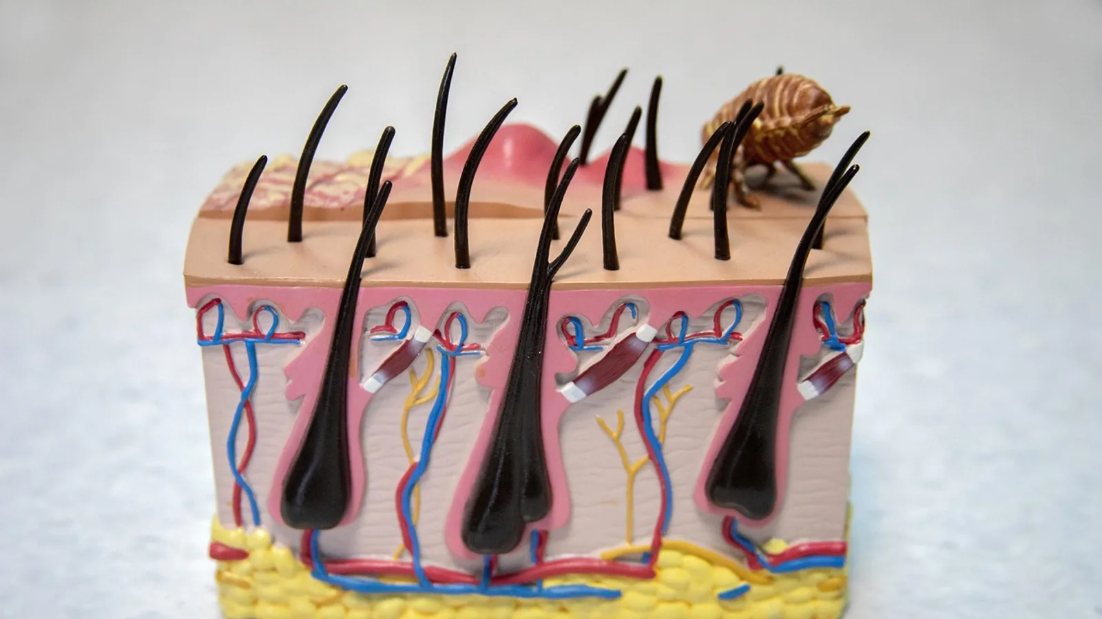
Alopecia means hair loss that leaves skin visible, and it is a clinical sign rather than a diagnosis. In dogs and cats, the most common underlying causes fall into five broad groups: parasites such as mites, fungal infections such as ringworm, allergic skin disease, endocrine disorders such as hypothyroidism and hyperadrenocorticism, and inherited or acquired hair follicle disorders. The single most useful first step is not a test. It is a careful history and physical examination that establishes whether the hair loss is itchy or non-itchy, symmetrical or patchy, inflammatory or quiet.
That distinction drives everything that follows. Symmetrical, non-inflammatory alopecia on the trunk points toward an endocrine or follicular dysplasia problem. Asymmetric, crusted, red, or itchy alopecia points toward infection, allergy, or an immune-mediated process. Your veterinarian uses this split to decide which tests to run first and which to defer.
Triage summary for owners: If your pet is losing hair and scratching, chewing, or rubbing the area, book a veterinary visit within a few days. If the hair loss is patchy with scaling or crusting, keep the pet away from young children and immunocompromised people until ringworm is ruled out, because some causes of alopecia are contagious to humans. If your pet has sudden widespread hair loss with lethargy, vomiting, or collapse, seek care the same day.
This article is educational and is not a substitute for veterinary diagnosis or treatment.
At a Glance: Lesion Pattern and What It Suggests
The table below links the pattern seen on examination to the most likely differentials and the next test a veterinarian would typically reach for. It is a starting framework, not a diagnosis.
| Lesion pattern | Itch level | Skin appearance | Top differentials | Next test |
|---|---|---|---|---|
| Symmetrical trunk and flank hair loss | None to mild | Smooth, thin, often hyperpigmented | Hypothyroidism, hyperadrenocorticism, flank alopecia, alopecia X | Blood work including thyroid and adrenal testing |
| Symmetrical hair loss with scaling, no redness | None | Dry, scaly, sometimes greasy | Follicular dysplasia, color dilution alopecia, ectodermal dysplasia | Trichogram, then skin biopsy |
| Patchy, circular, scaling or crusted | None to mild | Broken hairs, scale, follicular casts | Dermatophytosis (ringworm) | Fungal culture, tape impression |
| Patchy with intense itch and redness | Moderate to severe | Erythema, excoriation, secondary infection | Sarcoptic or Demodex mites, flea allergy, atopic dermatitis, adverse food reaction | Skin scrape, then allergy workup |
| Hair loss on ears, face, neck, chin | Variable | Greasy brown scale, erythema | Malassezia overgrowth, allergic skin disease | Cytology of the skin surface |
| Hair loss with nodules or thickened skin | Variable | Masses, plaques, thickened folds | Neoplasia, paraneoplastic alopecia, calcinosis cutis | Biopsy |
| Hair loss at injection or heat sites | None | Irregular patches, sometimes pigmented | Injection reaction, chronic moderate heat dermatitis | History review, biopsy if unclear |
Why Hair Loss Happens: A Short Physiology Primer
Hair growth is cyclical. Each follicle moves through a growth phase, a transition phase, and a resting phase, then sheds and regrows. Anything that interrupts this cycle, damages the follicle, or destroys the hair shaft produces visible alopecia.
Veterinary dermatology divides alopecia into two large categories. Inflammatory alopecia involves redness, scaling, crusting, pustules, or itch, and the follicle is being attacked or irritated. Non-inflammatory alopecia shows relatively quiet skin with hair simply missing or thinning, and the problem usually lies in the hair cycle itself or in the hormonal signals that regulate it [1].
Non-inflammatory alopecia can be congenital, meaning the animal was born with a defect in follicle formation, or acquired later in life. Congenital forms often have a hereditary cause, and examples include ectodermal dysplasias linked to variants in the ectodysplasin A gene [1]. A case report in a cat described diffuse truncal alopecia with a completely absent undercoat, missing and malformed teeth, and a confirmed EDA gene variant, which is the first report of this specific ectodermal dysplasia in cats [2].
Acquired non-inflammatory alopecia is more common in general practice and is usually tied to an endocrinopathy, a breed-associated follicular dysplasia, or a stress or vascular problem affecting the follicle [1]. Because the hair follicle has a limited number of ways to respond to injury, different diseases can look similar on the surface. That is why history, examination, and targeted testing have to be combined rather than relying on appearance alone [1].
History: The Questions That Narrow the List Fastest
A veterinary history for alopecia is structured to answer four questions: is it itchy, is it seasonal, what does the pet eat, and is parasite control current.
Pruritus
Itch is the strongest single divider. Most dogs with an adverse food reaction are pruritic, and the itch is often generalized, with the ears, feet, and abdomen frequently involved while the perineum is less often affected [3]. Canine adverse food reactions most often present as recurrent bacterial skin infections, otitis externa, and atopic dermatitis rather than as pure hair loss [3].
Cats behave differently. Feline allergic skin disease commonly produces head, neck, or pinnal pruritus, miliary dermatitis, self-induced alopecia, and eosinophilic granuloma complex lesions [4]. Pollen allergy in cats is associated with bronchial asthma and cutaneous reactive patterns including symmetric self-induced alopecia [5]. Self-induced alopecia means the cat is overgrooming, so the hair loss is a behavior-driven sign rather than a primary follicle disease.
A pet with quiet, non-itchy, symmetrical hair loss rarely has an allergy as the primary driver.
Seasonality
Seasonal patterns matter in two directions. Allergic skin disease tied to pollen follows a seasonal calendar, and cats with dermatophytosis showed a significantly higher prevalence of positive fungal cultures in summer and autumn in one regional study [6]. Canine flank alopecia is described as a recurrent, seasonal, non-inflammatory disorder affecting several breeds, including Rhodesian ridgebacks [7]. A seasonal history therefore supports allergy or seasonal flank alopecia and argues against a year-round endocrine cause.
Diet
Diet history should record the current diet, any recent changes, treats, table food, supplements, and chew items. Adverse food reactions can begin at any age, and in dogs the reported age of onset ranged from less than one year to 13 years, with signs beginning by 6 or 12 months of age in 22 to 38 percent of dogs respectively [3]. Four breeds (German shepherd dogs, West Highland white terriers, Labrador retrievers, and golden retrievers) accounted for about 40 percent of affected dogs in the reviewed cases [3].
Parasite Control
Ask which flea and tick product is used, how often it is applied, and whether it is applied correctly. Flea allergy dermatitis is a major cause of itchy, asymmetric hair loss over the rear half of the body. A gap in coverage is a common and easily corrected reason for ongoing signs.
Other History Elements
Record any drugs given by injection or by mouth, because some medications can cause skin reactions. One case report described a suspected dermatologic adverse effect after subcutaneous furosemide in a cat, which is the first report of its kind in feline medicine [8]. Record the pet's environment, including heat sources. Chronic moderate heat exposure caused irregular alopecia and erythema in dogs and cats that rested repeatedly against a heat source, a condition called erythema ab igne [9].
Physical Examination: Reading the Pattern
The examination maps the distribution, symmetry, and degree of inflammation. This is where the differential list is built.
Symmetry
Symmetrical hair loss on the trunk, flanks, and tail head, with thin skin and often increased pigmentation, is the classic presentation of an endocrine disorder. Hyperadrenocorticism in dogs produces alopecia along with increased drinking and urination, abdominal distension, lethargy, weakness, an enlarged liver, and calcinosis cutis [10]. Middle-aged to older Poodles, Dachshunds, Boston Terriers, and Boxers are most commonly affected, and cats are rarely affected [10].
Asymmetric hair loss, especially with crusting, broken hairs, or redness, points toward infection, allergy, or immune-mediated disease.
Inflammation
Inflammatory skin is red, warm, swollen, scaly, crusted, or pustular. Non-inflammatory skin is smooth and quiet. This single observation separates the two diagnostic pathways and determines whether a skin scrape or a biopsy comes first.
Distribution
Certain distributions are strongly suggestive. Hair loss confined to the flanks in a breed known for flank alopecia supports that diagnosis [7]. Hair loss on the face, ventral neck, abdomen, ear canals, chin, pinnae, interdigital skin, and claw folds in an allergic cat raises suspicion for Malassezia overgrowth, which appears as multifocal alopecia, erythema, crusting, and greasy adherent brownish scale [11].
Hair and Coat Quality
Look at whether hairs break easily, whether the coat is dry or greasy, and whether the undercoat is present. A completely absent undercoat with diffuse truncal alopecia in a cat, along with missing or malformed teeth, should raise suspicion for ectodermal dysplasia [2].
Nodules and Masses
Any nodule, plaque, or thickened area within an area of alopecia changes the plan. Cutaneous mast cell tumors are among the most common skin tumors in cats and can present as multiple nodules on the head and trunk [12]. Paraneoplastic alopecia is a recognized syndrome in cats associated with internal malignancy, and recognition of such lesions can help detect cancer earlier [13].
The Diagnostic Pathway
The flowchart below shows the main decision path from history and examination through the first-line tests to biopsy.
flowchart TD
    A[History and physical exam] --> B{Is the skin itchy or inflamed}
    B -->|Yes| C[Skin scrape and cytology]
    B -->|No| D{Is hair loss symmetrical}
    C --> E[Treat mites or infection]
    D -->|Yes| F[Blood work for endocrine disease]
    D -->|No| G[Fungal culture and trichogram]
    F --> H[Trichogram and biopsy if blood work normal]
    G --> I[Biopsy if culture negative]
    E --> J[Recheck in 3 to 4 weeks]
    H --> K[Diagnosis and targeted treatment]
    I --> K
    J --> K
Step 1: Skin Scrape and Surface Cytology
A skin scrape is the first test when the skin is inflamed or itchy. It looks for Demodex and Sarcoptes mites and for surface bacteria and yeast. Surface cytology identifies Malassezia overgrowth, which in allergic cats appears as multifocal alopecia with erythema, crusting, and greasy brown scale, and which responds to azole therapy [11]. This step is inexpensive, fast, and often decisive.
Step 2: Trichogram
A trichogram is a microscopic examination of plucked hairs. It shows the stage of the hair cycle, whether hairs are broken or fractured, whether the cuticle is damaged, and whether the hair shaft itself is abnormal. Trichography has been formally characterized in normal dogs, and a systematic evaluation of hair samples from five body locations showed strong agreement between observers for most variables [14]. That study also described a round hair tip that was more common on the dorsal head, and noted that color change to the proximal hair shaft was common in black Dobermann pinschers [14]. Dobermanns are predisposed to color dilution alopecia and follicular dysplasia, so knowing what normal looks like in that breed matters [14].
A trichogram helps separate diseases that destroy the hair shaft from diseases that stop the follicle from cycling. It is a bridge test between the scrape and the biopsy.
Step 3: Fungal Culture for Dermatophytes
Dermatophytosis should be on the list for any alopecic, papular, or pustular lesion in dogs [15]. In a study of dogs and cats with alopecia and peripheral scaling, 23.3 percent of samples cultured positive, with 20.5 percent of dog samples and 28.2 percent of cat samples positive [6]. Microsporum canis was the most common isolate at 77.7 percent, followed by geophilic species [6]. Young animals under one year had a significantly higher prevalence of M. canis infection [6]. Male dogs were more affected, and Yorkshire terriers showed the highest positivity at 50 percent, mostly from M. canis [6]. Importantly, the presence of dermatophytes was not associated with itching [6].
Diagnostic accuracy varies by method. In one comparison of 30 dogs and 15 cats with alopecia or kerion, tape preparations identified dermatophytes in 82.2 percent of cases, hair plucks in 66.7 percent, and fungal culture in 80 percent [16]. In kerions, tape preparations and fungal culture both reached 90.9 percent sensitivity while hair plucks fell to 36.4 percent [16]. Sensitivity was higher in cats than in dogs for all three tests [16]. Wood's lamp fluorescence and microscopic examination were of limited value compared with culture, with only 45.5 percent of culture-positive M. canis samples fluorescing and 53.2 percent showing organisms on microscopy [6].
A newer molecular approach using PCR and sequencing of ribosomal DNA regions has improved identification of dermatophyte species, and one study reported the first isolation of Trichophyton indotineae in a dog in Africa, with younger animals under one year at significantly higher risk of infection [17]. In that study, 52.22 percent of dog samples and 70 percent of cat samples cultured positive, with M. canis the most prevalent isolate at 60 percent followed by Trichophyton mentagrophytes at 20 percent [17].
Serology has been explored as an adjunct. An ELISA for canine dermatophytosis caused by M. canis showed good sensitivity of 83.3 percent and high specificity of 95.2 percent, with a strong correlation between duration of infection and antibody concentration [15].
Step 4: Blood Work for Endocrine Disease
When the hair loss is symmetrical and non-inflammatory, the next step is blood testing. Hyperadrenocorticism is diagnosed using a combination of tests. The high-dose dexamethasone suppression test helps differentiate pituitary-dependent disease from adrenal tumors, and the low-dose dexamethasone suppression test, plasma ACTH levels, and the ACTH response test are additional diagnostic aids [10]. Routine blood work in affected dogs may show neutrophilia, lymphopenia, monocytosis, eosinopenia, and increased alkaline phosphatase, SGPT, cholesterol, sodium, and glucose, with decreased potassium and T4 levels [10].
Thyroid testing is also part of this panel. Because thyroid hormone affects the hair cycle, hypothyroidism is a classic cause of symmetrical non-inflammatory alopecia in dogs.
Step 5: Biopsy
Biopsy is reserved for cases where the pattern is unclear, where first-line tests are negative, or where a follicular dysplasia, alopecia X, or a paraneoplastic syndrome is suspected. Non-inflammatory alopecia is a common reason to take biopsies, and the diagnosis depends on combining detailed clinical history, thorough clinical examination including blood work, appropriate biopsy site selection, and detailed histological findings [1]. Because the hair follicle has limited responses to altered regulation and histopathology can change over the course of a disease, a single biopsy taken at the wrong time or the wrong site can mislead [1].
Biopsy is also the test that identifies paraneoplastic alopecia, a syndrome in cats associated with internal malignancy where recognizing the skin lesion can lead to earlier cancer detection [13].
Common Causes in Detail
Allergic Skin Disease
Allergic skin disease is one of the most common reasons pets present with hair loss, because itch drives self-trauma. Adverse food reactions in dogs most often present as recurrent bacterial skin infections, otitis externa, and atopic dermatitis, with generalized itch and frequent involvement of ears, feet, and abdomen [3]. Cats with adverse food reactions develop signs later than dogs, and there is no apparent breed or gender predisposition in cats [3].
Pollen allergy produces eczematous lesions in dogs, described as canine atopic dermatitis, while in cats it produces bronchial asthma and cutaneous reactive patterns including eosinophilic granuloma complex, head and neck pruritus, and symmetric self-induced alopecia [5]. In cats, detection of allergen-specific IgE in serum supports a diagnosis of feline atopy based on compatible history, clinical signs, and elimination of other pruritic dermatoses [4].
Malassezia Overgrowth
Malassezia overgrowth is a secondary problem in allergic cats. In a series of 18 allergic cats, 16 had atopic dermatitis and one had an adverse food reaction, and all were otherwise healthy with negative retroviral testing [11]. Lesions were multifocal alopecia, erythema, crusting, and greasy adherent brownish scale [11]. Cytology confirmed overgrowth on the face, ventral neck, abdomen, ear canal, chin, ear pinnae, interdigital skin, and claw folds [11]. After three to four weeks of treatment, substantial reduction of pruritus and lesions was seen in all 11 cats given combined therapy and in five of seven treated with azoles alone [11].
Dermatophytosis
Ringworm is a zoonotic infection, meaning it can spread to people. It should be considered in any alopecic, papular, or pustular lesion in dogs [15]. Young animals under one year are at significantly higher risk [6][17]. Cats housed indoors only had a significantly higher risk than cats with indoor and outdoor access in one study [17]. Because Wood's lamp and microscopy are unreliable compared with culture, fungal culture remains the practical confirmatory test [6].
Endocrine Disease
Hyperadrenocorticism produces alopecia along with increased drinking and urination, abdominal distension, lethargy, weakness, an enlarged liver, calcinosis cutis, testicular atrophy, and absence of estrus [10]. It affects middle-aged to older dogs, with Poodles, Dachshunds, Boston Terriers, and Boxers most commonly affected, and it is rare in cats [10]. Hypothyroidism produces a similar symmetrical, non-inflammatory pattern and is diagnosed with thyroid testing as part of the same blood panel.
Follicular Dysplasia and Breed-Associated Alopecia
Follicular dysplasia refers to a group of disorders where the hair follicle fails to regenerate normally. Some have a clear breed predilection and begin early in life, and a hereditary background is suspected though not proven in all cases [1]. Canine flank alopecia is a recurrent, seasonal, non-inflammatory disorder affecting several breeds including Rhodesian ridgebacks, and a genome-wide study identified candidate genes involved in cytoskeletal function, cell adhesion, neural signaling, and hair follicle cycling pathways including Wnt, BMP, MAPK, Notch, and Hedgehog signaling [7]. The authors concluded that flank alopecia is likely genetically heterogeneous with multiple loci and possible modifier variants rather than a simple single-gene disorder [7].
Congenital Alopecia
Congenital alopecia results from decreased formation or differentiation of the hair follicle or shaft before birth, often with a hereditary cause such as ectodermal dysplasia linked to ectodysplasin A gene variants [1]. A cat with a confirmed EDA variant had diffuse truncal alopecia with a completely absent undercoat, missing teeth, and abnormally shaped conical teeth [2]. The equivalent missense variant in humans is a recurring pathogenic variant, which supports the genetic finding [2].
Environmental and Drug-Related Alopecia
Chronic moderate heat dermatitis, or erythema ab igne, produces irregular alopecia and erythema at sites of repeated heat exposure, typically where the animal rests against a heat source [9]. Histology shows karyomegaly, keratinocyte atypia, apoptotic or vacuolated basal cells, mild mixed mononuclear dermatitis, adnexal atrophy, and wavy eosinophilic elastic fibers [9]. Removing the heat source prevents progression [9].
Drug reactions can also cause alopecia. A suspected dermatologic adverse effect was reported after subcutaneous furosemide in a cat, the first such report in feline medicine [8].
Paraneoplastic Alopecia
Paraneoplastic syndromes are noncancerous skin diseases associated with internal malignancy, and their recognition can help detect and treat underlying cancer earlier [13]. Feline paraneoplastic alopecia is one of the recognized veterinary paraneoplastic dermatoses, along with feline thymoma-associated exfoliative dermatitis, nodular dermatofibrosis, feminization syndrome associated with testicular tumors, superficial necrolytic dermatitis, and paraneoplastic pemphigus [13].
Neoplasia
Cutaneous mast cell tumors are among the most common skin tumors in cats and usually behave benignly, though multiple tumors, recurrence, or lymph node involvement carry a more guarded prognosis [12]. Surgery is the treatment of choice, and electrochemotherapy has been reported as an alternative for multiple or large tumors, with complete resolution in a small case series and alopecia and scar tissue noted after treatment [12].
Infectious and Parasitic Causes Beyond Ringworm
Leishmaniasis is an emerging disease in domestic cats in endemic regions, and infection has been confirmed in cats using serology and molecular methods on conjunctival swabs [18]. This is regionally important and should be considered in cats with compatible skin lesions that have lived in or traveled to endemic areas.
Otitis media-interna in cats can present with pruritus and alopecia alongside ear disease. In a study of 58 previously hoarded cats, pruritus or alopecia was present in 50 percent, otitis externa in 79 percent, and otitis interna in 40 percent [19]. Resolution of pruritus and alopecia occurred in a statistically significant proportion of cats after surgical treatment, though worsening of pruritus and alopecia occurred in nine cases [19]. This shows that ear disease and skin signs can be linked.
Evidence-Based Management Principles
Management follows the diagnosis. There is no single treatment for alopecia because alopecia is a sign, not a disease.
For allergic skin disease, management centers on identifying and avoiding the trigger where possible and controlling secondary infection. In cats with Malassezia overgrowth, azole therapy alone or combined with antibacterial and anti-inflammatory therapy produced substantial reduction of pruritus and lesions after three to four weeks [11].
For dermatophytosis, treatment typically combines topical and systemic antifungal therapy. In one study, treatment regimens included topical clotrimazole, systemic itraconazole, supportive therapy, and a dermatophyte vaccine [17]. Because ringworm is zoonotic, treatment also protects people in the household.
For endocrine disease, treatment targets the underlying hormone disorder. Medical treatment of hyperadrenocorticism has involved oral mitotane, and hypophysectomy has been used in pituitary-dependent cases with low reported mortality, while adrenalectomy is indicated for adrenal tumors [10]. These decisions are made by the veterinarian based on the specific diagnosis and the pet's overall health.
For follicular dysplasia and flank alopecia, management is often supportive because the underlying follicle disorder cannot be cured. Removing the heat source resolves erythema ab igne [9].
For paraneoplastic alopecia and cutaneous neoplasia, management depends on identifying and treating the underlying tumor [13][12].
A general principle applies across all causes. Veterinary management primarily depends on identifying and correcting the underlying disorder, and there is limited species-specific evidence for most alopecia treatments in dogs and cats [20]. Differences in follicular architecture, hair cycle kinetics, skin permeability, drug metabolism, coat characteristics, and grooming behavior mean that human hair loss treatments cannot be directly applied to pets [20].
Unsafe Home Remedies
Do not apply human hair growth products to pets. Human alopecia treatments are not formulated for animals, and differences in skin permeability, drug metabolism, and coat characteristics mean they cannot be directly translated to dogs and cats [20].
Do not use human antifungal creams on pets without veterinary guidance. The dose, vehicle, and duration differ, and some ingredients are toxic if licked off.
Do not assume hair loss is cosmetic. Alopecia can be the first visible sign of an internal disease such as hyperadrenocorticism or an internal malignancy [13][10].
Do not skip parasite prevention. Gaps in flea control are a common and preventable cause of ongoing itchy hair loss.
Do not use essential oils, tea tree oil, or over-the-counter itch sprays on pets. Cats in particular are sensitive to many topical products and can become seriously ill after grooming them off.
Prevention
Prevention depends on the cause. For allergic skin disease, keeping parasite control current, managing the environment, and following the veterinarian's plan reduces flare frequency. For dermatophytosis, limiting contact between infected and healthy animals and cleaning the environment reduces spread, and young animals under one year are at higher risk so they deserve closer monitoring [6][17]. For environmental alopecia such as erythema ab igne, removing or modifying the heat source prevents progression [9]. For hereditary conditions such as ectodermal dysplasia, genetic counseling and breed health screening are the relevant steps [1][2].
Prognosis
Prognosis varies widely. Allergic skin disease is usually manageable but often requires lifelong control. Dermatophytosis generally resolves with appropriate treatment, though it can take weeks and requires environmental decontamination. Endocrine disease carries a good prognosis when diagnosed and managed, though treatment is typically lifelong [10]. Follicular dysplasia and congenital alopecia are usually permanent, and management focuses on comfort and skin health [1]. Paraneoplastic alopecia carries a guarded prognosis because it signals underlying malignancy [13]. Cutaneous mast cell tumors in cats usually behave benignly and carry a good prognosis after complete treatment [12].
Emergency Red Flags
Seek immediate veterinary care if your pet has any of the following:
- Sudden widespread hair loss with lethargy, vomiting, diarrhea, or collapse
- Hair loss with open sores, bleeding, or signs of severe pain
- Hair loss with difficulty breathing, especially in a cat
- Hair loss with a rapidly growing mass or multiple new nodules
- Hair loss with increased drinking and urination and a distended abdomen
- Hair loss with fever, loss of appetite, or weight loss
Limitations and When to Contact a Veterinarian
This article provides a framework for understanding how veterinarians approach alopecia. It cannot replace an examination of your individual pet.
Contact a veterinarian promptly if your pet has hair loss that is spreading, itchy enough to cause self-trauma, or accompanied by scaling or crusting. Contact a veterinarian the same day if your pet has hair loss with lethargy, vomiting, breathing difficulty, a rapidly growing mass, or a distended abdomen. If anyone in the household is immunocompromised, pregnant, or very young, mention this to the veterinarian because ringworm is zoonotic and can spread to people.
Frequently Asked Questions
What is the most common cause of alopecia in pets?
There is no single most common cause because alopecia is a sign with many triggers. Allergic skin disease, parasites, fungal infection, and endocrine disease are the most frequent categories. The pattern of hair loss and the level of itch help the veterinarian narrow the list quickly.
Is alopecia in pets painful?
Alopecia itself is not usually painful, but the underlying cause can be. Itchy allergic skin disease and infected skin are uncomfortable. Quiet, symmetrical hair loss from an endocrine disorder is typically not painful.
Can alopecia in pets be cured?
Some causes can be cured, including ringworm, mites, and heat-related alopecia once the source is removed. Others, such as allergic skin disease, endocrine disease, and follicular dysplasia, are managed rather than cured.
How long does it take for hair to grow back after treatment?
Hair regrowth depends on the cause and on the hair cycle. Some pets regrow hair within weeks of treating an infection, while endocrine and follicle disorders can take months. Your veterinarian will set a realistic timeline at recheck.
Is ringworm the same as a worm?
No. Ringworm is a fungal infection of the skin, hair, and nails, not a parasitic worm. It is contagious to people and other animals, so diagnosis matters for the whole household.
Can food allergies cause hair loss in pets?
Yes. Adverse food reactions most often cause itch, and itch leads to self-trauma and secondary hair loss. In dogs, adverse food reactions commonly present as recurrent skin infections, ear infections, and atopic dermatitis.
Do pets get hair loss from stress?
Stress can contribute to hair loss, particularly in cats that overgroom. Symmetric self-induced alopecia in cats is a recognized pattern in allergic skin disease and can also reflect behavioral overgrooming. A veterinarian can help sort out which is happening.
When should I see a veterinarian for my pet's hair loss?
See a veterinarian if the hair loss is spreading, if your pet is scratching or chewing, if there is scaling or crusting, or if the hair loss is symmetrical on the trunk. See a veterinarian the same day if there is lethargy, vomiting, breathing difficulty, or a rapidly growing mass.
Related Articles
- Cat Hair Loss on Ears: Causes and What to Do
- Dog Hair Loss on Nose: Causes and When to Worry
- Cat Hair Loss on Tail: Causes and Solutions
- Dog Hair Loss After Puppies: Causes and What to Do
- Dog Hair Loss on Tail: Causes and Treatment
- Cat Hair Loss with Itching: Causes and Treatment
- Ascites in Pets: Causes and Diagnostic Steps
- Dog Digestive Issues: Causes, Symptoms, and When to Worry
- What Is Anthrax Disease? Causes and Symptoms
Sources
- Canine noninflammatory alopecia: An approach to its classification and a diagnostic aid.
- EDA Missense Variant in a Cat with X-Linked Hypohidrotic Ectodermal Dysplasia.
- Critically appraised topic on adverse food reactions of companion animals (7): signalment and cutaneous manifestations of dogs and cats with adverse food reactions.
- Serum allergen-specific immunoglobulin E in atopic and healthy cats: comparison of a rapid screening immunoassay and complete-panel analysis.
- Pollen Allergies in Humans and their Dogs, Cats and Horses: Differences and Similarities.
- The epidemiology of canine and feline dermatophytoses in southern Italy.
- Genetic insights into canine flank alopecia in Rhodesian ridgebacks: identifying candidate genes.
- Dermatologic adverse effect of subcutaneous furosemide administration in a cat.
- Chronic moderate heat dermatitis (erythema ab igne) in five dogs, three cats and one silvered langur.
- Diseases of the adrenal cortex of dogs and cats.
- Malassezia spp. overgrowth in allergic cats.
- Successful treatment of cutaneous mast cell tumors in cats using electrochemotherapy: A case series.
- Cutaneous paraneoplastic syndromes in dogs and cats: a review of the literature.
- Trichographic features of hair from normal black Dobermann pinscher dogs.
- Development of an enzyme-linked immunosorbant assay (ELISA) for the serodiagnosis of canine dermatophytosis caused by Microsporum canis.
- Comparison of Adhesive Tape Impression Cytology, Hair Plucks, and Fungal Culture for the Diagnosis of Dermatophytosis in Dogs and Cats.
- Molecular Diagnostics and Control of Zoonotic Dermatophytosis: First Detection of Trichophyton indotineae in a Dog in Africa.
- Diagnosis and epidemiology of Leishmania infantum in domestic cats in an endemic area of the Amazon region, Brazil.
- A Pandora's box in feline medicine: presenting signs and surgical outcomes in 58 previously hoarded cats with chronic otitis media-interna.
- Alopecia management from a comparative perspective: pharmacological and pharmaceutical insights in humans, dogs, and cats.