Giardia lamblia: Protozoan Biology and Life Cycle
By Dr. Zubair Khalid, DVM, MS, PhD ·
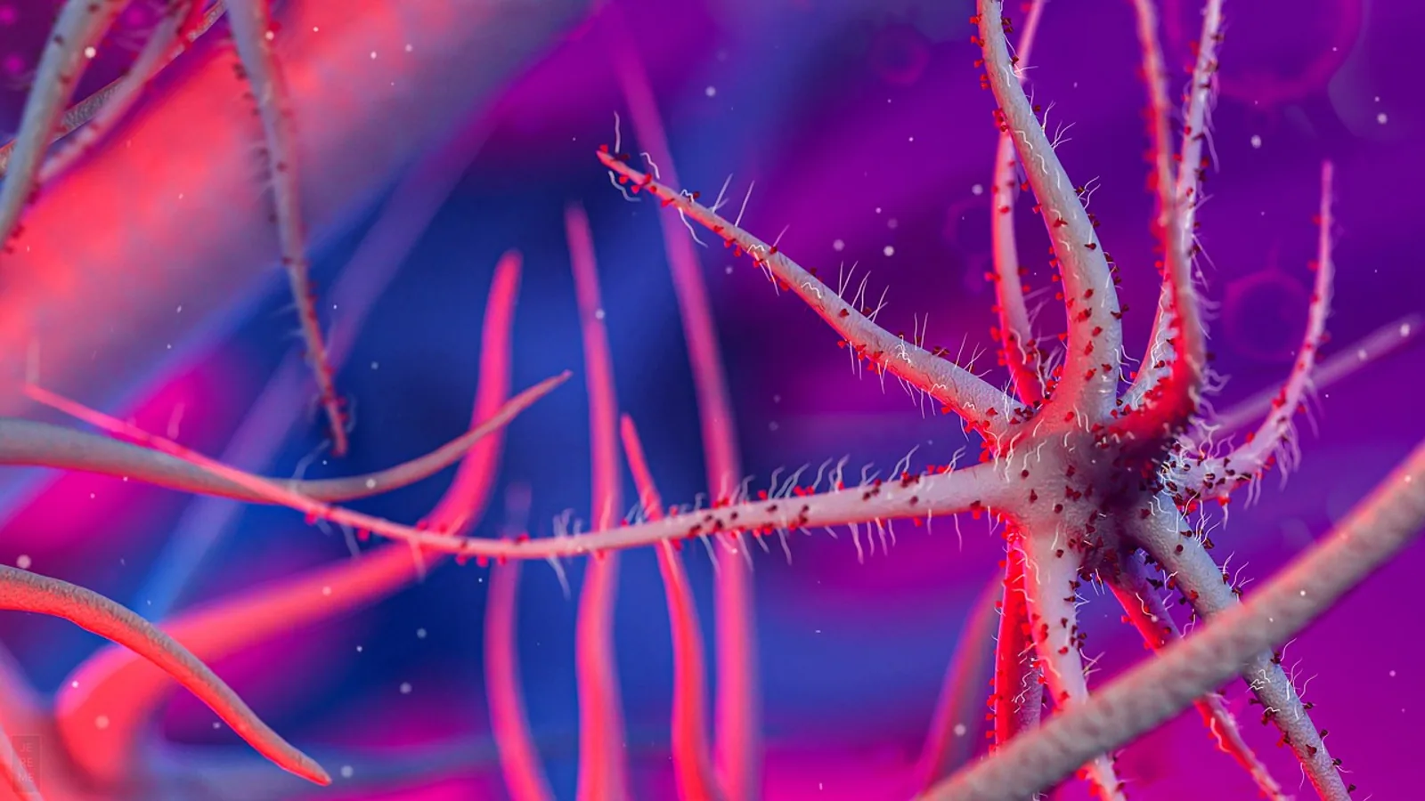
Giardia lamblia is a flagellated, single-celled intestinal protozoan parasite that infects mammals, including dogs, cats, cattle, birds, and humans. Its two-stage life cycle alternates between a motile, binucleate trophozoite that colonizes the host small intestine and an environmentally resistant oval cyst that is passed in feces and is infectious immediately on passage [1].
Giardiasis is one of the most common waterborne and fecal-oral parasitic infections worldwide, and it is a leading cause of epidemic and sporadic diarrhea in humans and animals [2]. In veterinary practice, the parasite is a routine finding in young dogs and cats with acute or chronic diarrhea, in shelter and kennel populations, and in livestock. Because the same species can circulate among wildlife, livestock, pets, and people, Giardia sits squarely inside a One Health framework, and its biology shapes how clinicians test, treat, and prevent infection [3][4].
This article is educational and is not a substitute for veterinary diagnosis or treatment.
Naming and Taxonomy: One Species, Many Names
The organism carries three widely used scientific names: Giardia lamblia, Giardia duodenalis, and Giardia intestinalis. These are synonyms for the same species, and modern molecular literature favors G. duodenalis [5][6]. Older textbooks and laboratory reports still use G. lamblia, and the name persists in clinical and parasitology teaching. When you read a diagnostic report that says Giardia lamblia and a research paper that says Giardia duodenalis, they are describing the same protozoan.
The genus Giardia belongs to the diplomonads, a group of flagellated protists with two nuclei and a reduced set of organelles. For more than a century, taxonomy within the genus was debated because parasites from very different hosts look nearly identical under the microscope [7]. That morphological uniformity hid real genetic diversity, and it delayed understanding of host specificity and zoonotic transmission. Molecular tools resolved much of the confusion by revealing distinct genetic groups, called assemblages, within G. duodenalis [7][8].
What the Name Does Not Tell You
A common student error is to treat Giardia lamblia as a single uniform organism. In reality, G. duodenalis is a species complex. The name identifies the organism to the species level, but the assemblage label (A through H) identifies the genetic lineage, and the lineage predicts host range and zoonotic potential far better than the species name alone [8][3].
The Two Life Stages
Giardia has exactly two stages in its life cycle, and every clinical and epidemiological feature flows from their differences. The trophozoite is the feeding, motile, disease-causing stage inside the host. The cyst is the dormant, transmission stage outside the host [1].
The Trophozoite
The trophozoite is a pear-shaped, bilaterally symmetrical cell with two nuclei, four pairs of flagella, and a striking ventral disc. The ventral disc is a spiral microtubule array made of roughly 50 parallel microtubules with associated microribbons and proteins, and it functions as the parasite's attachment organelle [1]. Attachment to the small intestinal epithelium is what allows the trophozoite to resist being flushed downstream by intestinal flow.
Trophozoites also possess an unusual endomembrane system that includes peripheral vesicles. These vesicles behave like endosomal and lysosomal compartments, accumulate ingested macromolecules, and show acid phosphatase activity [9]. Some peripheral vesicles mature into multivesicular bodies containing intraluminal vesicles, and the parasite releases microvesicles that contribute to giardiasis pathogenesis [9]. Extracellular vesicles derived from G. duodenalis help the parasite evade host immunity and trigger inflammatory cascades in the gut [10].
Trophozoites multiply by binary fission in the small intestine. A recently isolated canine strain of zoonotic assemblage A was shown to multiply faster in axenic culture than a long-cultured human reference strain, though it produced cysts at a lower rate [11]. That finding is a reminder that growth and encystation rates vary between strains and can shift with laboratory adaptation.
The Cyst
The cyst is oval, immotile, and surrounded by a protective wall. It forms when trophozoites undergo encystation, a differentiation program that involves major morphological and genetic changes and culminates in synthesis of cyst wall proteins [12]. Encystation is regulated in part by histone modifications. The enzyme lysine methyltransferase 2 is required for progression through late encystation, and reducing its expression arrests the process and lowers cyst output [12].
Cysts are the stage that leaves the host. They are environmentally resistant, which is why Giardia spreads so effectively through water, food, and contaminated surfaces. Two facts about cysts drive most transmission:
- Cysts are infectious immediately on passage. No maturation period in the environment is required.
- The infectious dose is very low. As few as 10 cysts can establish infection.
Trophozoites, by contrast, do not survive outside the host. They are fragile, do not tolerate desiccation or the external environment, and are not the transmission stage. If a fecal sample contains only trophozoites, the parasite has not yet produced the form that spreads to new hosts.
Summary Table: Trophozoite Versus Cyst
| Feature | Trophozoite | Cyst |
|---|---|---|
| Shape | Pear-shaped, bilaterally symmetrical | Oval |
| Motility | Motile, four pairs of flagella | Immotile |
| Nuclei | Two | Two to four |
| Location | Small intestine of host | Environment, feces, water, food |
| Attachment | Ventral disc | None |
| Function | Colonization, feeding, replication, disease | Survival, transmission |
| Environmental survival | Does not survive outside host | Resistant, persists in cool moist conditions |
| Infectivity | Not the transmission stage | Infectious immediately on passage |
| Infectious dose | Not applicable | As few as 10 cysts |
| Detection | Fecal smear, duodenal aspirate, antigen test | Fecal flotation, antigen test, immunofluorescence |
The Life Cycle Step by Step
The following numbered sequence describes the full cycle from ingestion to shedding.
- Ingestion. A susceptible host swallows infectious cysts from contaminated water, food, soil, or fomites, or through direct fecal-oral contact.
- Excystation. In the small intestine, the cyst wall breaks down and releases trophozoites. Excystation methodology has been standardized in research settings, which reflects how consistent this step is across strains [2].
- Colonization. Trophozoites attach to the small intestinal epithelium using the ventral disc and begin feeding and multiplying by binary fission [1].
- Disease or carriage. Infection may cause acute or chronic diarrhea, malabsorption, and failure to thrive, or it may remain asymptomatic [7][5].
- Encystation. As trophozoites move toward the colon, they differentiate into cysts through a regulated program that builds the protective cyst wall [12].
- Shedding. Cysts are passed in feces. They are infectious the moment they leave the host, so the next host can be infected by the same bowel movement that contaminated the environment.
- Environmental persistence. Cysts survive in cool, moist conditions and contaminate water sources, which is why Giardia is a significant cause of waterborne outbreaks worldwide [8].
flowchart TD
    A[Cyst ingested by host] --> B[Excystation in small intestine]
    B --> C[Trophozoite attaches to gut wall]
    C --> D[Binary fission and colonization]
    D --> E{Symptoms develop}
    E --> F[Diarrhea or malabsorption]
    E --> G[Asymptomatic carriage]
    F --> H[Encystation in colon]
    G --> H
    H --> I[Cysts shed in feces]
    I --> J[Cysts infectious immediately]
    J --> K[Contaminate water food or surfaces]
    K --> A
Host Assemblages A Through H
Molecular studies recognize eight assemblages within G. duodenalis, labeled A through H, with varying host specificity and zoonotic potential [8][3]. Assemblages A and B are the lineages most frequently reported in humans and are considered the zoonotic assemblages [8][13]. The remaining assemblages are largely host-adapted.
| Assemblage | Primary host association | Zoonotic relevance |
|---|---|---|
| A | Humans, dogs, cats, livestock, wildlife | Yes, zoonotic |
| B | Humans, dogs, cats, wildlife, birds | Yes, zoonotic |
| C | Dogs | Low |
| D | Dogs | Low |
| E | Ruminants, pigs, horses | Low |
| F | Cats | Low |
| G | Rodents | Low |
| H | Marine mammals | Low |
Assemblage A is subdivided into sub-assemblages AI, AII, and AIII. AII is closely associated with humans, while AI has been found in diverse animals and humans, which supports its cosmopolitan and zoonotic nature [14]. A study of non-human primates in the Brazilian Amazon identified a new assemblage A genotype using high-resolution multilocus sequence typing, which shows that assemblage diversity is still being mapped [14].
Host-adapted assemblages dominate in companion animals. In a molecular survey of dogs and cats in central Spain, assemblages C and D predominated in dogs and assemblage F predominated in cats, while zoonotic assemblages A and B occurred at moderate frequencies [4]. A study of owned dogs in central Vietnam detected only the canine-specific assemblage D [15]. In Ireland, assemblages C and D appeared in canines as expected, but assemblage E, normally linked to ruminants, was also detected across most host species tested [3].
Zoonotic Potential: Real but Usually Host-Specific
The zoonotic question has a nuanced answer. Molecular and epidemiological studies over four decades support the conclusion that zoonotic transmission of G. duodenalis is possible [7]. At the same time, most Giardia infections are host-specific, which makes zoonotic transmission relatively uncommon [7]. From a public health standpoint, the rare events still matter, because children and immunocompromised people can develop severe outcomes such as chronic diarrhea, malabsorption, and failure to thrive [7].
A well-documented field case illustrates how transmission can occur. A fieldworker contracted G. duodenalis shortly after collecting fecal samples from wild Norwegian reindeer, nearly half of which showed heavy infections with assemblage AI. Molecular comparison showed identical sequences between the reindeer samples and the worker's infection at the loci that amplified successfully [16]. The worker had no prior history of infection, had intense exposure during sampling, and had no other known risk factors, which made the reindeer the most plausible source [16].
Species Affected
Giardia duodenalis infects a broad range of vertebrates. Documented hosts in the recent literature include:
- Dogs. Assemblage D is common in owned dogs, and assemblages C and D predominate in clinical canine cases [15][4].
- Cats. Assemblage F is the most frequent feline lineage, with zoonotic A and B occurring less often [4].
- Cattle and other ruminants. Assemblage E is typically associated with ruminants, and it has been detected in multiple host species in veterinary diagnostic work [3].
- Horses. Assemblages A, B, and E have been identified in horses [5].
- Birds. Zoonotic assemblage B has been identified in pigeons, including wood pigeons and rock pigeons [17].
- Humans. Assemblages A and B are the predominant human lineages, with AII strongly associated with people [13][14].
- Wildlife. Assemblage A has been found in anteaters, non-human primates, reindeer, and other wildlife, which reflects its broad host range [14][16].
Fish and shellfish can act as biological hosts or mechanical concentrators of Giardia cysts, and zoonotic assemblages A and B have been detected in both [18]. This matters for food and water safety, particularly where wastewater management is inadequate [18].
Transmission Routes
Giardia spreads by the fecal-oral route. The specific routes that matter in practice are:
- Waterborne transmission. Contaminated surface water and drinking water are major sources, and G. duodenalis is a significant cause of waterborne outbreaks worldwide [8]. Cysts survive in cool water and resist routine chlorine disinfection at standard concentrations.
- Foodborne transmission. Contaminated produce and food handling can transmit cysts, and the low infectious dose makes even minor contamination relevant.
- Direct fecal-oral contact. This route dominates in kennels, shelters, catteries, and multi-pet households, and it is the main route in young animals.
- Environmental and fomite transmission. Cysts persist on surfaces, in soil, and in standing water. Shared bowls, bedding, and grooming equipment can all serve as vehicles.
- Wildlife and livestock spillover. Wildlife-associated transmission is documented, including the reindeer case, and livestock can carry zoonotic assemblages [16][3].
Risk factors identified in dog populations include direct contact with other dogs, which was strongly associated with infection in a study of owned dogs in central Vietnam [15]. This finding supports the standard advice to isolate and test new or diarrheic animals before introducing them to a group.
Giardia Versus Cryptosporidium
Giardia and Cryptosporidium are frequently discussed together because both are environmentally resistant protozoan parasites transmitted by the fecal-oral route, often through contaminated water or food [4]. They are different organisms with different biology, and the differences affect testing and management.
| Feature | Giardia duodenalis | Cryptosporidium spp. |
|---|---|---|
| Classification | Flagellated protozoan, diplomonad | Apicomplexan protozoan |
| Infective stage | Cyst | Oocyst |
| Life stages | Trophozoite and cyst | Multiple stages including meronts and gamonts |
| Motility of infective stage | Cyst immotile, trophozoite motile | Oocyst immotile |
| Location in host | Small intestine | Small intestine, primarily |
| Common species in dogs | Assemblages C, D, and sometimes A or B | Cryptosporidium canis |
| Common species in cats | Assemblage F, sometimes A or B | Cryptosporidium felis |
| Zoonotic species | Assemblages A and B | C. parvum, C. hominis |
| Detection | Fecal flotation, antigen tests, immunofluorescence, PCR | Fecal flotation, immunofluorescence, PCR |
| Environmental resistance | High | High |
In a molecular survey of dogs and cats in central Spain, Cryptosporidium canis and Cryptosporidium felis were the predominant species in dogs and cats respectively, while C. parvum and C. hominis were detected only sporadically [4]. In owned dogs in central Vietnam, Giardia was detected in 8.0% of animals while Cryptosporidium DNA was not detected at all [15]. These patterns show that the two parasites do not always co-circulate at the same rate, and they should be tested for separately.
The practical distinction matters because the two organisms differ in treatment response and in zoonotic risk profile. A fecal test that identifies "protozoan cysts" without further characterization is not enough to guide management.
How Giardia Is Detected in Practice
Diagnosis combines clinical signs with laboratory confirmation. The main approaches are:
- Fecal flotation. Cysts can be identified on flotation, though shedding is intermittent, so a single negative result does not rule out infection.
- Direct smear. Trophozoites may be seen in fresh, watery feces, but they degrade quickly outside the host, so timing matters.
- Antigen tests. Fecal antigen tests are widely used and are convenient for in-clinic screening.
- Immunofluorescence. Direct immunofluorescence is used in both clinical and research settings and can detect cysts and oocysts with good sensitivity [17].
- Molecular testing. PCR and multilocus genotyping identify the assemblage and are the tools used in epidemiological studies to distinguish zoonotic from host-adapted lineages [3][4][15].
In research settings, isolation and purification methods vary widely and often involve two phases, an initial separation step such as filtration or centrifugation followed by density gradient purification for fecal samples or immunomagnetic separation for water samples [2]. This variability limits direct comparison between studies, but it does not change the clinical approach.
Clinical Relevance
Infection can range from asymptomatic carriage to severe diarrheal disease. In animals, clinical signs include acute or chronic diarrhea, weight loss, and poor growth, and in humans the parasite can cause growth stunting and malabsorption [5]. In children and immunocompromised individuals, even infrequent zoonotic events can produce chronic diarrhea, malabsorption, and failure to thrive [7].
The clinical picture in dogs and cats often involves young animals in group housing, and the parasite should be on the differential list for any puppy or kitten with persistent diarrhea. Because shedding is intermittent and the infectious dose is low, a single negative fecal test in a symptomatic animal does not close the case.
Misconceptions Students Commonly Hold
- "Giardia is a bacterium." It is a protozoan, a eukaryote with nuclei, and antibacterial drugs do not treat it.
- "Trophozoites spread the infection." Trophozoites do not survive outside the host. Only cysts transmit.
- "Cysts need time in the environment to become infectious." Cysts are infectious immediately on passage.
- "You need to swallow many cysts to get infected." As few as 10 cysts can establish infection.
- "All Giardia is zoonotic." Assemblages A and B are zoonotic, but most infections are host-specific, and host-adapted assemblages C, D, and F dominate in dogs and cats [7][4].
- "Giardia and Cryptosporidium are the same thing." They are different organisms with different infective stages and different zoonotic species.
- "A negative fecal test means no Giardia." Intermittent shedding means repeat testing is often needed.
Quick Review
- Giardia lamblia, G. duodenalis, and G. intestinalis are synonyms for one species.
- The life cycle has two stages: a motile binucleate trophozoite in the small intestine and an oval cyst passed in feces.
- Cysts are infectious immediately on passage, and as few as 10 cysts can cause infection.
- Trophozoites do not survive outside the host and are not the transmission stage.
- Eight assemblages (A through H) exist. A and B are zoonotic, while C, D, and F are largely host-adapted to dogs and cats.
- Transmission is fecal-oral, mainly through water, food, direct contact, and contaminated surfaces.
- Giardia and Cryptosporidium are distinct protozoa and require separate testing.
Limitations and When to Contact a Veterinarian
This article describes the biology and life cycle of Giardia lamblia. It does not replace an individual veterinary evaluation, because diagnosis and treatment depend on the specific animal, its history, and its test results.
Contact a veterinarian promptly if your dog or cat has diarrhea lasting more than a few days, bloody or black stool, repeated vomiting, refusal to eat, marked lethargy, or signs of dehydration such as tacky gums or skin that does not spring back when gently pinched. Puppies and kittens with diarrhea should be seen quickly because they dehydrate faster than adults. Animals in shelters, kennels, or multi-pet homes with an ongoing diarrhea problem need veterinary involvement to break the transmission cycle. If a person in the household develops diarrhea at the same time as a pet, tell the physician about the animal contact so that both can be evaluated.
Frequently Asked Questions
What is the difference between a Giardia trophozoite and a cyst?
The trophozoite is the motile, feeding stage that attaches to the small intestine and causes disease. The cyst is the immotile, environmentally resistant stage that is passed in feces and transmits infection to new hosts.
Can you get Giardia from your dog or cat?
Zoonotic transmission is possible, particularly with assemblages A and B, but most Giardia infections are host-specific, so transmission from a healthy pet is relatively uncommon [7]. Children and immunocompromised people are at higher risk of severe outcomes if transmission does occur [7].
How long do Giardia cysts survive in the environment?
Cysts are resistant and persist in cool, moist conditions, which is why contaminated water is a major transmission route [8]. They are infectious immediately on passage, so fresh fecal contamination is a real risk.
Do Giardia trophozoites survive outside the body?
No. Trophozoites do not survive outside the host and are not the transmission stage. Only cysts spread infection.
Is Giardia the same as Cryptosporidium?
No. They are different protozoan parasites with different infective stages and different zoonotic species. Giardia has cysts and trophozoites, while Cryptosporidium has oocysts and a more complex life cycle [4].
Which Giardia assemblages affect dogs and cats?
Assemblages C and D are most common in dogs, and assemblage F is most common in cats, while zoonotic assemblages A and B occur at lower frequencies [4][15].
Related Articles
- Fleas on Dogs: Life Cycle Environmental Control and Prevention
- Toxoplasmosis in Cats: Life Cycle, Transmission, and Zoonotic Implications
- Chicken Coccidiosis: Etiology, Life Cycle, and Control in Poultry
- Toxoplasmosis in Cats: Life Cycle, Zoonotic Risk, and Prevention
- Heartworm Disease in Dogs: Life Cycle Prevention and Melarsomine Treatment
- Common Skin Parasites in Chickens: Identification, Life Cycle, and Treatment
- Guinea Worm Disease (Dracunculiasis): Life Cycle
- Lemmings: Biology, Behavior, and Population Cycles
- Do Fish Sleep? Fish Rest and Activity Cycles
Sources
- A detailed, hierarchical study of Giardia lamblia's ventral disc reveals novel microtubule-associated protein complexes.
- Exploring Methodologies from Isolation to Excystation for Giardia lamblia: A Systematic Review.
- Characterisation of Giardia duodenalis assemblages from veterinary clinical cases in Ireland.
- Molecular characterization and zoonotic potential of Giardia and Cryptosporidium infections in dogs and cats in Central Spain.
- Prevalence and Genetic Characterization of Giardia duodenalis and Blastocystis sp. in Horses in Shanxi Province, North China.
- Identification of an uncharacterized protein as a novel regulator of Giardia lamblia virus (GLV) infection in Giardia duodenalis.
- Regarding the Zoonotic Transmission of Giardia duodenalis Infection.
- Zoonotic risk of transmission of Giardia duodenalis from water resources; Worldwide molecular and network analyses.
- The peripheral vesicles gather multivesicular bodies with different behavior during the Giardia intestinalis life cycle.
- miR-375 Regulates Extracellular Vesicle Secretion From Giardia duodenalis via Targeting Rab1a.
- Morphological and physiological characteristics of a virulent and zoonotic assemblage A Giardia duodenalis canine strain.
- Lysine methyltransferase 2 plays a key role in the encystation process in the parasite Giardia lamblia.
- Molecular identification of Giardia duodenalis and Cryptosporidium species in newly arrived migrants and asylum seekers in Greece: Implications for public health.
- Giardia duodenalis assemblage A: new genotype in non-human primates from the Brazilian Amazon region revealed by high-resolution MLST.
- Molecular detection of Giardia duodenalis and the absence of Cryptosporidium spp. in owned dogs from Central Vietnam.
- From the field: a case of zoonotic transmission of Giardia duodenalis from wild reindeer?
- Zoonotic Cryptosporidium and Giardia infections in pigeons (Columba spp.) at a wildlife hospital in France: Occurrence and molecular identification.
- Fish and shellfish as sentinels of aquatic contamination: Global distribution and One Health implications of zoonotic Cryptosporidium and Giardia.