Botfly in Dogs: Cuterebra Signs and Treatment
By Dr. Zubair Khalid, DVM, MS, PhD ·
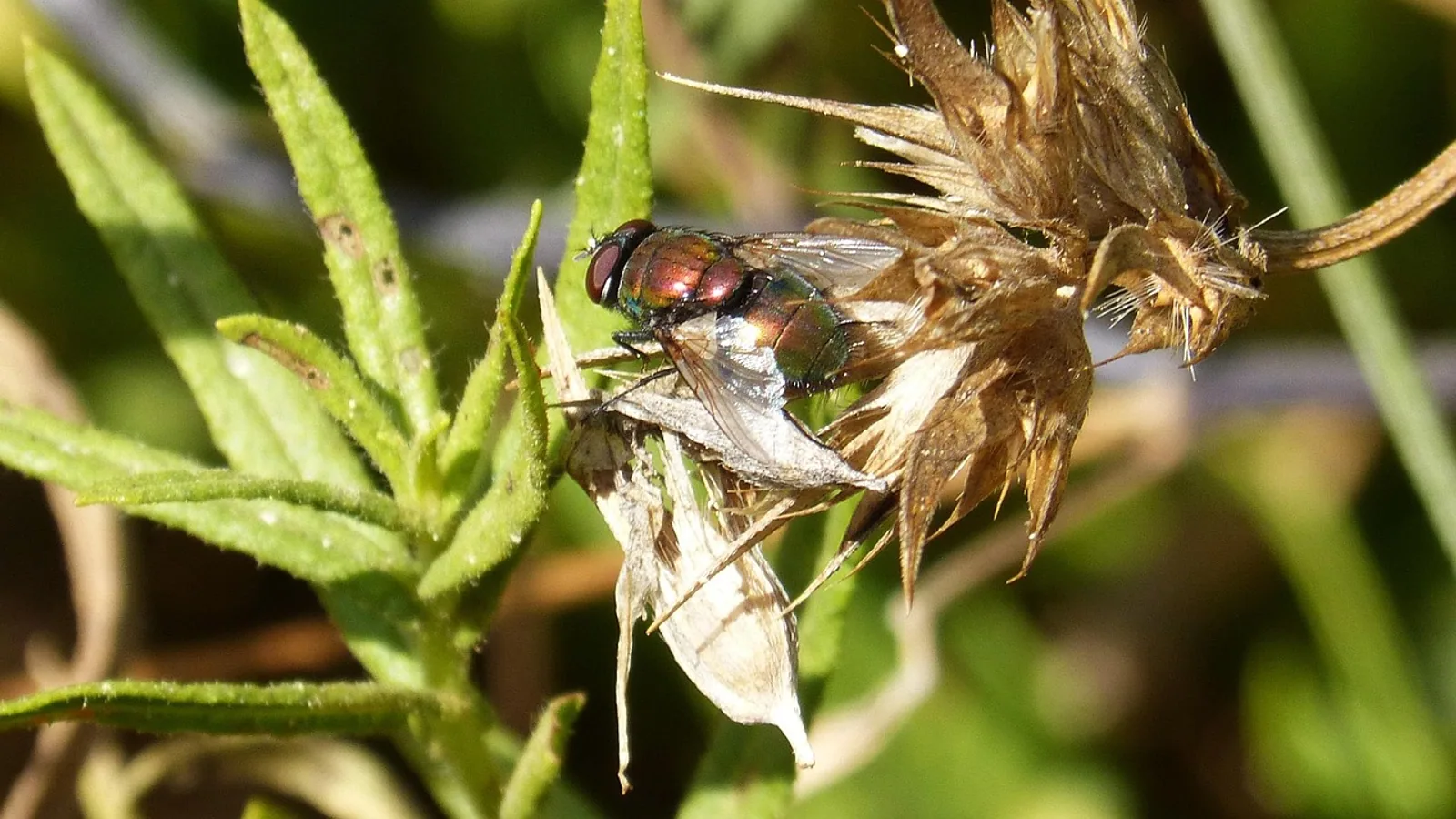
A botfly in a dog most often shows up as a firm, round swelling under the skin with a small dark hole in the center. That hole is the larva's breathing pore, and the swelling is a warble. The parasite is a Cuterebra larva, a botfly maggot that develops inside a pocket under the skin of dogs and cats. The lump can look like an abscess, a tumor, or a foreign body reaction, so a veterinary examination matters. Do not squeeze it. Pressure can rupture the larva inside your dog, release its contents into the tissues, and trigger a severe allergic reaction. The standard treatment is removal by a veterinarian under local or general anesthesia, flushing of the wound, and antibiotics when the site is infected or contaminated [1][2][3].
This article explains how to recognize cuterebriasis, why home extraction is risky, how veterinarians diagnose and treat it, and which signs mean your dog needs urgent care.
This article is educational and is not a substitute for veterinary diagnosis or treatment.
At a Glance: What Owners Need to Know
| Question | Short answer |
|---|---|
| What is it? | Furuncular myiasis caused by a Cuterebra botfly larva living under the skin |
| Typical look | Firm subcutaneous swelling with a central breathing pore, often with crust or discharge |
| Common locations | Head, neck, trunk, and sometimes the eye or nasal area [1][4] |
| Who is at risk | Dogs and cats with outdoor access in North America, especially in late summer and fall [5][2] |
| Do not do this | Never squeeze, cut, or pull the larva out at home |
| Why not | Larval rupture can cause anaphylaxis and leave fragments behind |
| Standard treatment | Veterinary removal under local or general anesthesia, wound flushing, antibiotics if needed [1][2] |
| Prognosis | Most skin cases resolve well after removal, but secondary infection is common |
| Emergency signs | Facial swelling, hives, collapse, difficulty breathing, seizures, or a suddenly quiet dog |
What Is a Botfly in a Dog?
A botfly is a fly in the family Oestridae. The genus Cuterebra includes the botflies that most often infest dogs and cats in North America. Adult flies look like large, heavy-bodied bees. They do not bite or sting. The female fly lays eggs near the openings of rodent burrows, along trails, or on vegetation. When a dog or cat passes by, the eggs hatch and the tiny first-stage larvae attach to the coat or skin. They then enter the body through the nose, mouth, or a small break in the skin [2][6].
Once inside, the larva migrates. In the most common form of infestation, it settles in a pocket under the skin and grows there. This produces the classic warble, a swelling with a central pore that the larva uses to breathe. The scientific term for this pattern is furuncular myiasis. The word furuncular refers to a boil-like nodule. Myiasis means infestation of living tissue by fly larvae [7][2].
Cuterebra larvae are host-specific in their normal life cycle. Different species develop in rodents, rabbits, and other small mammals. Dogs and cats are accidental hosts. The larva cannot complete its life cycle in a dog, but it can still cause significant local and sometimes systemic disease while it survives [8][2].
Why Botfly Larvae Target Dogs
The adult fly does not hunt dogs. Infestation happens when a dog enters an environment where eggs or larvae are waiting. Dogs that roam wooded areas, fields, farms, or yards near rodent burrows face higher exposure. Cats that hunt or spend time outdoors face the same risk, which is why cat botfly cases follow a similar seasonal pattern [5][2].
Cuterebra activity in North America is strongly seasonal. Most clinical cases appear from mid-summer through early fall. The larvae develop over weeks, so a lump found in September may have started in July. In cats with central nervous system disease, cases cluster from July through September [5].
Body size appears to influence how sick a dog becomes. In a retrospective study of 42 dogs and cats with Cuterebra infection, most of the affected dogs weighed 10 pounds or less. Yorkshire Terriers made up 40 percent of the dogs in that series. Small-breed dogs were also more likely to develop severe systemic illness, and all six dogs that died weighed 10 pounds or less and had systemic inflammatory response syndrome, disseminated intravascular coagulation, or both [9]. That finding does not mean large dogs are safe. It means small dogs may decompensate faster.
How Cuterebra Larvae Cause Disease
The larva causes disease in three main ways.
Local skin disease
The larva creates a subcutaneous cavity. The skin over it thickens and forms a nodule. The central pore drains a small amount of serum or pus. The site is often painful and itchy. Secondary bacterial infection is common because the pore is open to the environment and the larva's activity damages local tissue [7][2].
Aberrant migration
Sometimes the larva does not stop in the skin. It can migrate through the eye, the nasal cavity, the brain, or the spinal cord. These are called aberrant migrations because they occur in locations where the larva cannot develop. Ophthalmomyiasis, or fly larva infestation of the eye, has been reported in dogs and cats. In one dog, a Cuterebra larva was found embedded in the iris, with uveitis and retinal migration tracks. The larva was removed surgically and the dog remained visual [10]. In another dog, a larva was removed from the vitreous by vitrectomy, also with preservation of vision [11]. A puppy had a larva anchored at the lateral canthus of the eye, and removal under general anesthesia allowed the eyelid to heal normally [1].
Central nervous system disease
Cuterebra larvae can enter the brain or meninges. This is uncommon but serious. Reported signs include seizures, blindness, abnormal mentation, circling, head tilt, tremors, and progressive weakness [12][5][13][14][15]. In cats, central nervous system cuterebriasis often follows a period of upper respiratory signs, which suggests larvae can migrate from the nasal cavity toward the brain [5]. In dogs, cerebral cuterebrosis has been confirmed at necropsy after rapid neurologic decline [13][15]. One dog survived with medical management after a presumed parasitic tract was seen on magnetic resonance imaging and a larva was found in the vomitus [14]. Another dog with presumed central nervous system migration and concurrent protein-losing nephropathy improved after treatment with ivermectin, antihistamines, anticonvulsants, and tapering glucocorticoids [16].
Systemic inflammation can develop even when the larva stays in the skin. In the 42-case series, half of the dogs had systemic inflammatory response syndrome at initial evaluation, and 40 percent had disseminated intravascular coagulation. The overall mortality rate was 17 percent, but it was 30 percent in dogs compared with 4.5 percent in cats [9]. These numbers come from referral and necropsy cases, so they likely overstate the risk for a typical uncomplicated warble. They do show that cuterebriasis is not always a minor skin problem.
Recognizing a Botfly on a Dog: Signs and Appearance
The classic presentation is a single swelling, though some animals have more than one. In a group of zoo animals with Cuterebra infestation, only one animal had multiple lesions [17]. In dogs with Dermatobia hominis, a related botfly that causes furuncular myiasis in Latin America, lesions can be single or multiple, with each nodule containing one larva [7].
What the lesion looks like
- A firm, raised nodule under the skin, often 1 to 3 centimeters across
- A small central pore, sometimes dark or crusted
- A thin discharge of serum or pus from the pore
- Hair loss over the swelling
- Redness and warmth of the surrounding skin
- Pain when the area is touched
- Itching, rubbing, or licking at the site
Owners sometimes see movement inside the swelling or notice a small gray or dark structure at the pore. That structure is the larva. It has rows of black spines that help it anchor in the cavity [13].
Where lesions appear
Cuterebra warbles are most often found on the head, neck, and trunk. In zoo animals, lesions were on the thorax or the lateral aspect of a hind leg [17]. In dogs with eye involvement, the larva was at the canthus or within the globe [10][1][11]. Nasal infestation can cause sneezing, nasal discharge, or a visible larva in the nostril [4].
Signs that the problem is deeper
Some signs point away from a simple skin warble and toward migration into the eye, nose, or nervous system:
- Sudden blindness or a cloudy eye
- One-sided facial swelling
- Persistent sneezing or bloody nasal discharge
- Seizures, tremors, or chewing motions
- Circling, head tilt, or loss of balance
- Depression, stupor, or confusion
- Weakness or paralysis
A dog with any of these signs needs immediate veterinary assessment. Central nervous system cuterebriasis can progress quickly [5][13].
Why You Should Never Squeeze a Botfly Larva
The single most important piece of advice for a botfly on a dog is simple. Do not squeeze it.
Owners often assume a warble is a boil or an abscess and try to pop it. That is dangerous for several reasons.
- Larval rupture. The larva is a living organism with a fragile body. Pressure can tear it open inside the tissue. Its contents, including proteins and bacteria, spill into the surrounding skin and bloodstream.
- Anaphylaxis. A ruptured larva can trigger a severe allergic reaction. Signs include facial swelling, hives, vomiting, difficulty breathing, and collapse. This is a medical emergency.
- Retained fragments. If the larva breaks apart, pieces can stay in the cavity. Retained fragments cause ongoing inflammation, infection, and delayed healing.
- Secondary infection. Squeezing pushes bacteria deeper into the wound and can turn a contained lesion into a spreading infection.
- Incomplete removal. Even if the larva comes out, the cavity may still need flushing and the larva's anchoring spines can leave tissue damage behind.
Veterinarians sometimes remove larvae by applying controlled pressure after the larva is no longer viable, but that is done with sedation, monitoring, and the ability to manage a reaction [7]. At home, you do not have those safeguards.
There is also a practical problem. A larva that is disturbed can retreat deeper into the cavity, making removal harder. The breathing pore may close over, and the swelling can become more painful.
What Not to Do at Home
Home remedies for botfly larvae range from ineffective to harmful. Avoid all of the following:
- Squeezing, pinching, or pressing the swelling
- Cutting the skin open with a knife, scissors, or needle
- Pulling the larva with tweezers or forceps
- Applying petroleum jelly, oil, or ointment to smother the larva
- Pouring alcohol, hydrogen peroxide, or other irritants into the pore
- Covering the pore with a tight bandage
- Giving human medications or leftover pet medications
Some of these methods appear in older folklore or online forums. None of them replace veterinary removal. Smothering agents can kill the larva, but a dead larva still has to come out, and it may disintegrate during removal. That leaves fragments behind. The safest path is to keep your dog calm, prevent licking or scratching, and call your veterinarian.
If you see a larva protruding from the skin, do not grab it. Cover the area loosely with a clean cloth and go to the clinic.
How Veterinarians Diagnose Cuterebra Infestation
Diagnosis is often straightforward when the classic pore is visible. The veterinarian will examine the swelling, look for the central opening, and may see larval movement or a small portion of the larva at the pore [7][2].
Physical examination
The veterinarian will:
- Palpate the swelling gently to assess size, depth, and mobility
- Look for a central pore and any discharge
- Check for additional lesions elsewhere on the body
- Examine the eyes, nose, and mouth for larvae
- Assess your dog's overall condition, including temperature, heart rate, and mentation
- Look for signs of systemic inflammation such as lethargy, poor perfusion, or abnormal bleeding
Diagnostic testing
Testing depends on the presentation. For a simple skin warble, no laboratory testing may be needed before removal. If your dog is systemically ill, the veterinarian may recommend:
- Complete blood count and chemistry panel
- Coagulation testing if bleeding or bruising is present
- Imaging such as ultrasound, magnetic resonance imaging, or computed tomography for suspected deep or nervous system migration
- Cerebrospinal fluid analysis for suspected central nervous system disease
In dogs with central nervous system signs, magnetic resonance imaging has shown tubular tracts consistent with larval migration [14][16]. Cerebrospinal fluid analysis may show inflammation, but results are not consistently diagnostic [5]. In one cat with posterior segment ophthalmomyiasis, the larva was identified by histopathology after euthanasia [18]. In a dog with an intraocular larva, the organism was confirmed as Cuterebra after surgical extraction [10].
Larval identification
Definitive species identification usually requires the larva itself. Morphology can confirm the subfamily, and DNA sequencing of mitochondrial genes can identify the species [13][8]. This level of testing is mainly for surveillance and research. It does not change treatment for a routine skin case.
Differential Diagnoses: What Else Looks Like a Warble
A swelling with a pore has a short list of look-alikes. The table below compares the most common possibilities.
| Condition | Typical appearance | Key difference from Cuterebra |
|---|---|---|
| Cutaneous abscess | Painful, warm, fluctuant swelling | No true breathing pore. Pus drains when lanced. Often follows a bite or wound |
| Foreign body reaction | Firm nodule, sometimes with a draining tract | No larva. May follow a plant awn, splinter, or suture |
| Neoplasia | Firm or irregular mass, slow or rapid growth | No pore unless ulcerated. Does not move or drain like a warble |
| Other myiasis flies | Wound infestation, nasal infestation, or eye infestation | Different fly species and life cycle. Oestrus ovis can cause nasal botfly signs [19] |
| Cyst or seroma | Soft to firm fluid-filled swelling | No pore, no larva, usually nonpainful |
| Insect bite or sting | Acute swelling, redness, itching | Rapid onset, no pore, resolves with supportive care |
The distinction matters because treatment differs. An abscess needs drainage and antibiotics. A tumor needs staging and possibly surgery. A warble needs careful larval removal.
Other botfly species can also affect dogs. Oestrus ovis, the sheep nasal botfly, has been reported in a dog that expelled a mature larva after violent sneezing [19]. Dermatobia hominis causes furuncular myiasis in dogs in Latin America and produces similar nodules [7]. In North America, Cuterebra is the main concern.
Veterinary Treatment: Removal, Flushing, and Aftercare
Treatment has three goals: remove the larva completely, clean the cavity, and prevent or treat infection.
Anesthesia and removal
Most veterinarians remove Cuterebra larvae under local or general anesthesia. Anesthesia allows a controlled approach, reduces pain, and protects the airway if the larva ruptures. The veterinarian widens the pore or makes a small incision, then removes the larva intact. In some cases, the larva can be extracted through the existing pore with gentle pressure or forceps [1][2].
For eye involvement, removal may require specialized surgery. A puppy with a larva at the lateral canthus had manual removal under general anesthesia and repair of the eyelid margin [1]. A dog with an intraocular larva underwent keratotomy and extraction, with an uneventful recovery [10]. Another dog had a larva removed from the vitreous by pars plana vitrectomy after photocoagulation failed [11]. These are referral-level procedures.
For nasal infestation, endoscopic removal has been described in a puppy [4]. This approach avoids open surgery and allows direct visualization.
Wound flushing and debridement
After removal, the veterinarian flushes the cavity to remove debris, larval fragments, and bacteria. Flushing also helps assess whether the wound is clean or infected. Dead tissue may be debrided. The wound is usually left open to drain or closed loosely, depending on its size and location.
Antibiotics and anti-inflammatory treatment
Antibiotics are commonly prescribed because secondary infection is frequent. In the puppy with an eyelid larva, systemic and topical antibiotics plus systemic anti-inflammatory treatment were used, and the lesion healed completely within 10 days [1]. Antibiotic choice depends on the wound and your dog's history. Never use leftover antibiotics from a previous illness.
Anti-inflammatory medication can reduce swelling and pain. If your dog has systemic signs, more intensive care may be needed. The 42-case series found that some dogs developed systemic inflammatory response syndrome and disseminated intravascular coagulation, which require hospitalization and aggressive management [9].
Follow-up
Recheck timing depends on the wound. A simple warble may need one recheck in 7 to 14 days to confirm healing. A larger or infected wound may need more frequent visits. If your dog had systemic illness, follow-up blood work may be needed to confirm that inflammation has resolved.
Medications That Kill the Larva Before Removal
In some cases, veterinarians use medication to kill the larva before extraction. This can reduce inflammation and discomfort and make removal less traumatic [7]. Isoxazolines are a class of ectoparasiticides with larvicidal activity. In a study of 10 dogs with Dermatobia hominis infestation, sarolaner killed all 75 larvae retrieved from the dogs, with no live larvae observed 24 hours after treatment [7].
This approach does not replace removal. A dead larva still has to come out. The advantage is that a nonviable larva is less likely to resist extraction and less likely to release irritating contents if it ruptures. Your veterinarian will decide whether this step is appropriate for your dog.
Do not give your dog an isoxazoline or any other antiparasitic medication for a suspected warble without veterinary guidance. The dose, product, and timing matter, and some products are not appropriate for every dog.
Prognosis for Dogs with Cuterebra Infestation
For uncomplicated skin warbles, the prognosis is good after complete removal and wound care. Most lesions heal within one to two weeks. Secondary infection is common but treatable [1][2].
The prognosis is more guarded when the larva migrates. Central nervous system cuterebriasis can be fatal. In the 42-case series, the overall mortality rate was 17 percent, with a higher rate in dogs than cats [9]. Some dogs survive with medical management, including anti-inflammatory drugs and antiparasitic treatment [14][16]. Others deteriorate rapidly despite care [13].
Eye involvement can be treated successfully with surgery in some cases, with preservation of vision [10][11]. The outcome depends on how much damage occurred before removal.
Preventing Botfly Infestation in Dogs and Cats
Prevention focuses on reducing exposure to adult flies and their eggs.
- Keep dogs on a leash or in a secure yard during peak fly season.
- Avoid walking dogs through tall grass, brush, and areas with visible rodent burrows.
- Check your dog's coat and skin weekly during summer and fall, especially the head, neck, and trunk.
- Groom regularly to find small lumps early.
- Ask your veterinarian about year-round parasite prevention, including products with activity against fly larvae.
- Keep cats indoors if they have a history of cuterebriasis or if you live in a high-risk area.
No prevention method is perfect. Adult flies are fast and eggs are small. Early detection is the best backup.
Emergency Red Flags
Call your veterinarian immediately or go to an emergency clinic if your dog has any of these signs:
- Facial swelling, hives, or swelling around the eyes or muzzle
- Difficulty breathing, noisy breathing, or open-mouth breathing
- Collapse, weakness, or inability to stand
- Vomiting, diarrhea, or excessive drooling after touching the swelling
- Seizures, tremors, or abnormal chewing movements
- Sudden blindness or a bulging eye
- A swelling that bursts and bleeds heavily
- A larva that is partially out and cannot be removed gently
- Lethargy, confusion, or a body temperature that feels very high or very low
These signs can indicate anaphylaxis, systemic inflammation, or nervous system involvement. They are not wait-and-see problems.
Limitations and When to Contact a Veterinarian
This article provides general information about Cuterebra infestation in dogs. It cannot replace an examination by a veterinarian who can see your dog, assess the lesion, and decide on the right treatment.
Contact a veterinarian if:
- You see a swelling with a central pore anywhere on your dog
- Your dog has more than one lump
- The lump is growing, draining, or painful
- Your dog is licking, chewing, or scratching at the site
- Your dog seems tired, off food, or uncomfortable
- You are unsure whether the lump is a warble, an abscess, or something else
- You have already tried to remove the larva and the area is now bleeding, swollen, or worsening
If your dog has neurologic signs, eye signs, or breathing difficulty, seek emergency care. Do not wait for a scheduled appointment.
Frequently Asked Questions
What does a botfly look like on a dog?
A botfly larva under the skin looks like a firm lump with a small central hole. The hole is the larva's breathing pore. You may see a thin discharge, crusting, or movement inside the swelling.
Can I remove a botfly larva from my dog at home?
No. Home removal risks rupturing the larva, which can cause a severe allergic reaction and leave fragments behind. A veterinarian should remove it under local or general anesthesia.
Is a botfly on a dog contagious to other pets or people?
Cuterebra infestation is not spread directly from dog to dog or dog to person. The larva comes from eggs laid by an adult fly in the environment. Other pets in the same area could be exposed separately.
How do veterinarians treat a botfly in a dog?
The veterinarian removes the larva under anesthesia, flushes the wound, and prescribes antibiotics if the site is infected. Anti-inflammatory medication may be used to reduce swelling and pain.
What happens if a botfly larva is left untreated?
Some skin warbles resolve on their own when the larva exits, but secondary infection is common. Larvae that migrate to the eye, nose, or nervous system can cause serious and sometimes fatal disease.
Can a botfly larva make a dog seriously ill?
Yes. Cuterebra infection can cause systemic inflammatory response syndrome and disseminated intravascular coagulation, especially in small dogs. Central nervous system migration can be fatal.
Do cats get botflies too?
Yes. Cats can develop skin warbles and central nervous system cuterebriasis. Outdoor cats are at highest risk, especially in late summer and early fall.
How long does it take for a botfly wound to heal?
Most uncomplicated wounds heal within one to two weeks after removal and appropriate care. Larger or infected wounds may take longer and need follow-up visits.
flowchart TD
    A[Lump with central pore] --> B{Is the dog stable}
    B -->|No| C[Go to emergency clinic]
    B -->|Yes| D[Do not squeeze or cut]
    D --> E[Call your veterinarian]
    E --> F[Examination and diagnosis]
    F --> G{Larva confirmed}
    G -->|No| H[Evaluate other causes]
    G -->|Yes| I[Remove under anesthesia]
    I --> J[Flush wound]
    J --> K[Antibiotics if infected]
    K --> L[Recheck and monitor healing]
Related Articles
- Feline Hyperthyroidism: Signs, Diagnosis, and Treatment
- Dog Ear Infection Signs Treatment
- Sugar Glider Dental Disease: Signs and Treatment
- Chinchilla Respiratory Infections: Causes, Signs, and Treatment
- Cat Skin Infection: Signs and Treatment Options
- Guinea Pig Respiratory Infections: Causes, Signs, and Treatment
- Liver Worm in Dogs and Cats: Signs and Treatment
- Cat Anal Gland Issues: Signs and Treatment
- Dog Eyelid Infection: Causes, Signs, and Treatment
Sources
- Ophthalmomyiasis externa in a puppy due to Cuterebra infestation.
- Cuterebra Infestation in Small Animals - Integumentary System - Merck Veterinary Manual
- Cuterebra or 'Warbles' in Dogs - VCA Animal Hospitals
- Case report: Endoscopic removal of a nasal Cuterebra larva from a puppy.
- Clinical and clinicopathologic features in 11 cats with Cuterebra larvae myiasis of the central nervous system.
- Cuterebriasis in Dogs and Cats - Today's Veterinary Practice
- Effectiveness of sarolaner in the clinical management of furuncular myiasis in dogs naturally infested with Dermatobia hominis (Diptera: Cuterebridae).
- Molecular identification of two species of myiasis-causing Cuterebra by multiplex PCR and RFLP.
- Systemic manifestations of Cuterebra infection in dogs and cats: 42 cases (2000-2014).
- Ophthalmomyiasis interna anterior in a dog: keratotomy and extraction of a Cuterebra sp. larva.
- Pars plana vitrectomy for the treatment of ophthalmomyiasis interna posterior in a dog.
- Cuterebra encephalitis in a dog.
- Fatal cerebral cuterebrosis in a dog.
- Survival of a suspected case of central nervous system cuterebrosis in a dog: clinical and magnetic resonance imaging findings.
- Cerebral cuterebrosis in a dog.
- Presumptive central nervous system cuterebriasis and concurrent protein-losing nephropathy in a dog.
- Cuterebra myiasis in a group of red kangaroos (Megaleia rufa), a Bennett's wallaby (Macropus rufogriseus fruticus), and a Gunther's dik dik (Maloqua guentheri smithi).
- Ophthalmomyiasis (interna posterior) of the posterior segment and central nervous system myiasis: Cuterebra spp. in a cat.
- Oestrus ovis infestation of a dog in the UK.