Developmental Orthopedic Disease in Pets
By Dr. Zubair Khalid, DVM, MS, PhD ·
A young dog that limps is not simply going through a phase. Developmental orthopedic disease is the umbrella term for a group of skeletal and joint conditions that appear while an animal is still growing. These disorders are a common cause of pain and lameness in young dogs, and most of them occur in large-breed dogs with rapid growth rates [1]. The practical consequence for an owner is direct. A puppy with a persistent limp, a stiff gait, or a swollen limb needs a veterinary examination rather than a wait-and-see approach.
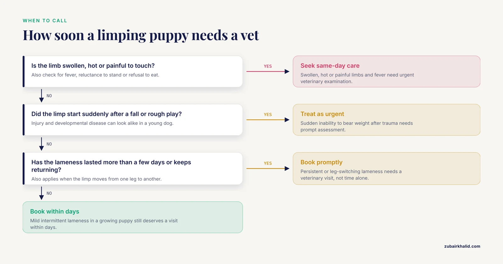
Owner triage summary
- Mild, intermittent lameness in a growing puppy: book a visit within days, not weeks.
- Lameness that lasts more than a few days, recurs, or shifts between legs: book promptly.
- A swollen, hot, or painful limb, reluctance to stand, fever, or poor appetite: seek same-day care.
- Sudden inability to bear weight after a fall or rough play: treat as urgent, because injury and developmental disease can look alike in a young dog.
This article is educational and is not a substitute for veterinary diagnosis or treatment.
At a Glance: The Major Developmental Orthopedic Diseases
| Condition | Typical patient | Joints or bones usually involved | What owners notice | First clinical step |
|---|---|---|---|---|
| Osteochondrosis and osteochondritis dissecans | Immature, rapidly growing large-breed dogs | Shoulder, elbow, stifle and tarsus are the joints most often affected [2] | Lameness and pain that may be subtle at first [2] | Orthopedic examination plus imaging of the affected joint |
| Hip dysplasia | Young dogs across many breeds, heaviest burden in large breeds | Coxofemoral joint | Hind-limb stiffness, reluctance to rise, weak push-off | Hip laxity assessment and pelvic radiographs [3] |
| Elbow dysplasia and medial coronoid disease | Large-breed adolescents | Elbow | Forelimb lameness, often worse after activity | CT and referral for arthroscopy [4] |
| Hypertrophic osteodystrophy | Growing large and giant breed puppies | Metaphyses of long bones | Painful, swollen limb segments, reluctance to walk | Same-day veterinary examination [1] |
| Panosteitis | Growing large-breed dogs | Long bones | Lameness that shifts from one leg to another | Examination and radiographs [1] |
| Legg-Calve-Perthes disease | Young dogs | Hip | Progressive hind-limb lameness | Pelvic radiographs [1] |
| Pes varus and other angular deformities | Growing dogs | Hind limb | Visibly crooked limb, abnormal stance | Orthopedic assessment [1] |
What Developmental Orthopedic Disease Covers
Developmental orthopedic disease is not one diagnosis. A standard clinical review groups hypertrophic osteodystrophy, panosteitis, osteochondrosis, Legg-Calve-Perthes disease, hip dysplasia, elbow dysplasia, and pes varus under the heading [1]. The conditions differ in which tissue fails, which joint is affected, and how the disease behaves over months and years. What they share is timing. They emerge during skeletal growth, while cartilage is being converted to bone and the joint surfaces are taking on adult load.
More than one can appear in the same dog. Hip dysplasia and elbow dysplasia can coexist, and each contributes independently to the osteoarthritis that tends to follow.
The Biology: Why Growing Bone Fails
Endochondral ossification and its blood supply
Long bones lengthen at the growth plate through endochondral ossification. Cartilage cells multiply and enlarge, the matrix around them calcifies, blood vessels invade, and bone replaces cartilage. The sequence is orderly, and it depends on a blood supply reaching the right place at the right time. When blood supply to a focal area of epiphyseal growth cartilage fails, the result is osteochondrosis, a focal disturbance of endochondral ossification [2].
Work in puppies identified cartilage channels crossing the epiphyseal cartilage. Where those channels disrupted ossification in the secondary center of ossification, the cartilage layer thickened, and the change was found most often at the sites where osteochondrosis typically develops, especially the head of the humerus [5]. Those channels were identified more often in male animals and in large breeds [5].
The subchondral foundation beneath the cartilage
A second line of evidence points below the joint surface. In a study of large and giant breeds, the primary lesion in osteochondrosis was an acquired pattern of osteopenic, biomechanically weak subchondral spongiosa that could not provide adequate bony support for the articular cartilage [6]. Once the cartilage loses that support, normal loading becomes excessive loading, and the chondrocytes in the developing joint surface suffer secondary disturbances in nutrition, metabolism, function, and viability [6].
The same work found that the epiphyseal spongiosa of rapidly growing dogs from larger breeds is inherently less dense and less strong per unit area than that of smaller breeds. Overnutrition exaggerates the tendency by speeding skeletal growth and cancellous bone remodeling. Great Dane puppies fed ad libitum or fed a restricted diet until 6 months of age were part of that experimental investigation [6].
Calcium, vitamin D, and the speed of growth
Growth rate changes how the body handles calcium. Great Danes and Miniature Poodles raised on the same balanced diet under identical conditions diverged sharply. The Great Danes gained 17 times more body weight, grew longer radii and ulnae, and had thicker growth plates. They also had higher plasma growth hormone and IGF-I concentrations and higher plasma 1,25-dihydroxycholecalciferol, produced by reduced clearance rather than increased production [7]. Growing giant-breed dogs are more susceptible to skeletal disorders when raised on diets with deficient or excessive calcium content [7].
Put together, these findings describe a puppy whose skeleton is being built fast, on a scaffold that is temporarily less dense than adult bone, while mechanical load is rising every week. Small errors in supply or timing have more room to cause damage at that stage than at any later point in life.
The Conditions in Detail
Osteochondrosis and Osteochondritis Dissecans
Osteochondrosis most often affects the shoulder joint, followed by the elbow, tarsal, and stifle joints [2]. A population study of insured Swedish dogs covering just over 600,000 dogs identified 685 with appendicular osteochondrosis and calculated an incidence rate of 3.77 cases per 10,000 dog-years at risk. Stifle osteochondrosis was more common than tarsal osteochondrosis in that population [2]. Prognosis varies by joint [2].
Tarsocrural lesions in Labrador Retrievers show measurable change in the bone beneath them. CT osteoabsorptiometry found significantly lower subchondral bone density at the lesion site, particularly on the medial trochlear ridge, with a higher-density rim surrounding the low-density area. The opposite joint showed higher overall mineral density, which points to altered loading across the two limbs [8]. The lower density in the affected joint suggests abnormal bone turnover, which may contribute to secondary osteoarthritis later [8].
Hip Dysplasia
Canine hip dysplasia is the most common orthopedic developmental condition in dogs, and early hip laxity is its main risk factor [3]. The condition is regarded as a pre-osteoarthritis disease. Left untreated, it progresses to osteoarthritis, and in dogs that progression can occur within one to two years of age, much faster than in humans with the comparable human diagnosis [9].
Prevalence is not uniform across breeds. A French screening program covering 27,710 dogs from 10 breeds between 1997 and 2017 found prevalence ranging from 5% in the Siberian Husky to 51.9% in the Cane Corso. Prevalence decreased over time in six breeds, with a significant decrease in four of them [10].
Laxity is the target of early detection. Passive hip laxity can be measured with distraction radiography, and a comparison of two certified distraction devices across 104 dogs found close agreement between them [3].
Genetics is being mapped. A model combining genome-wide association and candidate gene data using seven single nucleotide polymorphisms produced an area under the ROC curve of 0.85 for hip dysplasia in Labrador Retrievers and was validated in an independent group of 114 dogs [11]. A breed-specific tool of this kind supports individualized decisions, and it does not replace the physical examination.
Elbow Dysplasia and Medial Coronoid Disease
Elbow dysplasia is a common cause of forelimb lameness in young large-breed dogs [1]. The medial coronoid process is a frequent site of disease. Micro-computed tomography of excised coronoid segments from 21 affected dogs identified fissure and fracture patterns in three orientations and revealed subchondral micro-fracturing associated with fragmentation. Canalicular impaction was associated with a higher trabecular number and reduced trabecular space, most notably in the radial incisure category [12].
In a prospective series of 15 dogs with elbow dysplasia lameness, CT was consistent with medial coronoid disease and a fissured or fragmented medial coronoid process in every dog. Median age at presentation was 14 months and median body weight was 31.6 kg [4]. These are adolescent dogs, not geriatric ones, and that timing is a clue in itself.
Hypertrophic Osteodystrophy and Panosteitis
Hypertrophic osteodystrophy and panosteitis complete the list in the standard clinical review [1]. Both cause pain and lameness in growing dogs. Both are diagnoses a veterinarian reaches by combining history, physical examination, and imaging, and both are managed differently from the joint-centered diseases described above. An accurate label changes the plan, which is why the workup matters even when the limp looks minor.
Legg-Calve-Perthes Disease
Legg-Calve-Perthes disease is a hip disorder grouped with the developmental orthopedic diseases [1]. It produces lameness in a young dog and is diagnosed from history, examination, and pelvic imaging. It belongs on the differential list whenever a young dog has a hip that is painful to manipulate.
Pes Varus and Angular Limb Deformity
Pes varus is an angular deformity of the hind limb that appears in the same clinical family [1]. The limb deviates and the stance changes. Correction, when it is needed, is a surgical decision made after an orthopedic assessment, and the timing of that decision is part of the assessment.
Cats and Other Growing Pets
The conditions described here are recognized mainly in dogs, and the standard clinical review is drawn from canine practice [1]. Cats can develop orthopedic problems during growth, and a lame kitten deserves a full examination rather than an assumption that it will outgrow the problem. The diagnostic pathway in a cat follows the same logic: locate the source of pain, image it, and treat what is found.
Risk Factors Owners Can and Cannot Change
Breed and growth rate
Most developmental orthopedic diseases occur in large-breed dogs with rapid growth rates [1]. Rapid growth is not a fault in itself. It is a period of vulnerability, and the larger the breed, the narrower the margin for nutritional or mechanical error, [6].
Genetics
Hip dysplasia has a multifactorial mode of inheritance, with multiple gene effects influenced by environmental factors [13]. Heritability estimates from a large United States registry were 0.22 for hip score and 0.17 for elbow score, with a weak genetic correlation of 0.12 between the two [14]. A weak correlation means a dog with excellent hips is not automatically a low-risk producer for elbows.
Sex
Cartilage channels that disturb ossification were found more often in male animals in the puppy study [5]. Sex is a small factor compared with breed, growth rate, and genetics, and it does not change the diagnostic approach.
Nutrition and body condition
Overnutrition that accelerates skeletal growth and remodeling is the mechanism most clearly linked to osteochondrosis in large and giant breeds [6]. Dietary calcium outside the balanced range is a documented risk in growing giant breeds [7]. Diet is one of the few levers an owner controls, and it has been a central focus in the discussion of how to reduce the burden of these diseases.
What Owners Notice at Home
The signs are often easier to see in motion than at rest. A puppy may be stiff after sleeping and loosen up after a few minutes. It may be reluctant to climb stairs, lag behind on walks, or sit instead of standing when it waits. Hind-limb lameness from hip dysplasia often shows as a bunny-hopping gait or a swaying rear end. Forelimb lameness from elbow disease tends to appear or worsen after activity and improve with rest.
Some conditions produce pain without obvious swelling. Panosteitis is one of them, and its signature is lameness that moves from one leg to another [1]. Others produce a limb that is visibly swollen and tender to the touch, which is a same-day problem rather than a watch-and-wait problem.
Subtle signs matter. A puppy that is simply less playful than its littermates, or that tires earlier than expected, may be limiting itself because of discomfort rather than laziness. Owners who notice a change in gait should record a short video of the dog walking and trotting from behind and from the side. That footage often shortens the diagnostic conversation.
The Veterinary Visit: Examination and Diagnostics
Physical and orthopedic examination
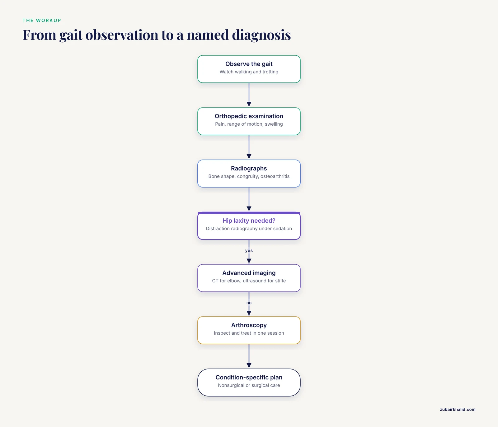
The examination starts with gait observation, then moves to hands-on assessment of each limb and joint. The veterinarian checks for pain on manipulation, range of motion, muscle symmetry, and joint swelling. A neurologic assessment is part of the picture, because spinal disease can mimic orthopedic lameness in a young dog.
Radiography and hip laxity
Standard radiographs show bone shape, joint congruity, and osteoarthritis. Hip laxity can be assessed separately with distraction radiography, which quantifies passive laxity as a distraction index and is directly relevant to early hip dysplasia screening [3]. Sedation or anesthesia is often used to get a relaxed, repeatable view.
Advanced imaging
Computed tomography has become the reference standard for elbow disease in referral practice. In the prospective series of 15 dogs, CT identified medial coronoid disease and a fissured or fragmented medial coronoid process in every case [4]. Micro-computed tomography goes further and reveals the internal fissure and fracture architecture of the coronoid process, which explains why some lesions are stable and others are not [12].
Ultrasonography is a useful, low-stress complement. It is cost-effective, non-invasive, and can be performed in conscious dogs, and it contributes diagnostic information in dogs with stifle lameness, including developmental disorders of that joint [15].
Arthroscopy
Arthroscopy allows direct inspection of the joint and, in the right cases, treatment at the same time. Small-bore needle arthroscopy has been evaluated as a way to explore the elbow and remove fragments with less surgical trauma. In the pilot series, needle arthroscopy allowed safe inspection of structures in the medial and caudal compartments before treatment [4]. A later case series of 41 affected elbows in 30 dogs treated with needle arthroscopy-assisted fragment removal reported lameness scores that fell from a median of 2 before surgery to 0 at 30 days afterward [16].
Genetic and screening tests
Breeders and owners of predisposed breeds increasingly have access to genetic risk information. The seven-marker model for Labrador Retrievers is one example [11]. Screening programs that combine radiographs with estimated breeding values remain the broader tool for population-level control [14]. A single hip score on one dog is a weaker predictor than an estimated breeding value that incorporates pedigree and relatives [13].
Evidence-Based Management
Treatment is condition-specific, which is why the veterinary workup comes before any plan. The general shape of care is the same across conditions: reduce pain, protect the joint from loads it cannot currently tolerate, preserve muscle, address the mechanical problem if it can be addressed, and monitor for progression.
Nonsurgical care
Conservative management generally combines veterinarian-prescribed pain relief, weight control, controlled exercise, and physical rehabilitation. Exercise restriction does not mean confinement to a crate for months. It means controlled, low-impact activity that keeps muscle mass without repeated high-impact loading. Swimming and leash walks are common examples, and the specific program should come from the treating veterinarian or a rehabilitation specialist.
Weight management matters at both ends of life. A lean growing puppy loads its joints less, and a lean adult with osteoarthritis carries less mechanical stress every day.
Surgical care
Surgery is considered when the mechanical problem can be corrected or when a fragment is causing pain and cartilage damage. For medial coronoid disease, needle arthroscopy-assisted fragment removal produced measurable short-term lameness improvement, and dogs still lame 30 days after surgery had higher cartilage lesion scores at the time of the operation [16]. Corrective surgery for hip dysplasia follows the same principle as surgical treatment of the comparable human condition, and outcomes in that human condition have improved substantially in recent decades [9].
The decision for or against surgery depends on the joint, the severity of the lesion, the age of the dog, the presence of osteoarthritis, and the owner's goals. It is a conversation, not a default.
Rehabilitation and long-term follow-up
Recheck timing depends on the procedure and the condition. After arthroscopic fragment removal, short-term assessment typically happens within the first month, with longer-term follow-up by owner questionnaire at a year or more [16]. Dogs managed medically are usually rechecked at intervals that track growth, weight, and comfort. The key long-term question is not whether the dog is currently lame. It is whether osteoarthritis is progressing on imaging and whether function is holding up at home.
Feeding a Growing Dog: What the Evidence Supports
The strongest dietary evidence concerns growth rate and calcium. Overnutrition that accelerates skeletal growth and remodeling is the pathway most clearly implicated in osteochondrosis in large and giant breeds [6]. Diets with deficient or excessive calcium are linked to skeletal disorders in growing giant breeds [7].
For an owner, that translates into a few practical rules.
- Feed a diet formulated for the dog's life stage and expected adult size, and feed to a body condition that keeps growth steady rather than maximal.
- Do not free-feed a large or giant breed puppy. The experimental work used ad libitum feeding as the overnutrition model [6].
- Do not add calcium or other supplements to a complete diet. Additional calcium pushes intake outside the balanced range that the diet was designed to deliver [7].
- Ask the veterinarian before changing diets, especially during a lameness workup.
Appetite and body condition are easier to monitor than growth rate. A puppy that is visibly gaining weight faster than its frame can support is a puppy to discuss with the veterinarian at the next visit.
Unsafe Home Remedies and Common Mistakes
The most common mistake is time. Owners wait for a puppy to outgrow a limp, and by the time imaging happens, cartilage damage and osteoarthritis have already had months to develop. A persistent limp is a reason to book a visit, not a reason to wait.
The second mistake is human pain medication. Never give a pet an over-the-counter or prescription pain medicine intended for people without explicit veterinary direction. Some common human analgesics are toxic to dogs and cats, and masking pain without treating the cause makes the underlying problem worse.
The third mistake is supplement stacking. Adding calcium, vitamin D, or joint products on top of a complete diet can unbalance intake, and in a growing large-breed puppy that is the wrong direction [7].
The fourth mistake is complete rest. Prolonged crate confinement weakens muscle and reduces joint support. Activity modification is a veterinary prescription, not an assumption.
Prevention: Growth Rate, Body Condition, and Breeding Choices
Prevention has two halves. The first is management of the growing puppy, which means controlled growth, balanced nutrition, and prompt attention to lameness [7], [6],. The second is genetic selection, which operates over generations rather than months.
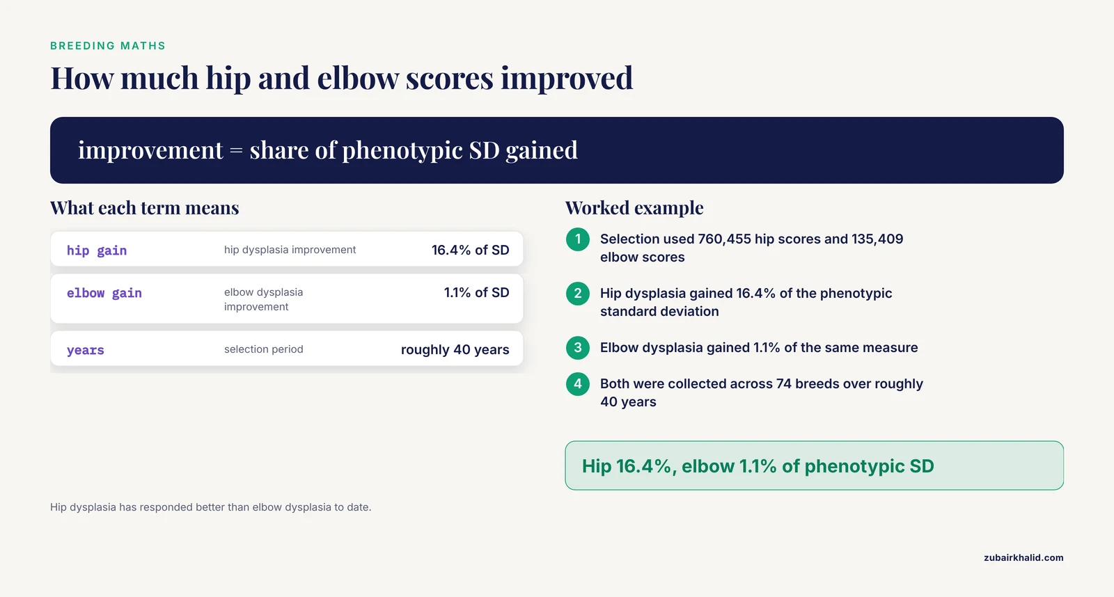
Selection based on estimated breeding values has produced measurable progress. An analysis of 1,264,422 dogs from 74 breeds, using 760,455 hip scores and 135,409 elbow scores collected over roughly 40 years, found total genetic improvement of 16.4% and 1.1% of the phenotypic standard deviation for hip and elbow dysplasia respectively [14]. Progress is real and it is slow, and hip dysplasia has responded better than elbow dysplasia to date [14].
Phenotypic screening alone has limits. The most successful implementations of traditional hip scoring have occurred where scoring is mandatory and registries are open, and several commentators have recommended that selection be based on estimated breeding values rather than on an individual dog's hip score [13]. Breeders should use both the radiograph and the pedigree information. A single excellent score on one dog is not a guarantee for its offspring.
Breed-level prevalence data are a useful context for buyers. Knowing that prevalence in a breed ranges from 5% to 51.9% depending on the breed helps an owner ask better questions about health testing.
Prognosis
Prognosis depends on the condition and the joint [2]. Some dogs do well for years with management of body weight, activity, and pain. Others develop osteoarthritis that progresses despite good care, particularly with hip dysplasia, which is considered a pre-osteoarthritis disease with a relatively fast progression timeline in dogs [9]. Subchondral bone changes around osteochondral lesions suggest that abnormal bone turnover may drive secondary osteoarthritis even after the cartilage lesion is addressed [8].
Honest expectations are part of good care. Most dogs with developmental orthopedic disease can be kept comfortable for a long time. Few are cured in the sense of returning to a completely normal joint.
Emergency Red Flags
Seek care immediately if a young pet shows any of the following.
- Sudden inability to bear weight on a limb, especially after a fall or rough play.
- A limb that is markedly swollen, hot, or painful to the touch.
- Reluctance or refusal to stand, or crying when moved.
- Fever, lethargy, or refusal to eat accompanying a limp.
- Lameness that worsens rapidly over hours rather than days.
- Any sign of weakness, wobbliness, or loss of bladder or bowel control.
Limitations and When to Contact a Veterinarian
This article describes patterns, not a diagnosis for an individual animal. Growing pets can have more than one problem at once, and the same limp can come from bone, cartilage, joint, nerve, or muscle. Only a veterinarian who examines the pet can sort that out.
Contact a veterinarian promptly if any of these apply.
- The lameness has lasted more than a few days, or it keeps returning.
- The lameness switches legs.
- The puppy is stiff after rest and slow to loosen up.
- The dog is reluctant to climb stairs, jump, or rise from lying down.
- There is visible swelling, muscle wasting, or a change in limb shape.
- The dog is limping and also losing weight, eating poorly, or acting subdued.
- A previously diagnosed developmental orthopedic disease seems to be worsening despite treatment.
For dogs with an established diagnosis, follow the recheck schedule set by the treating veterinarian. Ask directly what to watch for, what activity level is safe, and what would prompt a change in plan.
Related Guides
- Canine Hip Dysplasia Ramps: Incline Angles, Non-Slip Grip, and Car vs. Bed Accessibility: Ramps and incline angles help manage hip dysplasia at home
- Elbow Dysplasia in Cats: Symptoms and Care: Cat-specific elbow dysplasia symptoms and care guidance
- Bernese Mountain Dog Health Problems: Malignant Histiocytosis and Elbow Dysplasia: Breed-specific health concerns including elbow dysplasia
- Hip Dysplasia in Cats: Symptoms, X-rays, and Treatment Options: Hip dysplasia in cats: symptoms and treatment options
- Orthopedic Memory Foam Beds for Giant Breeds: Joint Pressure Relief, Foam Density, and Water Resistance: Memory foam beds for giant breeds with joint issues
- Are Orthopedic Dog Beds Good for Dogs?: Explains whether orthopedic beds benefit dogs with joint pain
- Saint Bernard Health Problems: Entropion Ectropion and Hip Joint Disease: Saint Bernard health issues, including hip joint disease
Frequently Asked Questions
Is developmental orthopedic disease the same as hip dysplasia?
No. Hip dysplasia is one member of a larger group that also includes osteochondrosis, elbow dysplasia, hypertrophic osteodystrophy, panosteitis, Legg-Calve-Perthes disease, and pes varus [1].
Can a puppy outgrow developmental orthopedic disease?
Some mild signs improve as growth finishes, but the underlying joint change does not always resolve. Hip dysplasia is considered a pre-osteoarthritis disease that can progress to osteoarthritis within one to two years of age in dogs [9]. A veterinarian should make the call rather than time alone.
At what age do these problems usually appear?
They appear during skeletal growth. Many dogs present in the first year or two of life, and one series of medial coronoid disease cases had a median age at presentation of 14 months [4].
Does diet cause developmental orthopedic disease?
Diet contributes rather than causes. Overnutrition that accelerates skeletal growth and calcium outside the balanced range have both been linked to osteochondrosis in large and giant breeds [6], [7].
Are these conditions limited to large breeds?
No. Most occur in large-breed dogs with rapid growth rates [1], but small dogs can be affected, and hip dysplasia prevalence varies widely by breed [10].
Does every dog with developmental orthopedic disease need surgery?
No. Treatment depends on the condition, the joint, and the severity. Some dogs are managed with activity control, weight management, and pain relief, while others benefit from fragment removal or corrective surgery [16].
Can a breeder reduce the risk in a line of dogs?
Yes, over time. Selection based on estimated breeding values has produced measurable genetic improvement in hip and elbow scores across four decades [14].
Will my dog develop arthritis?
Often, yes. Abnormal joint loading and cartilage damage can lead to secondary osteoarthritis, and hip dysplasia is regarded as a pre-osteoarthritis condition [9], [8]. Early diagnosis and good management give the best chance of slowing that progression.
Sources
- Developmental orthopedic disease.
- The epidemiology of osteochondrosis in an insured Swedish dog population.
- Comparison of Two Distraction Devices for Assessment of Passive Hip Laxity in Dogs.
- Evaluation of a small-bore needle arthroscope for diagnosis and treatment of medial coronoid disease in dogs: a pilot study with short-term assessment.
- [[Histological study of cartilage channels in the epiphyseal cartilage of young dogs and their relationship to that of osteochondrosis dissecans in the most frequently affected locations].](https://pubmed.ncbi.nlm.nih.gov/8964237/)
- Relationship between nutrition and bone growth in large and giant dogs.
- Hormonal regulation of calcium homeostasis in two breeds of dogs during growth at different rates.
- Subchondral bone density changes of the talus in dogs with tarsocrural osteochondrosis.
- Canine hip dysplasia: A natural animal model for human developmental dysplasia of the hip.
- Prevalence of canine hip dysplasia in 10 breeds in France, a retrospective study of the 1997-2017 radiographic screening period.
- A genetic predictive model for canine hip dysplasia: integration of Genome Wide Association Study (GWAS) and candidate gene approaches.
- Micro-CT Structural Analysis of the Canine Medial Coronoid Disease.
- Canine hip dysplasia: phenotypic scoring and the role of estimated breeding value analysis.
- Monitoring Hip and Elbow Dysplasia achieved modest genetic improvement of 74 dog breeds over 40 years in USA.
- [[Ultrasonographic examination of the stifle joint in the dog. Part 2: Impaired wound healing and neoplastic, congenital, developmental, degenerative and traumatic disorders].](https://pubmed.ncbi.nlm.nih.gov/25966794/)
- Short- and long-term outcomes after small-bore needle arthroscopic removal of fragmented portions of the medial coronoid process for treatment of dogs with medial coronoid process disease.