Foxtail Plant: Why It's Dangerous for Pets and Livestock
By Dr. Zubair Khalid, DVM, MS, PhD ·
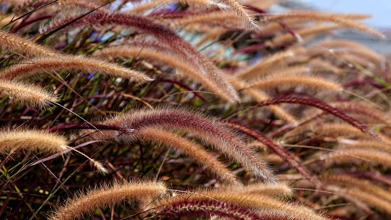
The foxtail plant is dangerous because its seed head carries barbed awns that drive forward into tissue and cannot back out. Each awn is covered in microscopic barbs that all point backward, toward the base of the seed, so every movement of the animal pushes the awn deeper and no movement pulls it free.
Grass awn migration is not a rare curiosity. In a one-year retrospective study of dogs and cats at a veterinary teaching hospital, grass awn problems accounted for 61 percent of all foreign body cases, and the external ear canal was the single most common site, involving 51 percent of affected animals [1]. Awns have been recovered from the brain, the spinal canal, the pericardium, the peritoneal cavity and the urinary bladder of dogs and cats [2][3][4][5]. For livestock, the same barbed seed heads lodge in the mouth, the interdigital cleft and the eye, and awns swept into baled hay are the classic route into the tissues of cattle, sheep and horses.
This article is educational and is not a substitute for veterinary diagnosis or treatment.
What Is a Foxtail Plant?
A foxtail is any grass whose seed head resembles the bushy tail of a fox. That common name covers several genera, and not all of them behave the same way. The veterinary literature most often names three groups:
- Hordeum species (barley grasses, including mouse barley, Hordeum murinum, and foxtail barley, Hordeum jubatum). These are the classic migrating awns of dogs, and they have been typed directly in brain lesions of affected animals [6].
- Setaria species (foxtails in the strict botanical sense, including yellow foxtail and green foxtail). These are common in lawns, pastures and disturbed ground, and their bristly seed heads are a frequent source of oral and interdigital injury in livestock and pets.
- Bromus species (brome grasses). These are tall pasture and roadside grasses whose awns vary from short to long depending on species.
A fourth grass, wild oat (Avena species), produces awns that migrate in soft tissue of dogs and behaves in the same mechanical way as the true foxtails [7]. Wild oat awns were the most common grass awn found in the soft tissue of dogs in one ultrasound case series [7]. For an owner standing in a field, the practical takeaway is that any grass with a bristly, barbed seed head in late spring and summer is a candidate, whatever the botanical label.
The veterinary significance of foxtails comes entirely from the shape of the seed dispersal unit. Grasses have evolved to bury their seeds in soil and in fur. The awn is the tool that does the work.
Species, Awn Morphology and Typical Presentation
| Grass group | Awn shape | Where it lodges | Typical presentation |
|---|---|---|---|
| Hordeum species (barley grasses) | Long, stiff, with prominent backward barbs along the whole length | Ear canal, interdigital skin, nasal cavity, trachea and lung, brain and spinal canal in chronic cases | Head shaking and ear pain, acute coughing, draining interdigital tract, progressive neurologic signs months after exposure [2][8][6] |
| Setaria species (yellow and green foxtail) | Dense bristly awns forming a fat, bushy head | Oral cavity, interdigital skin, conjunctiva, nostril | Drooling, pawing at mouth, lameness with a draining tract, unilateral ocular swelling |
| Bromus species (brome grasses) | Variable awn length, rougher texture, less sharp per awn but many per head | Interdigital skin, ear canal, nasal cavity, hay-related oral injury in livestock | Lameness, otitis externa, sneezing and nasal discharge |
| Avena species (wild oat) | Long, twisted awn with a sharp base | Soft tissue, subcutis, fistulous tracts | Firm swelling with a draining sinus, identified by ultrasound as a spindle-shaped echogenic interface [7] |
The Barbed Awn Mechanism Explained
An awn is a bristle extending from the seed coat of a grass. In foxtail species it is stiff, tapered and covered with microscopic barbs that point toward the base of the seed, away from the tip. Think of a fish hook or a porcupine quill. The geometry allows motion in one direction only.
Two forces drive that motion. The first is the animal itself. Every step, scratch, head shake, sneeze and swallow moves skin against the awn. Because the barbs slide smoothly in the forward direction and catch in the reverse direction, the seed advances a fraction of a millimeter with each movement. The second force is the hygroscopic awn itself. Foxtail awns twist and untwist as humidity changes, and this ratcheting motion, combined with the backward barbs, is the classic textbook mechanism for one-way migration in tissue. Awns can travel centimeters over days to weeks, and can cross tissue planes.
That is why a foxtail in the ear does not simply sit there. It ratchets deeper toward the tympanic membrane. A foxtail in the nasal cavity advances toward the choana and the pharynx, and can be inhaled into the trachea. The most dramatic migrations involve awns entering through the nose, the mouth, the conjunctiva or the skin between the toes, then tracking through soft tissue into the chest or abdomen.
An inhaled grass awn migrating from the lung through the diaphragm into the peritoneal cavity has been documented in a dog, and in that case radiographic enlargement of the sternal lymph node was the clue that pointed to intraperitoneal disease [9]. Grass awns in the pleural space can perforate the lung and cause spontaneous pneumothorax, which accounts for about 5 percent of spontaneous pneumothorax cases in dogs and about 22.5 percent of thoracic grass awn cases [10]. Computed tomography identified the perforation site in 21 of 22 dogs in that series, and the site was the caudal lung lobes in 20 of 22 cases [10].
Step 1: Local Irritation
The awn penetrates with about the same force as a splinter, but it keeps working deeper. Within hours to days, the surrounding tissue becomes inflamed, swollen and painful. This is the stage an owner notices: a dog shaking its head, a cat sneezing blood, a lame cow with a swollen foot.
Step 2: Bacterial Inoculation
Every awn carries a microbial load. In a study of 54 grass awns removed from the respiratory tracts of 41 dogs, one or more bacterial strains were isolated from every single awn, and fungal strains were isolated from 4 cases [11]. Staphylococcus species was the most frequent isolate from the upper respiratory tract at 36.8 percent, and Escherichia coli was the most frequent from the lower tract at 24.4 percent [11]. Fluoroquinolones and doxycycline were the most effective antibiotics tested, and resistance was common against gentamicin, cefapirin and clindamycin [11].
Step 3: Abscess and Migration
The combination of a moving foreign body and a bacterial inoculum produces an abscess. Within an abscess, the awn can continue to migrate. Published cases include an epidural abscess at the atlantoaxial region in a dog [12], an epidural empyema in the cervical spinal canal of a cat [3], a spinal empyema with a Hordeum awn in a dog [8], a caudal mediastinal abscess caused by a Hordeum awn in a cat [13], septic pericarditis with tamponade in a cat [5], septic peritonitis from an intraperitoneal awn in six dogs and one cat [14], a brain abscess in a Great Dane [2], and bacterial meningoencephalitis with ventriculitis in three dogs from Colorado, one of which had a floret of Hordeum jubatum in the occipital cortex [6].
The lesson from these cases is that the clinical signs can appear far from the point of entry, and the delay between exposure and diagnosis can be weeks to months. In one dog with a spinal empyema, the owner had removed a large number of barley grass seeds from the dog's coat and ears approximately 10 weeks before the dog lost the ability to walk [8].
flowchart TD
    A[Awn contacts fur or skin] --> B{Awn reaches tissue}
    B --> C[Ear canal]
    B --> D[Nasal cavity]
    B --> E[Interdigital skin]
    B --> F[Conjunctiva]
    C --> G[Head shaking and pain]
    D --> H[Sneezing and discharge]
    E --> I[Lameness and draining tract]
    F --> J[Ocular swelling]
    G --> K[Seek veterinary care]
    H --> K
    I --> K
    J --> K
    K --> L[Do not pull the awn]
Common Sites Where Foxtails Lodge
Ear Canal
The external ear canal is the most common site of grass awn localization in dogs and cats, accounting for 51 percent of cases in the 1983 case series [1]. The shape of the canal and the routine of sniffing and rolling in grass make this the classic exposure route. Signs include head shaking, a head tilt, pain when the ear is touched and a discharge from one ear. Awns are commonly removed with a scope under sedation or anesthesia, because the awn is often against or through the tympanic membrane by the time the animal is presented.
Nasal Cavity
Awns inhaled through the nose cause sudden, violent sneezing, often with blood. This is a true emergency from the migration standpoint, because the awn can be drawn into the pharynx and trachea. In the 41-dog respiratory foreign body study, the most frequent localizations were the left nostril and the right hemithorax [11]. Rhinoscopy under anesthesia is the usual route for removal.
Interdigital Skin
The webbing between the toes is a favorite destination for awns picked up while walking. The seed tip wedges in the thin skin, and every step drives it deeper. The result is a painful swollen paw with a draining tract, often mistaken for a foxtail foreign body that has already been removed. In the 1983 series, the interdigital webs were the second most common site after the ear canal [1]. Diagnosis is often made at exploratory surgery, because awns in the paw can be missed by radiographs.
Conjunctiva and Eye
Ocular involvement was present in 7 of 8 cats with grass awn problems in the 1983 series [1], so cats are disproportionately represented in this category. An awn can enter the conjunctival fornix behind the nictitating membrane and perforate the sclera. In one French bulldog, ultrasound and conventional MRI both failed to confirm a perforating foreign body, and a 12 mm grass awn was only localized using high-resolution MRI with a microscopy coil before surgical retrieval [15]. By then the eye had developed panuveitis, and a scleral perforation was strongly suspected [15].
Trachea, Lung and Beyond
Inhaled awns can lodge in the trachea or in a mainstem bronchus, where they cause a cough. A 3-year-old English Setter presented for acute coughing, and tracheobronchoscopy allowed removal of one awn while a second migrated awn was found in the left caudal lung lobe using transesophageal ultrasound [16]. Awns in the lung can perforate the visceral pleura and cause pneumothorax [10]. In one case an awn tracked from the tonsillar crypt into the pleural cavity of a mixed-breed dog [17]. Beyond the chest, migration has reached the retrobulbar space abutting the optic nerve [18], the bladder as the nidus of struvite uroliths in two male dogs [4], and even the arterial wall, where a migrating awn caused an esophageal perforation and a carotid artery pseudoaneurysm in a Kerry Blue Terrier [19].
What to Do If a Foxtail Is Embedded or Inhaled
The most important rule is that you must not pull the awn out if any part of it has entered the tissue. Pulling backward against backward-pointing barbs tears tissue and can snap the awn, leaving fragments behind. Follow this sequence:
- Do not pull. If the awn is on the surface of the fur, you can remove it with your fingers or a brush. If any part is in skin, in an ear, in a nostril or under the eyelid, leave it.
- Restrain the animal safely. Use a muzzle if the animal is painful, because a painful pet will bite even a familiar owner. For a horse or a cow, use a halter or a crush as appropriate.
- Do not flush the ear or nose with water unless your veterinarian tells you to. Flushing can drive the awn deeper.
- Call ahead and seek veterinary care the same day. Ear, nose, eye, paw and airway awns all need sedation or anesthesia for safe removal. Tell the clinic that you suspect a foxtail so they can prepare endoscopy or imaging.
- Describe the exposure. Tell the veterinarian which grass was involved, when it happened and which body part is affected. Grass awn localization corresponds to symptoms, so an accurate history speeds diagnosis.
- Expect imaging to be imperfect. Awns are organic material and can be invisible on plain radiographs. In the seven cases of intraperitoneal grass awn causing septic peritonitis, the vegetal foreign body was not identified in any case with ultrasound, and only suspected in one case with CT [14]. Ultrasonography is useful for soft tissue awns and typically shows a double or triple spindle-shaped echogenic interface within the tissue [7]. Where an awn in the chest is suspected, CT often identifies the perforation site, and a grass awn is directly visible in some but not all cases [10].
- Follow the treatment plan, including antibiotics if prescribed. The microbial load on awns is real, and culture-guided antibiotic selection is the standard of care in deep infections [11].
- Do not wait for the signs to resolve on their own. Migration is progressive. A swelling that improves for a few days and then worsens is a classic pattern.
Species Differences and Risk Factors
The 1983 retrospective study found an increased prevalence of grass awn problems in Springer Spaniels, Golden Retrievers, Brittany Spaniels and Airedale Terriers, and a decreased prevalence in German Shepherd Dogs, Miniature Poodles and Dachshunds [1]. These patterns reflect coat type, ear carriage, activity level and the amount of time spent in long grass. They do not change the recommendation. Any dog that runs through dry grass in summer is at risk.
Livestock exposure follows a different pattern. Awns enter the mouths of cattle and sheep grazing dry pasture and can lodge between the molar teeth, in the buccal mucosa or under the tongue. Awns swept up in hay bales are the classic route, and the same backward-pointing barbs make these awns migrate through the oral mucosa into the soft tissues of the head and neck. In the equine patient, awns embedded in hay can lodge between the cheek teeth and cause quidding, pain on chewing and weight loss. Interdigital awns occur in all hoofed species, and ocular awns occur whenever dry grass meets the eye at head height. The mechanism, the bacterial inoculation and the surgical principle are the same across species.
Clinical Relevance, Limitations and Common Mistakes
Four mistakes drive most of the bad outcomes. The first is removal by the owner. Pulling a foxtail out of an ear or a nostril almost always leaves a fragment behind, and the fragment keeps migrating. The second is delay. Owners often wait for a swollen paw to drain on its own, by which time the awn has tunneled several centimeters. The third is a false negative imaging result. A normal radiograph does not rule out a grass awn, and neither does a normal ultrasound [14]. A clinician who relies on imaging alone can miss the diagnosis. The fourth is antibiotic selection by habit. In vitro testing has shown marked resistance to several common agents [11], and deep grass awn infections need appropriate coverage.
The published case reports carry a strong message about why the awn should be physically retrieved whenever possible. In every reported case where the awn was surgically removed, the patient either recovered or improved substantially, and the cases in which retrieval was delayed or impossible had the worst outcomes. In the intraperitoneal series, all seven animals survived to discharge after surgical resection of the omental abscess, though one required partial pancreatectomy and splenectomy and another required partial gastrectomy [14]. That is an excellent outcome for a condition that had been missed on preoperative imaging.
One genuine limitation is that many published cases are single case reports, so the true incidence of each presentation is not known. However, the mechanism of migration is consistent and does not depend on which species is affected.
Frequently Asked Questions
What does a foxtail plant look like?
A foxtail has a brushy, tail-shaped seed head at the top of a grass stem. The head is made of tightly packed spikelets, each with one or more awns that give the head its bristly texture. Heads are green in early summer and turn yellow or tan as they dry.
Can a foxtail work its way out on its own?
No. The backward-pointing barbs make migration one way only, so the awn moves deeper and does not back out. Veterinary removal is required.
Why shouldn't I pull a foxtail out of my dog's ear or nose?
Pulling drags the backward-pointing barbs through the tissue, causing pain, bleeding and tissue damage, and it often snaps the awn so that fragments stay behind. Removal under sedation with a scope allows the veterinarian to withdraw the whole awn along the path it entered, or to retrieve it surgically.
What are the first signs of a foxtail in a dog?
The signs depend on the site. Ear awns cause head shaking and ear pain. Nasal awns cause sudden violent sneezing, often with blood. Paw awns cause lameness and a swollen toe. Eye awns cause squinting, swelling and discharge. Any of these after time in dry grass should prompt a same-day veterinary visit.
Can a foxtail show up on an X-ray?
Often it does not. Grass awns are organic and can be invisible on plain radiographs. Ultrasound, CT and high-resolution MRI can each help in specific locations, but none is 100 percent reliable, and a normal scan does not rule out an awn [15][14][10].
Is the foxtail plant a problem for cattle and horses too?
Yes. Awns in hay and dry pasture lodge in the mouth, between the teeth, in the interdigital cleft and in the eye of cattle, sheep and horses. The same barbed mechanism drives them deeper, and the same surgical principle applies.
How long after exposure can signs appear?
Signs can appear within hours for an ear or nasal awn. For migrating awns that reach the chest, the spine or the abdomen, signs can appear weeks to months later, and owners may have forgotten the original exposure by then [8].
Which animals are most at risk?
Dogs with long coats and drop ears that run through tall dry grass in summer are at highest risk, and specific breeds have shown increased prevalence in case series [1]. Cats are affected less often, but when they are, the eye is a common site [1].
Related Articles
- Can You Give Your Dog Human Antibiotics? Why It's Dangerous
- Are Tarantulas Dangerous to Dogs?
- Why Is My Dog Sneezing repeatedly? Foxtail, Allergies & Nasal Mites
- Is Dog Reverse Sneezing Dangerous
- Dog Reverse Sneezing: Is It Dangerous?
- Are Praying Mantises Dangerous to Dogs?
- Label a Plant Cell: Diagram and Parts Explained
- Botany Definition: The Study of Plants
Sources
- Grass awn migration in dogs and cats: a retrospective study of 182 cases.
- Surgical management of a brain abscess due to plant foreign body in a dog.
- Successful treatment of cervical spinal epidural empyema secondary to grass awn migration in a cat.
- Mouse barley awn (Hordeum murinum) migration induced cystolithiasis in 2 male dogs.
- Septic pericarditis caused by a migrating grass awn in a cat.
- Bacterial meningoencephalitis and ventriculitis due to migrating plant foreign bodies in three dogs.
- Ultrasonographic features of grass awn migration in the dog.
- Use of magnetic resonance imaging in the diagnosis of spinal empyema caused by a migrating grass awn in a dog.
- Imaging diagnosis: pneumothorax and focal peritonitis in a dog due to migration of an inhaled grass awn.
- CT is helpful for the detection and presurgical planning of lung perforation in dogs with spontaneous pneumothorax induced by grass awn migration: 22 cases.
- Microbial Isolates from Vegetable Foreign Bodies Inhaled by Dogs.
- Atlantoaxial epidural abscess secondary to grass awn migration in a dog.
- Caudal mediastinal abscess due to a grass awn (Hordeum spp) in a cat.
- Septic peritonitis secondary to intra-peritoneal grass awn migration: 7 cases (2014-2021).
- The localization of a conjunctivoscleral foreign body via high-resolution microscopy coil magnetic resonance imaging in a dog.
- Imaging diagnosis--transesophageal ultrasound-guided removal of a migrating grass awn foreign body in a dog.
- Treatment of intrathoracic grass awn migration with video-assisted thoracic surgery in two dogs.
- Migrating grass awn within the intraconal part of the retrobulbar space in a dog.
- Computed Tomography Findings of Migrating Grass Awn Leading to Esophageal Perforation and Carotid Artery Erosion in a Dog.