Label a Plant Cell: Diagram and Parts Explained
By Dr. Zubair Khalid, DVM, MS, PhD ·
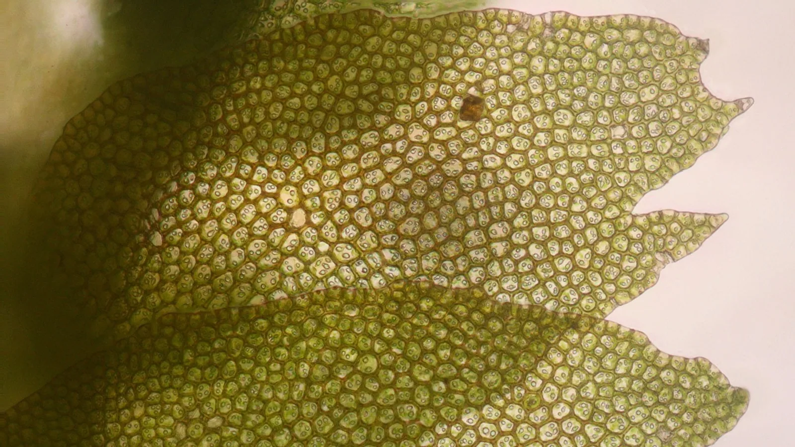
By the end of this guide you will be able to take a blank plant cell outline and correctly place every major label, from the cell wall on the outside to the nucleolus deep inside the nucleus. You will know what each part does, how the plant cell differs from an animal cell, and how to check your own work.
You do not need a microscope or a lab bench. You need one printed or on-screen outline of a plant cell, a pen, and this page open beside it. If you are studying for a quiz, keep a second blank copy nearby so you can test yourself after reading.
The single most common mistake in labeling a plant cell is confusing the cell wall with the plasma membrane, or forgetting that the chloroplast has two named internal compartments. This guide fixes both problems by walking through the structure in a logical order, outside to inside, and by naming the parts that students most often miss.
What a Plant Cell Actually Is
A plant cell is a eukaryotic cell, which means it has a true nucleus enclosed by a membrane. It is surrounded by a rigid cell wall and contains several structures that animal cells lack. Those extra structures are exactly the ones examiners ask about, so they deserve careful attention.
The defining features of a plant cell are a cellulose-based cell wall, chloroplasts for capturing light energy, a large central vacuole that occupies most of the cell volume, and plasmodesmata that connect neighboring cells. Each of these appears on a standard labeled plant cell diagram.
A typical plant cell measures roughly 10 to 100 micrometers across, which places it at the larger end of the eukaryotic range. Textbook figures exaggerate the size of organelles so they can be drawn clearly. Real proportions differ. The vacuole in a mature cell can take up 80 percent or more of the internal volume, pushing the nucleus and cytoplasm to the edges.
That volume fact matters for labeling. On many diagrams the vacuole looks like a large empty circle in the middle. It is not empty. It is a membrane-bound sac filled with cell sap, a solution of water, sugars, ions, and pigments.
A Labeled Diagram: Organelles in the Correct Order

.svg)
The list below is a labeled diagram written in words, ordered from the outside of the cell to the inside. Use it as a checklist when you place your own labels.
- Cell wall
- Plasma membrane
- Plasmodesmata
- Cytoplasm
- Central vacuole
- Chloroplast
- Nucleus
- Nucleolus
- Mitochondria
- Endoplasmic reticulum
- Golgi apparatus
- Ribosomes
- Peroxisomes
1. Cell Wall
The cell wall is the outermost layer of a plant cell. It sits outside the plasma membrane and gives the cell its shape, strength, and protection. Plant cell walls are built mainly from cellulose, a long-chain polysaccharide, along with pectin and proteins.
The wall is not a solid shell. It is a dynamic structure that is constantly remodeled. Research on banana fruit ripening shows that enzymes in the xyloglucan endotransglucosylase/hydrolase (XTH) family drive cell wall remodeling during ripening, and that fruit firmness falls as this remodeling proceeds [1]. The same class of enzymes appears in rice roots, where cell wall components respond to metal stress [2].
Other work shows how responsive the wall is to its environment. In rice seedlings exposed to chromium, cell wall thickness changed measurably, and adding proline helped restore thickness and reduced electrolyte leakage across the membrane [3]. In a study of aluminum and iron stress in Commelina communis, cell wall pectin and hemicellulose content dropped substantially [4].
For labeling purposes, remember two things. The wall is outside the plasma membrane, and it is not the same as the membrane. Many students lose marks by switching them.
2. Plasma Membrane
The plasma membrane is a phospholipid bilayer that sits just inside the cell wall. It controls what enters and leaves the cell. The membrane is selectively permeable, meaning small molecules and certain ions pass through while larger or unwanted molecules are blocked or transported by protein channels.
Proline-rich proteins (PRPs) are structural components of the cell wall that rely on proline as a biosynthetic precursor, and their abundance tracks with proline accumulation in stressed rice seedlings [3]. Membrane integrity is often measured by electrolyte leakage, which rises when the membrane is damaged. In the rice chromium study, electrolyte leakage increased under stress and dropped when proline was applied [3].
On a labeled plant cell diagram, the plasma membrane is usually drawn as a thin line pressed against the inside of the cell wall.
3. Plasmodesmata
Plasmodesmata are narrow channels that pass through the cell wall and connect the cytoplasm of one plant cell to the cytoplasm of the next. They allow water, ions, small molecules, and some proteins and RNA to move directly between cells.
This is a plant-only feature. Animal cells do not have plasmodesmata. They have gap junctions, which serve a similar communication role but are structurally different.
Plasmodesmata turn a plant tissue into a connected network. A diagram often shows them as short tubes crossing the wall at a few points.
4. Cytoplasm
The cytoplasm is the jelly-like material that fills the space between the plasma membrane and the nucleus. It contains the cytosol (the fluid) plus all the organelles suspended in it. Almost every organelle on this list sits within the cytoplasm.
When you label a plant cell, the cytoplasm is usually labeled with a leader line pointing to open space between organelles, not to any single structure.
5. Central Vacuole
The central vacuole is the largest organelle in a mature plant cell. It is bound by a membrane called the tonoplast and filled with cell sap. In a fully grown cell it can occupy most of the cell volume, which is why the nucleus and cytoplasm appear pushed toward the edges.
The vacuole stores water, ions, nutrients, and pigments. It maintains turgor pressure, the internal pressure that keeps plant tissues firm. When a plant wilts, the vacuoles have lost water and turgor pressure has dropped.
Its large size is one of the three headline differences between plant and animal cells. Vacuoles are present in animal cells too, but they are small and numerous rather than one giant central sac.
6. Chloroplast
The chloroplast is the organelle that carries out photosynthesis, capturing light energy and converting it to chemical energy. It gives plants their green color because it contains chlorophyll.
A chloroplast has an outer and inner membrane, and inside it two named compartments that students must know.
- Thylakoids are flattened, membrane-bound sacs. They stack into structures called grana.
- Stroma is the fluid that surrounds the thylakoids.
The thylakoid stacking pattern in land plants is distinctive. Photosystem II and its antenna system concentrate in the stacked grana regions, while Photosystem I and ATPase are found in the grana end membranes and the stroma membranes connecting grana [5]. The light-harvesting complex subfamily Lhcb controls this architecture. Removing the antenna system serving Photosystem II completely abolished grana formation in a genetic study, and deleting specific Lhcb subgroups reduced stacking by up to 40 percent [5]. Lhcb5 alone was enough to partially restore stacking [5].
Chloroplast structure also shows how sensitive organelles are to their environment. In a study of Commelina communis, combined aluminum and iron stress caused chlorosis, reduced photosynthetic pigments, and altered photochemical parameters, all signs of chloroplast damage [4]. In melon, loss of the gene CmMAIL2 caused impaired photosynthetic performance and disrupted thylakoid organization [6].
Chloroplasts also contain complex redox regulatory machinery. Plant chloroplasts carry more than 20 thioredoxins (Trxs), small proteins that catalyze disulfide reduction, drawing reducing power from two sources: photosynthetically reduced ferredoxin and NADPH [7]. This machinery is far more elaborate than the two-component systems found in non-photosynthetic organisms [7].
For labeling, chloroplastic detail usually means marking the outer membrane, the stroma, a thylakoid, and a granum stack.
7. Nucleus
The nucleus is the control center of the cell. It stores the cell's DNA and coordinates gene expression, growth, and division. It is enclosed by a double membrane called the nuclear envelope, which has pores that allow molecules to move between the nucleus and the cytoplasm.
The nucleus contains chromatin, the packaged form of DNA. It also contains one or more nucleoli.
8. Nucleolus
The nucleolus is a dense region inside the nucleus. It is where ribosomal RNA is made and where ribosomal subunits begin assembly. It is not surrounded by its own membrane, which is why it is described as a region rather than a true organelle.
On a labeled diagram, the nucleolus is drawn as a solid circle inside the nucleus.
9. Mitochondria
Mitochondria are the sites of cellular respiration, the process that releases usable energy from sugars. They have a double membrane. The inner membrane is folded into cristae, which increase surface area for the reactions that generate ATP.
Mitochondria are present in both plant and animal cells, so they do not distinguish a plant cell from an animal cell. However, they are affected by the same stress conditions that damage chloroplasts. In transgenic tobacco expressing a fungal arsenic transporter, both chloroplast and mitochondrial ultrastructure were better preserved than in wild-type plants under arsenate stress [8].
10. Endoplasmic Reticulum
The endoplasmic reticulum (ER) is a network of membrane-bound tubules and flattened sacs connected to the nuclear envelope. It comes in two forms.
- Rough ER is studded with ribosomes and is involved in protein synthesis and folding.
- Smooth ER lacks ribosomes and is involved in lipid synthesis and other metabolic tasks.
Transcriptome analysis of banana fruit during ripening found that the "Protein processing in endoplasmic reticulum" pathway was enriched among differentially expressed genes, reflecting how much protein traffic flows through the ER during a major developmental transition [1].
11. Golgi Apparatus
The Golgi apparatus is a stack of flattened membrane sacs. It modifies, sorts, and packages proteins and lipids that arrive from the ER, then sends them to their destinations. In plant cells the Golgi also produces polysaccharides for the cell wall.
It sits near the ER in most diagrams. Label the stack of sacs, not the vesicles around it.
12. Ribosomes
Ribosomes are the machines that build proteins. They read messenger RNA and assemble amino acids into polypeptide chains. They are not membrane-bound, and they are found both free in the cytoplasm and attached to the rough ER.
Ribosomes are found in all cells, plant and animal. In high-resolution images they look like tiny dots.
13. Peroxisomes
Peroxisomes are small, membrane-bound organelles that carry out oxidative reactions. In plants they are involved in breaking down fatty acids and in managing reactive oxygen species. They are also found in animal cells.
They are often drawn as small circles with a slightly granular interior, distinct from the smooth outline of a vacuole.
Table: Organelle Function at a Glance
| Organelle | Main Function | Where It Sits | Plant Cell Only? |
|---|---|---|---|
| Cell wall | Shape, support, protection | Outermost layer | Yes |
| Plasma membrane | Selective barrier, transport control | Just inside the wall | No |
| Plasmodesmata | Direct channel between adjacent cells | Through the wall | Yes |
| Cytoplasm | Holds organelles, site of many reactions | Fills the cell interior | No |
| Central vacuole | Stores water and solutes, maintains turgor | Large central sac | Yes (large form) |
| Chloroplast | Photosynthesis, contains thylakoids and stroma | Cytoplasm | Yes |
| Nucleus | Stores DNA, controls gene expression | Cytoplasm | No |
| Nucleolus | Makes ribosomal RNA, assembles ribosome subunits | Inside the nucleus | No |
| Mitochondria | Cellular respiration, ATP production | Cytoplasm | No |
| Endoplasmic reticulum | Protein and lipid synthesis, transport | Around the nucleus | No |
| Golgi apparatus | Modifies, sorts, packages proteins and lipids | Near the ER | No |
| Ribosomes | Protein synthesis | Free or on rough ER | No |
| Peroxisomes | Oxidative reactions, fatty acid breakdown | Cytoplasm | No |
Key Differences From Animal Cells
Four features separate a plant cell from an animal cell on a standard diagram. Three are structures that animal cells lack or have in a different form. The fourth is a connection system.
Cell Wall
Plant cells have a rigid cell wall outside the plasma membrane. Animal cells do not. An animal cell is bounded only by its plasma membrane, which is flexible. This is why animal cells can change shape and move, while plant cells are fixed in place.
The wall is not inert. Research on metal stress shows it actively binds and sequesters ions. In rice, proline application increased the sequestration of chromium within root cell walls and reduced its distribution in the cytoplasm and organelles of shoot cells [3]. In Commelina communis, aluminum and iron mutually inhibited binding to root tips, and cell wall pectin and hemicellulose decreased under stress [4].
Chloroplasts
Plant cells contain chloroplasts. Animal cells do not. This is the feature that makes plants photosynthetic.
Chloroplasts contain thylakoids and stroma, and the thylakoid stacking pattern is controlled by specific light-harvesting proteins [5]. Redox regulation inside chloroplasts involves more than 20 thioredoxins, far more than the simple two-component systems in non-photosynthetic organisms [7].
Large Central Vacuole
Mature plant cells have one large central vacuole that occupies most of the cell volume. Animal cells have small, scattered vacuoles instead. This single structural difference is often the fastest way to tell a plant cell diagram from an animal cell diagram at a glance.
Plasmodesmata
Plant cells connect to each other through plasmodesmata, channels that pass through the cell wall. Animal cells use gap junctions instead, and those are not visible on a typical plant cell diagram because they are not part of the plant cell's own structure.
How to Check Your Labeled Diagram
After you place your labels, run through this checklist. Each item catches a mistake that costs marks.
- Is the cell wall outside the plasma membrane everywhere, not just in one spot?
- Is the central vacuole drawn as one large sac, not several small ones?
- Does the chloroplast have both a thylakoid and a stroma label, or at least a granum stack?
- Is the nucleolus inside the nucleus, not floating in the cytoplasm?
- Are the ribosomes shown both free in the cytoplasm and on the rough ER?
- Do the plasmodesmata cross the wall rather than sit inside the cell?
- Is the Golgi labeled as a stack, not as a single sac?
- Are the mitochondria drawn with an inner folded membrane?
If you can answer yes to all eight, your label of a plant cell is structurally sound.
Worked Example: Building Your Own Labels
If you are building a diagram from scratch for a class or a study sheet, this sequence keeps the proportions realistic. This is a text plan, not an image.
Step 1: Draw a rounded rectangle for the cell wall. Draw a second line just inside it for the plasma membrane, leaving a small gap between them.
Step 2: Fill the interior with light shading for the cytoplasm. Leave a large open central area for the vacuole.
Step 3: Draw the central vacuole as one large rounded shape that occupies roughly 70 to 80 percent of the interior. This matches the real volume in a mature cell.
Step 4: Push the nucleus to one side, near the edge. Draw it as a circle with a smaller solid circle inside for the nucleolus.
Step 5: Scatter five to eight chloroplasts around the cytoplasm, each drawn as an oval with small stacked lines inside for the grana.
Step 6: Add three or four mitochondria as ovals with wavy internal lines for cristae.
Step 7: Draw the ER as wavy lines near the nucleus, some with dots (rough ER) and some without (smooth ER).
Step 8: Add the Golgi as a stack of three or four curved sacs.
Step 9: Scatter small dots for free ribosomes and small circles for peroxisomes.
Step 10: Draw two short channels crossing the cell wall for plasmodesmata.
Step 11: Add leader lines and labels for all thirteen structures.
The order matters because it forces you to respect scale. Draw the largest structure first and the smallest last.
Common Mistakes and Limitations
The wall and membrane swap. The cell wall is outside, the plasma membrane is inside. If you label the outer boundary as the plasma membrane, everything else shifts.
The vacuole is drawn too small. In a mature cell the central vacuole dominates the interior. A small circle in the middle does not match reality.
The chloroplast is labeled as one undifferentiated blob. It has named internal parts. Label the thylakoids and the stroma, or at least the grana stack.
The nucleolus is placed outside the nucleus. It is a region within the nucleus, not a separate organelle floating in the cytoplasm.
Ribosomes are shown only on the rough ER. They also float free in the cytoplasm, and both locations matter.
Plasmodesmata are drawn inside the cell. They cross the wall. If they do not touch the wall on both sides, the label is wrong.
The Golgi is confused with the ER. The Golgi is a discrete stack of sacs. The ER is a connected network.
Scale is ignored. Textbook diagrams enlarge organelles for clarity. A real chloroplast is only a few micrometers long, and a real ribosome is about 20 to 30 nanometers across.
This guide covers structure. It does not cover the biochemistry of photosynthesis or the mechanics of cell division, both of which are separate topics with their own diagrams. If you are using this page for a specific course, check whether your instructor expects the tonoplast, the nuclear envelope, and the cristae as separate labels. Some do.
Individual diagrams from different textbooks vary in how much internal detail they show. A high school diagram may stop at the organelle level. A college diagram may require the thylakoid, stroma, granum, cristae, and nuclear pore. Match the depth of your labels to the level of your course.
Self-Quiz: 5 Fill-in-the-Blank Questions
Answer each question before checking the key below.
- The outermost layer of a plant cell, made mainly of cellulose, is the __________.
- The two named compartments inside a chloroplast are the thylakoids and the __________.
- The large organelle that stores water and maintains turgor pressure, occupying most of a mature plant cell's volume, is the __________.
- The channels that pass through the cell wall and connect the cytoplasm of adjacent plant cells are called __________.
- The dense region inside the nucleus where ribosomal RNA is made is the __________.
Answer Key
- cell wall
- stroma
- central vacuole
- plasmodesmata
- nucleolus
Score yourself honestly. If you missed the stroma or the plasmodesmata, those are the two most commonly dropped labels on student diagrams. Review those sections before your next attempt.
For a harder version, cover the table above and try to name one function for each of the thirteen structures without looking.
Frequently Asked Questions
What is the difference between a cell wall and a plasma membrane?
The cell wall is the rigid outer layer made mainly of cellulose, and the plasma membrane is the thin selectively permeable bilayer just inside it. The wall provides shape and support while the membrane controls what enters and exits the cell.
Does a plant cell have a cell membrane?
Yes. Every plant cell has a plasma membrane, also called the cell membrane, located between the cell wall and the cytoplasm. It is present in all cells, plant and animal.
Why is the central vacuole so large in a plant cell?
It stores water, ions, and nutrients and maintains turgor pressure that keeps tissues firm. In a mature cell it can occupy most of the internal volume, which pushes the nucleus and cytoplasm to the edges.
What are thylakoids and stroma?
Thylakoids are flattened membrane sacs inside a chloroplast that stack into grana. Stroma is the fluid surrounding the thylakoids. Together they form the two internal compartments of the chloroplast.
Where are plasmodesmata found?
Plasmodesmata cross the cell wall and connect the cytoplasm of one plant cell to the next. They are a plant-specific feature and are not found in animal cells.
Do plant cells have mitochondria?
Yes. Plant cells have mitochondria that carry out cellular respiration, just as animal cells do. Mitochondria are not a feature that distinguishes plant cells from animal cells.
What is the nucleolus and where is it located?
The nucleolus is a dense region inside the nucleus where ribosomal RNA is made and ribosomal subunits begin assembly. It is not enclosed by its own membrane.
How can I tell a plant cell diagram from an animal cell diagram quickly?
Look for three things: a rigid cell wall, green chloroplasts, and one large central vacuole. If all three are present, the diagram is a plant cell.
Related Articles
- Nucleotide Diagram: Structure, Parts, and How to Draw It
- Ribosome Diagram: Structure, Function, and Labeled Parts
- Microscope Parts and Functions: A Visual Guide to Labeled Diagrams
- Cryopreservation Labels for Cells: A Complete Guide
- Cancer Cell Diagram: Understanding the Hallmarks of Cancer
- Diagram of Yeast Cell: Structure, Function, and Labeling Guide
- Match Each Label to Its Correct Cell Type
Sources
- Transcriptomics and Gene Family Identification of Cell Wall-Related Differentially Expressed Genes Reveal MaXTH32.5 Involved in Fruit Firmness During Banana Ripening.
- Effect of Different Forms of Antimony (Sb) on Genes Encoding Functions Associated with Root Morphology, Physiology and Root Cell Wall in Rice (Oryza sativa).
- Exogenous Proline Maintains Cell Wall Structure and Membrane Integrity in Rice Seedlings Under Cr(VI) Stress Associated with Regulation of Proline-Rich Proteins.
- Aluminum and iron interactions in commelina communis: additive chloroplast toxicity versus synergistic internal and rhizosphere detoxification.
- Specific light-harvesting complexes mediate grana stacking and prevent energy spillover between photosystems in plant chloroplasts.
- Loss of CmMAIL2 compromises chloroplast function but permits developmental progression in melon.
- Why is the chloroplast redox regulatory machinery so complex?
- Insights into the arsenic resistance mechanisms conferred by Coprinellus disseminatus ACR3 in transgenic tobacco: Arsenic excretion, chloroplast protection, and cell wall thickening.