Fainting Goats: Myotonia Congenita Explained
By Dr. Zubair Khalid, DVM, MS, PhD ·
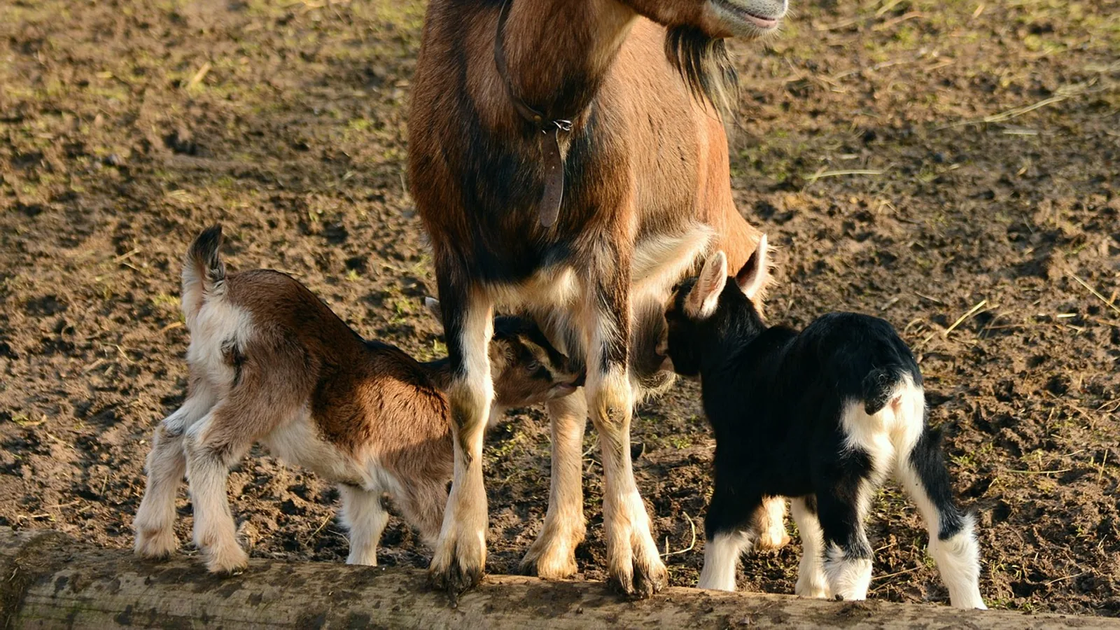
Fainting goats do not faint. When startled, a goat with myotonia congenita stiffens and may fall over while fully conscious, then recovers within seconds as the muscle relaxation delay passes.
Myotonia congenita is a skeletal muscle channelopathy, meaning a disorder caused by a defect in an ion channel protein. In these goats the defect sits in the CLCN1 gene, which encodes the main voltage-gated chloride channel of skeletal muscle. The channel opens poorly near the muscle resting potential, chloride conductance falls, and the muscle membrane becomes hyperexcitable. The result is a burst of repetitive electrical discharges that keeps the muscle contracted after the triggering movement has ended. This article explains the mutation, why the goat stiffens rather than faints, how the condition compares across species, and what it means for the animals' welfare.
This article is educational and is not a substitute for veterinary diagnosis or treatment.
What Actually Happens During a "Fainting" Episode
The word fainting describes a loss of consciousness caused by reduced blood flow to the brain. A myotonic goat never loses consciousness. The animal is awake, aware, and usually looking around during the entire episode. What the observer sees is a sudden failure of muscle relaxation.
The sequence is consistent. A sudden stimulus arrives, such as a handler clapping, a dog approaching, a feed bucket dropping, or the goat being startled by a sudden movement. The goat attempts to run or brace. The muscles that initiated the movement do not relax on schedule. The limbs lock, the neck and back stiffen, and the goat may topple onto its side with legs extended. The stiffness typically lasts a few seconds. As the repetitive electrical activity in the muscle fibers subsides, relaxation returns, and the goat stands and walks away normally.
Several features distinguish this from a true faint:
- Consciousness is preserved. The goat's eyes track movement and the animal responds to its surroundings.
- The trigger is a sudden voluntary or reflex contraction, not a drop in cerebral perfusion.
- Recovery is rapid and complete, usually within seconds.
- The episode can often be interrupted or shortened by handling the goat, which is why experienced owners sometimes steady an animal before it falls.
The historical name "fainting goat" is a misnomer that has stuck. The breed is also called the Tennessee fainting goat, the Tennessee meat goat, the nervous goat, and the stiff-leg goat. The term "nervous goat" is closer to the truth, because the trigger is a startle response and the animal's nervous system is intact.
The Biology: Why the Muscle Cannot Relax
Chloride Conductance Sets Muscle Stability
Skeletal muscle fibers maintain a resting membrane potential of roughly -80 to -90 millivolts. In a normal fiber, most of the resting membrane conductance is carried by chloride ions moving through ClC-1 channels. Chloride conductance acts as an electrical stabilizer. When the membrane starts to depolarize, chloride current opposes the change and pulls the membrane back toward rest.
When chloride conductance is low, that brake is weak. A small depolarization can trigger an action potential, and the resulting after-depolarization can trigger another, and another. The muscle fiber fires a train of action potentials rather than a single clean twitch. The mechanical consequence is a prolonged contraction because the fiber cannot repolarize and relax.
The Goat Mutation
The molecular basis was identified in 1996. Beck and colleagues found a single nucleotide change in the goat muscle chloride channel gene, gClC-1, that substitutes proline for a conserved alanine residue in the carboxyl terminus of the channel protein [1]. When the mutant channel was expressed in a heterologous system, the voltage dependence of activation shifted by about +47 millivolts. In practical terms, the channel needs a much stronger depolarization to open, so its open probability near the normal resting membrane potential is very low [1]. Less open channel means less chloride conductance, which is exactly the defect predicted by earlier physiological work.
That earlier work was foundational. Bryant and Morales-Aguilera measured cable parameters in isolated external intercostal muscle bundles from normal and myotonic goats and found that resting chloride conductance in myotonic fibers ranged from 0 to 147 micromhos per square centimeter, compared with 376 to 951 in normal fibers [2]. The same study showed that applying monocarboxylic aromatic acids to normal goat muscle blocked chloride conductance and reproduced the myotonic behavior, which established chloride conductance as the central variable [2]. Bryant and Conte-Camerino later showed that protein kinase C activation could block up to 76 percent of chloride conductance in normal goat fibers and induce myotonic hyperexcitability, while the nearly absent conductance of congenitally myotonic fibers could not be restored by kinase inhibitors, cAMP-raising agents, clofibric acid enantiomers, or taurine [3]. The congenital defect is structural, not a reversible regulatory state.
Why the Discharge Persists
Adrian and Bryant worked out the repetitive discharge mechanism directly. Myotonic goat fibers respond to an injected constant current with a train of action potentials, and if the train exceeds about 10 to 15 spikes, stopping the current does not stop the firing [4]. Normal goat fibers behave the same way when bathed in chloride-free Ringer solution, which confirms that chloride conductance is the missing brake in both cases. The authors attributed the self-sustaining discharge to potassium accumulation in the transverse tubules combined with the low chloride conductance of myotonic fibers [4].
This explains the clinical timing. A single twitch may look almost normal, because one action potential does not generate enough tubular potassium accumulation. A train of contractions, or a sudden strong contraction, does. In myotonic mouse diaphragm, myotonia was easily demonstrated during train stimulation but not during single twitch stimulation or during train stimulation preceded by a series of twitches [5]. The goat startle response is exactly the kind of sudden, strong, multi-fiber contraction that provokes the discharge.
What the Muscle Looks Like
Muscle biopsies from myotonic goats show structural differences from normal goat muscle. Atkinson and colleagues found increased periodic acid-Schiff positive material within myotonic fibers after diastase digestion, and increased alizarin red S staining, which indicates more calcium in the tissue [6]. Electron microscopy showed increased density of the transverse tubules, electron-dense material within the tubules, proliferation and dilatation of the sarcotubular elements, and abnormal mitochondria [6]. Membrane-specific stains showed increased density of the sarcolemma and transverse tubules [6]. These findings give a morphological basis for the abnormal contraction-relaxation behavior.
One negative finding matters for how we think about the disease. Erythrocytes from myotonic goats showed no significant differences from normal goat erythrocytes using electron paramagnetic resonance and saturation transfer electron paramagnetic resonance spin labeling with three fatty acid probes [7]. That result argues against a generalized membrane fluidity defect and supports the view that myotonia congenita is a specific channel defect rather than a whole-cell membrane disorder.
Genetics and Inheritance
Autosomal Dominant in Goats
In the myotonic goat, the condition behaves as an autosomal dominant trait. A single copy of the mutant allele is enough to produce the phenotype. This is consistent with the electrophysiology. The mutant channel has a dominant-negative effect on the channel complex, so co-expression of normal and mutant subunits still produces poorly functioning channels.
The recessive and dominant forms of myotonia congenita in humans are both caused by CLCN1 mutations, and the difference in inheritance reflects how severely the mutation disrupts channel function. Mutations that abolish function tend to behave recessively because a single normal allele can supply enough working channels. Mutations that produce a dysfunctional subunit capable of interfering with normal subunits behave dominantly. The goat mutation falls into the latter category.
The Same Gene, Different Species, Different Inheritance
The table below compares the three best-characterized species. It is worth reading carefully because the inheritance pattern is not the same across species, and that difference has real consequences for breeding programs.
| Species | Gene | Inheritance | Typical trigger | Key reference |
|---|---|---|---|---|
| Goat (myotonic goat) | CLCN1 (gClC-1) | Autosomal dominant | Sudden startle or sudden voluntary contraction | [1], [2] |
| Human (myotonia congenita) | CLCN1 | Both recessive (Becker) and dominant (Thomsen) forms exist | Sudden movement after rest, especially after inactivity | [8], [9] |
| Dog (miniature Schnauzer) | CLCN1 (CIC-1) | Autosomal recessive | Sudden movement, often after rest | [10] |
| Horse (New Forest pony) | CLCN1 | Recessive in the reported family | Episodes of recumbency and stiffness | [11] |
| Cat (random bred, Winnipeg) | CLCN1 | Not fully established in the report | Palpebral reflex testing, jaw movement | [12] |
| Sheep (Rasa Aragonesa) | CLCN1 | Recessive carrier state identified in rams | Sudden contraction in newborn lambs | [13] |
| Water buffalo (Murrah) | CLCN1 | Hereditary, pattern described as hereditary myotonia | Muscle stiffness and hypertrophy | [14] |
The human literature is the anchor here. Lipicky, Bryant, and Salmon compared external intercostal muscle from normal volunteers and patients with myotonia congenita and found that the human disease shares the same basic abnormality as the goat disease: higher membrane resistance, lower internal resistivity, and a higher resting potential [8]. That study explicitly noted the similarity between human myotonia congenita and hereditary myotonia in goats. The goat was, in effect, the animal model that helped define the human disease.
The dog provides the cleanest example of recessive inheritance. Rhodes and colleagues identified a mutation in canine CIC-1 that replaces a threonine residue in the D5 transmembrane segment with methionine in miniature Schnauzers [10]. Functional expression showed a profound reduction in channel open probability at voltages near the resting potential. When heterodimeric channels containing one wild-type and one mutant subunit were expressed together, the dysfunction was greatly diminished, which supports the recessive inheritance seen in affected pedigrees [10]. This is the opposite of the goat situation, where one mutant allele is enough.
The cat report adds a clinical picture that is useful for recognizing myotonia in any species. Five affected cats had a protruding tongue, limited jaw motion, drooling, prominent neck and proximal limb musculature, blepharospasm on palpebral reflex testing, and a short-strided gait [12]. Electromyography showed myotonic discharges at a mean frequency of 300 Hz, described as resembling a swarm of bees [12]. The mutation disrupted a donor splice site downstream of exon 16 and was predicted to truncate the protein and remove part of a conserved domain critical for ion transport [12]. The same report notes a worldwide human population prevalence of myotonia congenita of about 1 in 100,000 [12].
The horse, sheep, and buffalo reports confirm that CLCN1 myotonia is not a goat-only curiosity. A New Forest pony foal with episodes of recumbency and stiffness was homozygous for a missense mutation in a conserved CLCN1 domain, and the mutation showed a recessive pattern in that family [11]. In a Spanish Rasa Aragonesa sheep flock, a non-synonymous single nucleotide variation in exon 2 of CLCN1 was associated with congenital myotonia affecting about 1 percent of newborn lambs, and removing heterozygous rams eliminated new cases in subsequent lambing seasons [13]. In Murrah water buffalo, affected animals showed muscle hypertrophy and stiffness with myotonic discharges on electromyography, caused by aberrant splicing that deleted the last 43 nucleotides of exon 3 and shifted the reading frame [14].
Clinical Signs and Diagnosis
What Owners See
The classic presentation is a goat that stiffens and falls when startled. Beyond that, owners may notice:
- A stiff, short-strided gait that improves with exercise (the "warm-up" phenomenon).
- Difficulty rising after rest, especially in cold weather.
- Prominent muscle bulk, particularly in the hindquarters and proximal limbs.
- A startled expression during an episode, with the head held up and eyes open.
- Normal behavior, appetite, growth, and reproduction between episodes.
The warm-up phenomenon is important. Muscles that have been active for a few minutes relax more normally, which is why a myotonic goat that has been walking may appear almost normal. This is also why the condition is sometimes mistaken for a lameness or a neurological problem.
Diagnostic Approach
Diagnosis rests on three pillars: history and clinical signs, electromyography, and genetic testing.
Electromyography is the most direct functional test. Myotonic discharges appear as high-frequency repetitive electrical activity, the same "swarm of bees" sound described in the feline report [12]. The discharges are spontaneous or provoked by needle movement and by voluntary contraction.
Muscle biopsy is not usually necessary for diagnosis in goats, but if performed it may show the histochemical and ultrastructural changes described above: increased PAS-positive material, increased calcium staining, and tubular abnormalities [6].
Genetic testing for the goat CLCN1 mutation is available and is the most specific test. It is also the most useful tool for breeding decisions.
Differential Diagnoses
Several conditions can look like myotonia congenita in a goat:
- Hypocalcemia. Periparturient hypocalcemia causes muscle stiffness and recumbency, but the timing, the response to calcium therapy, and the metabolic profile distinguish it.
- Tetanus. Tetanus causes sustained rigidity and a characteristic facial expression, but the rigidity is constant rather than episodic and the history usually suggests a wound.
- White muscle disease. Nutritional myopathy from selenium or vitamin E deficiency causes weakness and stiffness, often with elevated muscle enzymes.
- Trauma or orthopedic injury. A goat that falls repeatedly may be assumed to have a musculoskeletal problem when the underlying issue is myotonia.
- Other neurological disease. Cerebellar or vestibular disease can cause falling, but the falling is not preceded by muscle stiffening and the animal does not recover in seconds.
A veterinarian should evaluate any goat with episodic collapse, because the treatment and prognosis differ substantially across these conditions.
Welfare Implications
Is Myotonia Congenita Painful?
The condition itself is not painful. The goat is not experiencing a cramp in the human sense and is not losing consciousness. The muscle stiffness is the mechanical result of repetitive electrical discharges, and the animal is aware throughout. Owners who handle myotonic goats consistently report that the animals are calm during episodes and resume normal activity immediately afterward.
The welfare concern is not pain from the myotonia. It is injury risk from the falls.
Injury Risk
A goat that stiffens and falls can land on uneven ground, against a fence, into a water trough, or down a slope. The risk is highest in young animals that fall more often, in animals housed on concrete or steep terrain, and in situations where the goat is repeatedly startled.
Practical risk reduction measures include:
- Keep the goat on level, well-bedded ground where possible.
- Avoid housing myotonic goats next to dogs, noisy equipment, or busy traffic areas.
- Introduce changes in routine gradually so the animal is not repeatedly startled.
- Provide sturdy fencing that will not injure an animal that falls against it.
- Supervise handling during transport and restraint, because restraint itself can be a trigger.
- Avoid breeding decisions that increase the number of affected animals if the goal is a low-stress herd.
Anesthesia and Malignant Hyperthermia
There is a specific clinical question about whether myotonic goats are at risk for malignant hyperthermia under anesthesia. A study by Newberg, Lambert, and Gronert exposed six goats with myotonia congenita to 1 percent halothane for one hour plus a single intravenous injection of suxamethonium in an attempt to induce malignant hyperthermia [15]. No evidence of malignant hyperthermia occurred. Suxamethonium did produce a myotonic response lasting 10 to 20 seconds in each goat, accompanied by transient changes in blood gases consistent with increased aerobic metabolism, but there was no metabolic acidosis [15]. The authors concluded that myotonia congenita in goats does not predispose to malignant hyperthermia susceptibility [15]. This is useful information for veterinarians planning anesthesia, but it does not mean that standard ruminant anesthesia precautions can be skipped.
Breeding and Genetic Management
Because the goat trait is autosomal dominant, an affected goat has a high probability of passing the mutation to offspring. A carrier or affected animal bred to a normal animal will produce affected offspring at a substantial rate. Breeders who want to reduce the number of affected animals should use genetic testing to identify carriers before breeding.
The sheep example is instructive. When heterozygous rams were identified and replaced with wild-type homozygous young males, no additional congenital myotonia cases were detected in subsequent lambing seasons [13]. That is a clean demonstration that genetic testing plus selective breeding works.
Clinical Relevance, Limitations and Common Mistakes
Myotonia congenita is a well-characterized channelopathy with a clear molecular basis, but several points are commonly misunderstood.
The first mistake is treating the condition as a form of epilepsy or syncope. It is neither. There is no loss of consciousness and no seizure activity in the brain. Antiepileptic drugs are not indicated, and the use of human anesthesia protocols or sedative protocols designed for other species is inappropriate without veterinary guidance.
The second mistake is assuming the goat is in pain. The available evidence does not support that conclusion. The stiffness is a mechanical consequence of the channel defect, and the animal recovers fully within seconds.
The third mistake is underestimating the injury risk. A fall is a fall. Even a brief episode can cause a fracture, a head injury, or a drowning if the animal falls into water. Housing and handling should be designed around that risk.
The fourth mistake is confusing the inheritance pattern across species. The goat form is dominant, the dog form is recessive, and the human form has both. A breeding program designed for one species does not transfer to another.
The fifth mistake is assuming that because the goat looks normal between episodes, no veterinary assessment is needed. A veterinarian should confirm the diagnosis and rule out the differentials listed above, because some of them are treatable and some are life-threatening.
Individual animals vary in how often they stiffen and how severely they fall. A veterinarian who knows the individual animal is the best source of advice on management.
Frequently Asked Questions
Do fainting goats actually faint?
No. They remain fully conscious during an episode. The stiffness is caused by delayed muscle relaxation from a chloride channel defect, not by a loss of consciousness.
Why do goats who faint fall over?
The sudden startle triggers a burst of repetitive electrical activity in the muscles. The muscles cannot relax on schedule, so the goat loses its balance and falls. Recovery is usually within seconds.
What gene causes myotonia congenita in goats?
The CLCN1 gene, which encodes the skeletal muscle chloride channel. A single nucleotide change substitutes proline for a conserved alanine in the channel protein and shifts the voltage dependence of activation by about +47 millivolts [1].
Is myotonia congenita in goats dominant or recessive?
It is autosomal dominant in the myotonic goat. One copy of the mutant allele is enough to produce the condition.
Is myotonia congenita the same in dogs and humans?
The gene is the same, but the inheritance differs. The miniature Schnauzer form is autosomal recessive [10], and humans have both recessive and dominant forms [8], [9].
Is the condition painful for the goat?
The condition itself is not painful. The main welfare concern is the risk of injury from falling.
Can a fainting goat be used for breeding?
Yes, but because the trait is dominant, affected animals are likely to pass it on. Genetic testing before breeding is the responsible approach.
Do fainting goats need special anesthesia?
Myotonic goats are not predisposed to malignant hyperthermia, based on a study that failed to induce it with halothane and suxamethonium [15]. Standard ruminant anesthesia precautions still apply, and the veterinarian should be informed of the diagnosis.
Related Articles
- Who Discovered Dna
- Why Is My Dog collapsing suddenly? Cardiac Arrest Fainting and Hypoglycemia
- Gel Electrophoresis Troubleshooting: Smears, Faint Bands, and Uneven Migration
- FeLV Vaccine for Cats: Who Needs It and Leukemia Protection Efficacy
- How to Learn Bioinformatics from Scratch: A Complete Roadmap (From a Biologist Who Couldn't Code)
- The World Health Organization (WHO) and Global Genomic Surveillance
- Damascus Goat: Shami Breed Traits and Health
Sources
- Molecular basis for decreased muscle chloride conductance in the myotonic goat.
- Chloride conductance in normal and myotonic muscle fibres and the action of monocarboxylic aromatic acids.
- Chloride channel regulation in the skeletal muscle of normal and myotonic goats.
- On the repetitive discharge in myotonic muscle fibres.
- Genetic CLC-1 chloride channel deficiency modifies diaphragm muscle isometric contractile properties.
- Myotonia congenita. A histochemical and ultrastructural study in the goat: comparison with abnormalities found in human myotonia dystrophica.
- Electron paramagnetic resonance and saturation transfer electron paramagnetic resonance studies on erythrocytes from goats with and without heritable myotonia.
- Cable parameters, sodium, potassium, chloride, and water content, and potassium efflux in isolated external intercostal muscle of normal volunteers and patients with myotonia congenita.
- Skeletal muscle ClC-1 chloride channels in health and diseases.
- A missense mutation in canine C1C-1 causes recessive myotonia congenita in the dog.
- A missense mutation in the skeletal muscle chloride channel 1 (CLCN1) as candidate causal mutation for congenital myotonia in a New Forest pony.
- A novel mutation in CLCN1 associated with feline myotonia congenita.
- Ovine congenital myotonia associated with a mutation in the muscle chloride channel gene.
- Clinical and molecular study of a new form of hereditary myotonia in Murrah water buffalo.
- Failure to induce malignant hyperthermia in myotonic goats.