Horse Hooves: Care, Trimming, and Disease Guide
By Dr. Zubair Khalid, DVM, MS, PhD ·
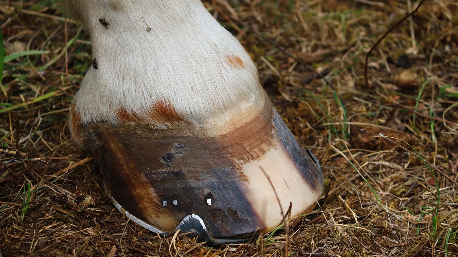
Horse hooves reward routine. Pick every hoof daily, keep the frog and sole clean and dry, and schedule a farrier every four to six weeks, though that interval shifts with hoof growth, work level, and individual conformation. Most hoof problems owners see are preventable with those three habits. The problems that are not preventable split into two groups. Some, like thrush and simple cracks, are managed with a farrier and good hygiene. Others, like laminitis, are medical emergencies that need a veterinarian the same day. Knowing which is which is the single most useful skill an owner can build.
This article is educational and is not a substitute for veterinary diagnosis or treatment.
At a Glance: Daily and Routine Hoof Care
| Task | Frequency | What to look for |
|---|---|---|
| Pick all four hooves | Daily, before and after work | Packed dirt, stones, manure, odor, discharge |
| Clean and dry the sole and frog | Daily in wet conditions | Soft, ragged frog, black greasy debris, deep sulci |
| Inspect the hoof wall and coronary band | Daily | New cracks, rings, heat, swelling, sensitivity |
| Check shoes and clenches | Daily for shod horses | Loose or risen nails, sprung or shifted shoe |
| Farrier trim or reset | Every 4 to 6 weeks on average | See the interval section below |
| Veterinarian exam | Any acute severe lameness, or a hoof that is hot and painful | See the emergency section below |
Why Hoof Care Matters
The hoof is a load-bearing structure built from keratin, the same structural protein found in hair and nails. Routine trimmings generate a steady, traceable supply of that keratin, which is why researchers now study horse hoof trimmings as a renewable biopolymer feedstock rather than waste [1]. From the horse's side, the hoof is a living organ with two very different layers. The outer hoof wall is hard, avascular keratin with no nerve supply. The sensitive laminae underneath are richly innervated and vascularized, and they are the tissue that hurts when something goes wrong.
That anatomical split explains a lot of clinical behavior. A horse can have a visibly cracked, ugly hoof wall and show no pain, because the wall itself has no nerves. The same horse will be violently lame from a tiny abscess or a laminitic insult because the sensitive laminae are inflamed. The lamellar junction is a dermo-epidermal interface where epithelial cells of the primary and secondary laminae interlock with the underlying dermis [2]. When that interface is disrupted, the distal phalanx (the coffin bone, also called P3) can lose its suspension and rotate or sink.
The lamellar tissue is also metabolically active. Progenitor cells reside in the hoof, including in the coronary corium, and they respond to inflammatory signaling [3]. Inflammatory interleukins change the behavior of hoof progenitor cells in culture, which points to how inflammation at the lamellar junction can impair normal healing [4]. This is the biological reason laminitis is treated as a systemic disease rather than a local foot problem.
Daily Hoof Care: Step by Step
Step 1: Pick out every hoof, every day
Use a hoof pick from heel toward toe, working along the sulci on either side of the frog. Clear packed material from the sole and around the frog. Daily picking does three things. It removes the moist, anaerobic debris that thrush organisms need. It lets you notice a stone or a nail before it works deeper. It gets your hands on the hoof so you feel heat, swelling, or a loose shoe early.
Step 2: Look at what comes out
Normal debris is dry dirt and manure. Abnormal findings include black, greasy, foul-smelling material in the frog sulci, which suggests thrush, and any discharge, blood, or a visible tract, which suggests a puncture or abscess. Puncture wounds of the hoof are common and potentially life-threatening, and in a large retrospective series of 96 cases the foreign body was still in place in about one in five horses [5]. In that same series, the penetration site was in the frog region far more often than in the sole, and the dorsal frog, central frog, and palmar or plantar frog regions were all commonly involved [5]. A horse that steps on a nail and then stands on it is a genuine emergency, not a wait-and-see problem.
Step 3: Assess the hoof capsule
Run your hand down the hoof wall and coronary band. Note heat, swelling, new horizontal rings, vertical cracks, or a depression at the coronary band. Rings and bilateral lameness are recognized clinical features associated with equine metabolic syndrome and past or ongoing laminitis [6]. A single new ring after a fever or a severe illness is a historical marker. Multiple rings with soreness are a reason to call a veterinarian.
Step 4: Check shoes and clenches
For shod horses, press each shoe and look at the clench line. A risen clench, a shoe that moves under thumb pressure, or a shoe that has shifted toward the heel means a farrier call, not a ride. Riding on a loose shoe can tear hoof wall and drive a nail into sensitive tissue.
Step 5: Keep the environment right
Standing in mud and urine is the single biggest environmental driver of thrush and softened soles. Dry footing, clean bedding, and turnout on well-drained pasture do more for hoof health than any topical product. Hoof trimming itself measurably changes foot conformation and gait, so the farrier's work is not cosmetic. Routine trimming and shoeing significantly changed coronary band length and height, dorsal hoof wall length, heel length, heel overhang, and hoof angle in a study of 15 horses, even though total head and pelvis movement did not change at the trot [7]. The takeaway for owners is that a trim changes the foot's geometry, which is why consistency of interval matters more than any single appointment.
Farrier Intervals: Why 4 to 6 Weeks Is a Starting Point
The commonly quoted four to six week interval is a default, not a rule. The right interval depends on how fast that horse's hoof grows, how much work the horse does, the surface it works on, and its conformation. A horse in light work on soft ground may go six weeks comfortably. A horse in hard daily work, or one with a long toe and low heel, may need four weeks or less. A horse with a hoof wall disorder may need a shorter cycle coordinated with the veterinarian.
Because trimming changes hoof angle and heel length [7], stretching the interval lets the toe run long and the heel underrun, which alters loading. That change in loading is not trivial. In an experimental model, prolonged preferential weight bearing and reduced ambulation produced elongation of secondary epidermal lamellae, cell death in parabasal keratinocytes, and basal cell proliferation, with the most severe lesions in the supporting limb that carried extra load [8]. In plain terms, uneven or excessive loading damages lamellar tissue. A late trim is a loading problem.
Use these signals to adjust the interval rather than following a fixed calendar:
- The hoof wall at the toe is visibly longer than at the last appointment.
- The white line is stretching or widening at the toe.
- Shoes are worn thin or clenches are rising before the next visit.
- The horse feels "footy" on hard ground in the days before the next trim.
- The farrier comments that the feet are running forward.
Coordinate the farrier and the veterinarian on the same page for any horse with laminitis, a hoof wall disorder, or a puncture wound. Farriery is a core part of equine practice, and veterinarians who engage directly with farriers and clients on foot health get better outcomes than those who delegate in silence [9].
Thrush: Prevention and Early Management
Thrush is a degeneration of the frog and adjacent sulci in a wet, oxygen-poor environment. The classic signs are a black, greasy, foul-smelling exudate in the central or collateral sulci, a ragged frog, and sometimes sensitivity to pressure. It rarely causes severe lameness on its own, but it can progress into sensitive tissue and it is a warning that the environment is wrong.
Prevention is mechanical and environmental:
- Pick hooves daily and remove all packed material from the sulci.
- Keep stalls and paddocks dry. Fix muddy gateways and standing water.
- Pick out hooves after washing or after working in wet conditions.
- Have the farrier trim away ragged, undermined frog tissue at each visit.
- Address any hoof deformity that traps debris, such as a contracted heel or a deep central sulcus.
If the frog is painful, bleeding, or the horse is lame, have a veterinarian examine the foot. Painful frog disease can resemble other conditions, and a diagnosis should be made before treatment. Do not pack a deep, painful sulcus with caustic material. That can damage sensitive tissue and obscure the problem.
Abscess, Laminitis, and Navicular Syndrome: Three Different Problems
Owners often lump these together because all three cause forelimb lameness. They are not related in cause, timeline, or urgency.
Hoof abscess
An abscess is a walled-off pocket of infection in the sensitive tissue of the foot. It typically presents as acute, severe lameness in one foot, often with heat, a strong digital pulse, and pain on hoof tester pressure over a focal area. The horse may be three-legged lame within hours. Abscesses often track and eventually drain at the coronary band, the heel bulb, or the sole, and drainage usually brings rapid relief.
An abscess is a localized problem. It is not a systemic disease. A veterinarian or farrier can locate and open the tract, and the horse usually improves quickly once it drains. The main reason to involve a veterinarian first is to rule out a puncture wound with foreign body retention, which is a different and far more serious diagnosis [5]. Puncture wounds frequently involve the frog, and synovial structures such as the navicular bursa can be penetrated, which requires surgical debridement and lavage [5][10]. In the 96-case series, most horses required general anesthesia for surgical treatment, and the majority survived to discharge, which shows how much the outcome depends on prompt, correct management [10].
Laminitis
Laminitis is a systemic, inflammatory, and vascular disease of the lamellar junction. It is a medical emergency. The hallmark is failure of the attachment between the epidermal laminae of the hoof wall and the dermal laminae over the distal phalanx, which allows the distal phalanx to rotate or displace distally within the hoof capsule. A case report of an 11-year-old Quarter Horse mare with bilateral forelimb lameness documented rotation and distal displacement of the third phalanx on radiographs, confirming chronic laminitis [11]. That is the structural end point of the disease.
Laminitis is associated with equine metabolic syndrome, which presents with obesity or regional fat deposition, insulin dysregulation, and bilateral lameness or hoof rings from current or previous laminitis [6]. Endocrine disease is a common underlying driver, which is why laminitis is managed as a whole-horse problem. Research into the lamellar junction in laminitic hooves has identified altered expression of cannabinoid receptors and inflammatory markers, including cyclooxygenase-2, which supports the view that laminitis involves active inflammation and vascular change rather than simple mechanical overload [12][2][13].
The clinical message is simple. Laminitis is not a trimming problem. It requires a veterinarian, radiographs, pain management, dietary and endocrine management, and often a coordinated farrier plan. A farrier alone cannot treat laminitis.
Navicular syndrome
Navicular syndrome is a chronic, performance-limiting lameness associated with the navicular bone and its supporting structures in the heel region. It typically presents as a gradual, bilateral, or shifting forelimb lameness that worsens with hard footing and improves with rest. It is not an emergency. Diagnosis involves a veterinarian, nerve blocks, and imaging. Management is long-term and often combines farriery changes with veterinary care.
The distinction that matters most for an owner is timeline and severity. An abscess is acute and focal. Laminitis is acute or chronic, systemic, and an emergency. Navicular syndrome is chronic and progressive. Treating one as another wastes critical time.
Common Hoof Conditions: Signs, Cause, and Who to Call
| Condition | Typical signs | Underlying cause | Who to call first |
|---|---|---|---|
| Thrush | Black greasy discharge, foul odor, ragged frog, sulcus pain | Wet, anaerobic environment and debris retention | Farrier for trimming, veterinarian if painful or lame |
| Hoof abscess | Sudden severe one-foot lameness, heat, digital pulse, focal pain | Localized infection in sensitive tissue | Veterinarian, especially if a puncture is possible |
| Puncture wound | Visible tract or foreign body, acute lameness, frog or sole involvement | Penetrating object, often in the frog region | Veterinarian immediately |
| Laminitis | Bilateral forelimb lameness, heat, hoof rings, reluctance to move, rocked-back stance | Systemic inflammatory and vascular disease, often endocrine | Veterinarian immediately, farrier later as part of the plan |
| Navicular syndrome | Gradual heel-region lameness, worse on hard ground | Chronic disease of the navicular bone and supporting structures | Veterinarian |
| Hoof wall cracks | Vertical or horizontal cracks, sometimes with instability | Mechanical stress, wall defects, nutritional or toxic insult | Farrier, veterinarian if deep or unstable |
| Hoof wall disintegration | Rough fragile wall, deep rings, transverse cracks | Toxic or nutritional insult, for example selenium toxicity | Veterinarian |
| White line disease | Widening or crumbling white line, cavity under the wall | Opportunistic organisms in the white line | Farrier and veterinarian together |
| Canker | Chronic proliferative, moist, foul tissue | Chronic proliferative hoof disease | Veterinarian |
| Coronary band injury | Swelling, discharge, defect at the hairline | Trauma or infection at the coronary band | Veterinarian |
A note on hoof wall disintegration. In a documented outbreak of chronic selenium poisoning in Thoroughbred racehorses, the typical abnormality was a deep circumferential ring or transverse cracks parallel to the coronet on all feet, and one horse had a rough, very fragile wall without rings and was euthanized because of permanent foot pain [14]. Energy-dispersive X-ray fluorescence confirmed markedly higher selenium deposition in the damaged wall than in uninjured wall [14]. This is a rare cause, but it shows that a hoof wall problem can be a sign of a systemic or toxic exposure, not just a local issue.
Nutrition, Toxins, and the Hoof Wall
The hoof wall is a keratin structure, and its quality reflects months of prior growth. A defect at the coronary band today reflects an insult from weeks or months ago, because the wall grows downward from the coronet. That delay is why owners sometimes cannot connect a hoof ring to an event until they count backward.
Two categories of problem show up in the wall. Mechanical problems, such as cracks from uneven loading or a long toe, are addressed with trimming and shoeing. Systemic problems, such as toxic exposure, show up as rings, cracks, or a fragile wall across multiple feet [14]. The selenium case is the clearest documented example, but the general principle is that a wall abnormality affecting all four feet deserves a veterinary workup rather than a cosmetic fix.
Nutrition is a legitimate topic for the veterinarian and the farrier to discuss together. Farriery in equine practice includes nutritional and health counseling as part of foot care, alongside radiographic guidance and imaging [9]. Ask your veterinarian whether your horse's diet, body condition, and metabolic status are appropriate before adding any supplement. Do not add a hoof supplement on the assumption that the wall is deficient.
Hoof Anatomy in Plain Terms
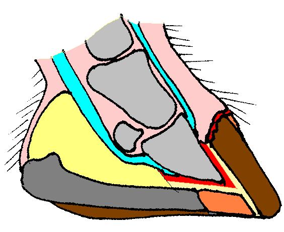

Understanding four structures prevents most owner confusion.
The hoof wall is the hard outer keratin shell. It has no nerve supply. It grows from the coronary band downward. A crack in the wall is not painful by itself.
The sole is the underside of the hoof. It protects the distal phalanx and is sensitive to pressure when inflamed.
The frog is the V-shaped structure at the heel. It aids circulation and traction and is the most common site of puncture wounds [5].
The sensitive laminae are the interlocking layers that suspend the distal phalanx inside the hoof capsule. They are innervated and vascularized. They are the tissue damaged in laminitis and the tissue that hurts in an abscess.
Imaging helps veterinarians assess the spatial relationship between the distal phalanx and the hoof capsule. Transmural ultrasonography can define normal hoof tissue measurements and the position of the distal phalanx, which is useful for comparing normal and abnormal feet [15]. Radiographic measurements of foot conformation have been described in Miniature Horses and Ponies, where reliable reference values are important for podiatric management [16]. These tools exist because the internal relationship between bone and capsule is what determines outcome in laminitis.
Cooling and Supportive Care in Laminitis
Veterinarians use distal limb cooling as part of laminitis management. In a study of eight healthy horses, high-volume and low-volume ice water immersion significantly decreased lamellar tissue temperature over four hours, while an ice pack system and an ice sleeve did not [17]. The same study estimated lamellar temperature from hoof wall temperature with good concordance, and in horses free in a stall for eight hours, immersion methods produced large sustained temperature reductions compared with control [17]. This is why veterinarians recommend standing a laminitic horse in ice and water rather than applying wraps. It is a clinical decision, and the method matters.
Mechanical support is also part of chronic laminitis management. In one case, wooden shoes were used successfully to manage lameness in a horse with radiographic rotation and distal displacement of the third phalanx [11]. This is a veterinary and farrier decision, not something an owner should improvise. Other research has explored hoof progenitor cells as a potential future therapy for laminitis, and those cells show distinct metabolic and inflammatory responses compared with other stem cell populations [3]. These are research directions, not current owner-level treatments.
The Decision Path: Farrier or Veterinarian
The fastest way to get this right is to sort the problem by urgency and by which tissue is involved. The flowchart below traces the main decision path from a hoof concern to the right professional.
flowchart TD
    A[Notice a hoof problem] --> B{Is the horse severely lame}
    B -->|Yes| C{Is one foot or both front feet affected}
    C -->|One foot| D[Call veterinarian for abscess or puncture]
    C -->|Both front feet| E[Call veterinarian now for laminitis]
    B -->|No| F{Is there a wound or foreign body}
    F -->|Yes| G[Call veterinarian immediately]
    F -->|No| H{Is there odor or black discharge}
    H -->|Yes| I[Farrier for thrush trimming]
    H -->|No| J{Is the shoe loose or the wall cracked}
    J -->|Yes| K[Call farrier]
    J -->|No| L[Continue daily care and monitor]
Equipment and Supplies: What to Keep
You do not need a large kit. You need the right few items and the discipline to use them.
- A hoof pick with a stiff brush. The brush matters as much as the pick for cleaning the sulci.
- A stiff-bristled brush for the wall and sole.
- Clean, dry bedding and a plan for muddy areas.
- A hoof knife is a farrier tool. Owners should not carve the sole or frog. Amateur paring removes protective tissue and can open a tract that was not there.
- A thermometer and a basic first aid kit for wounds.
- A written record of farrier dates and any hoof changes, so you can show the veterinarian and farrier a timeline.
Selection criteria for topical products: choose products intended for equine hoof use, follow the label, and avoid caustic or bleach-based preparations on sensitive tissue. If the frog is painful or bleeding, stop and call a veterinarian instead of treating.
Common Mistakes Owners Make
- Skipping daily picking because the horse is not in work. Packed debris is the problem, not exercise.
- Letting the interval drift to eight or ten weeks because the horse "looks fine." The foot changes before the lameness appears.
- Treating a three-legged lame horse with a poultice for days before calling a veterinarian. A puncture wound with a retained foreign body can involve synovial structures and needs prompt surgical care [5][10].
- Assuming a hoof ring is cosmetic. Rings and bilateral lameness are recognized features of metabolic disease and laminitis [6].
- Paring the sole or frog with a hoof knife. This is a farrier and veterinary task.
- Riding a horse with a loose shoe. This can tear wall and drive nails into sensitive tissue.
- Adding a hoof supplement without a diagnosis. A wall problem can be systemic or toxic rather than nutritional [14].
- Treating laminitis as a farrier problem. Laminitis is a medical emergency that requires a veterinarian.
Welfare and Safety
Hoof pain is one of the most significant welfare problems in horses, and it is also a human safety problem. A horse in severe foot pain can become recumbent, difficult to handle, and dangerous to move. In the selenium toxicity report, one horse was euthanized because of permanent foot pain and an inability to stand [14]. That outcome is a reminder that hoof disease is not a minor issue.
Handle a lame horse calmly and minimize forced movement. Do not force a laminitic horse to walk for exercise during the acute phase without veterinary direction. Use safe restraint and never stand directly behind or beside a hind limb that is being picked up. If a horse is recumbent and cannot rise, call a veterinarian immediately.
When to Call a Veterinarian Instead of a Farrier
Call a veterinarian for any of the following:
- Sudden severe lameness in one foot, especially with heat or a strong digital pulse.
- Lameness in both front feet, a rocked-back stance, or reluctance to move.
- Any suspected puncture wound, especially in the frog, or a visible foreign body.
- A hoof that is hot, swollen, or painful to pressure.
- A wound or discharge at the coronary band.
- New hoof rings on multiple feet, or a rough fragile wall on all four feet.
- A horse that is recumbent or cannot rise.
- Any hoof problem that is not improving with basic care within a few days.
Call a farrier for routine trimming, shoe resetting, a loose shoe, a ragged frog in a comfortable horse, and a simple superficial crack. When in doubt, call the veterinarian first. The veterinarian can then coordinate with the farrier.
Limitations and When to Contact a Veterinarian
This guide covers general hoof care and the most common hoof conditions. It cannot diagnose your horse. Individual horses differ in conformation, metabolic status, work, and environment, and the same sign can have different causes in different animals. A veterinarian who examines your horse, and in some cases takes radiographs, is the only reliable way to sort a serious hoof problem.
Contact a veterinarian promptly for acute severe lameness, bilateral forelimb lameness, any suspected puncture wound, a hot and painful hoof, a coronary band injury, or a horse that cannot rise. Contact a veterinarian if a hoof problem persists beyond a few days of basic care or if you are unsure whether the problem is mechanical or systemic. Contact a farrier for routine trimming and shoeing and for a loose shoe, and coordinate both professionals for any horse with laminitis or a chronic hoof wall disorder.
Frequently Asked Questions
How often should horse hooves be trimmed?
Every four to six weeks is the usual starting interval. The correct interval for your horse depends on hoof growth rate, work level, footing, and conformation, so treat four to six weeks as a default and adjust with your farrier.
Can I trim my own horse's hooves?
Routine trimming is a farrier skill. Owners can pick hooves daily and keep them clean, but paring the sole or frog with a hoof knife removes protective tissue and can cause harm. Leave trimming to a farrier.
What is the difference between laminitis and an abscess?
An abscess is a localized infection in one foot that causes sudden severe lameness and usually improves once it drains. Laminitis is a systemic disease of the lamellar junction that often affects both front feet and requires immediate veterinary care.
Is laminitis an emergency?
Yes. Laminitis is a medical emergency. It involves failure of the lamellar attachment and can lead to rotation or displacement of the distal phalanx, so it needs a veterinarian the same day, not just a farrier visit.
What causes thrush in horse hooves?
Thrush develops when the frog and sulci stay wet and packed with debris, creating an oxygen-poor environment. Daily picking, dry footing, and regular farrier trimming prevent most cases.
Why does my horse have rings on its hooves?
Hoof rings can mark past episodes of illness, fever, or laminitis. Rings combined with bilateral lameness or body condition changes are a reason to have a veterinarian evaluate the horse for metabolic disease.
Should I call a farrier or a veterinarian for a lame horse?
Call a veterinarian first for any sudden severe lameness, bilateral forelimb lameness, or suspected puncture wound. Call a farrier for a loose shoe or routine trimming. When unsure, start with the veterinarian.
Can a horse have a hoof crack without pain?
Yes. The hoof wall has no nerve supply, so a crack in the wall itself is not painful. Pain comes from the sensitive laminae and other innervated tissue beneath, so a deep or unstable crack still needs evaluation.
Related Articles
- Horse Hoof Care: Anatomy, Trimming, and Common Problems
- Equine Hoof Care: Anatomy, Trimming, and Shoeing
- Miniature Horse Care Guide
- Senior Horse Care Guide
- Complete Horse Care Guide for New Owners
- Chinchilla Care: Overgrown Dental Spurs & Tooth Trimming Guide
Sources
- Horse Hoof Trimmings as an Untapped Resource for Sustainable Keratin Utilization.
- Distribution of endocannabinoid system receptors in the equine hoof: dysregulation as a potential therapeutic target for laminitis.
- Equine Hoof Progenitor Cells Display Increased Mitochondrial Metabolism and Adaptive Potential to a Highly Pro-Inflammatory Microenvironment.
- Interleukins decrease epithelial protein and gene expression by K14 + CD105+ equine hoof progenitor cells.
- [[Puncture wounds of the equine hoof - a retrospective study of 96 cases - Part 1: Prevalence, clinical findings and diagnostic investigations].](https://pubmed.ncbi.nlm.nih.gov/41921941/)
- Equine Metabolic Syndrome: A Complex Disease Influenced by Multifactorial Genetic Factors.
- The Immediate Effect of Routine Hoof Trimming and Shoeing on Horses' Gait.
- Lamellar cell death and proliferation are associated with restricted ambulation and preferential weight bearing in a model relevant to supporting-limb laminitis.
- The role and importance of farriery in equine veterinary practice.
- [[Puncture wounds of the equine hoof - a retrospective study of 96 cases - Part 2: Management and prognosis].](https://pubmed.ncbi.nlm.nih.gov/42624121/)
- Use of wooden shoes for the treatment of chronic laminitis in an American quarter horse.
- Cannabinoid and cannabinoid related receptors in fibroblasts, inflammatory and endothelial cells of the equine hoof with and without laminitis: novel pharmacological target.
- Cyclooxygenase-2 and von Willebrand factor-an immunohistochemical study of the equine foot with and without laminitis, post-mortem perfused with paraffin oil.
- Selenium deposition in an atypically disintegrated hoof wall in a Thoroughbred racehorse with alkali disease: Proof by energy-dispersive X-ray fluorescence analysis.
- Transmural Ultrasonography in the Evaluation of Horse Hoof Internal Structures: Comparative Quantitative Findings-Part 2.
- Radiographic measurements of Miniature Horse and Miniature Pony Feet.
- Distal limb immersion in ice and water is the most effective means of cooling the equine hoof lamellae under clinically relevant conditions.