Horse Face Anatomy: Structures and Function
By Dr. Zubair Khalid, DVM, MS, PhD ·
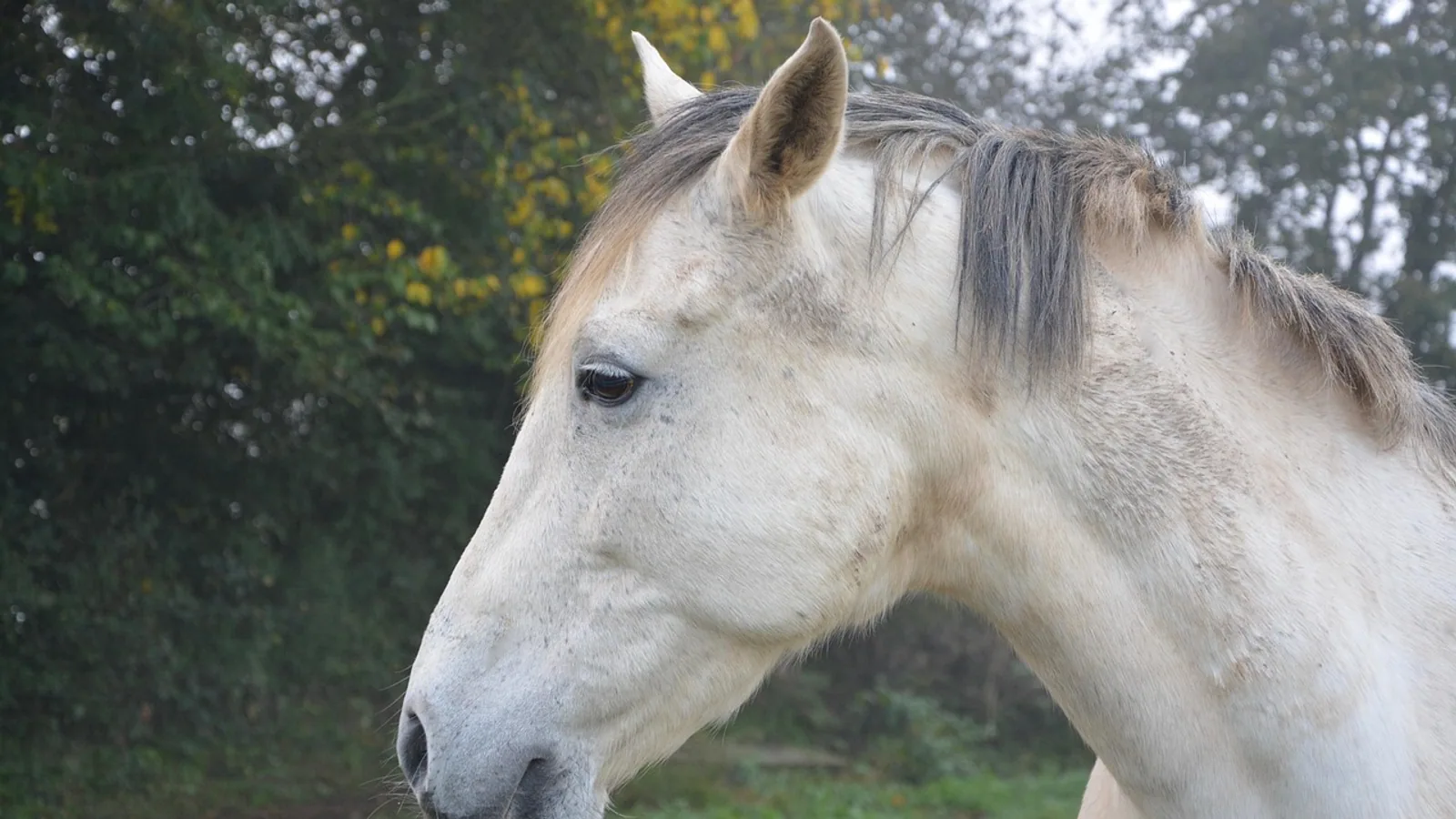
The horse's face is a load-bearing, air-filled, tooth-dense structure that supports grazing, breathing, vision, and social signaling all at once. Its bones, sinuses, and teeth are arranged so that the horse can grind tough forage for most of the day while keeping a large, lightweight nasal cavity for airflow and a wide visual field for detecting predators.
This article is educational and is not a substitute for veterinary diagnosis or treatment.
Why the Equine Face Is Built Differently
Domestic horses evolved as grazing animals that spend much of the day with their heads down. The face reflects that lifestyle. The skull is elongated, the nasal cavity is large, and the cheek teeth are deep-rooted grinders that wear down over a lifetime. The paranasal sinuses, a system of air-filled cavities inside the skull, reduce the weight of the head and warm and humidify inhaled air.
Veterinarians care about this anatomy for practical reasons. Sinus disease, dental disease, and nerve blocks for standing surgery all depend on knowing exactly where the bony landmarks and nerve paths sit on the horse's face. Modern imaging studies, including computed tomography (CT) and three-dimensional models, have refined that knowledge in recent years [1][2][3].
The Facial Bones of the Horse
The equine face is built from a set of flat and irregular bones that interlock at sutures. Each has a specific role in protecting soft tissue, anchoring teeth, or forming the walls of the sinuses.
Incisive Bone
The incisive bone (also called the premaxilla) forms the rostral part of the upper jaw and holds the upper incisor teeth. It meets the maxilla behind it. In the live horse, this is the bone you feel when you run your hand down the front of the face toward the upper lip.
Maxilla
The maxilla is the largest facial bone. It holds the upper cheek teeth (premolars and molars), forms the lateral wall of the nasal cavity, and contributes a large part of the maxillary sinus. The facial crest, a ridge running along the side of the face, is part of the maxilla. The facial crest is a palpable landmark and is used as a reference point for locating the infraorbital foramen and for surgical approaches to the sinuses [2].
Nasal Bone
The nasal bone forms the roof of the nasal cavity and the dorsal contour of the face. In the horse, the nasal bone is long and narrow, and its rostral end forms the nasal notch. The nasal bone is thin and can be fractured by kicks or collisions.
Lacrimal Bone
The lacrimal bone sits near the medial corner of the eye. It contains the lacrimal fossa and is associated with the nasolacrimal duct, which drains tears from the eye to the nasal cavity. The nasolacrimal duct is a palpable and clinically relevant structure because it can become obstructed or infected.
Zygomatic Bone
The zygomatic bone forms the lateral wall of the orbit and the caudal part of the facial crest. It articulates with the frontal, lacrimal, and maxillary bones. The zygomatic arch is the bony bridge you can feel behind the eye.
Frontal Bone
The frontal bone forms the forehead and the dorsal wall of the frontal sinus. It also contributes to the orbital rim. In the horse, the frontal bone is relatively thick and protects the frontal sinus, which is one of the larger sinus compartments [1][3].
The Paranasal Sinuses of the Horse
The paranasal sinuses are a bilaterally symmetrical system of air-filled cavities. In the horse, the system is usually described as eight compartments on each side, although the exact number depends on how the compartments are grouped [1]. The sinuses communicate with the nasal cavity through small openings, and that drainage is central to how sinus disease develops and resolves [4].
The equine sinus system is one of the most complex parts of the horse's body [1]. CT studies have shown that the sinuses are not simple balloons. They are overlapping, compartmentalized spaces that vary with age and breed [1][5][6].
Frontal Sinus
The frontal sinus sits under the frontal bone on the forehead. It is part of the caudal sinus system and communicates with the dorsal conchal sinus. In a CT study of 300 horses with sinonasal disease, the frontal sinus was affected in 26% of cases [3].
Caudal Maxillary Sinus
The caudal maxillary sinus is the largest sinus compartment by volume. In a study of Arabian horses, the caudal maxillary sinus had a mean volume of about 131.9 cm³, making it the largest of the sinuses measured [2]. It drains into the middle nasal meatus along with the frontal and dorsal conchal sinuses.
Rostral Maxillary Sinus
The rostral maxillary sinus is the more rostral and more dependent compartment. It was the most commonly affected sinus in a large CT study, involved in 284 of 300 horses (94.7%) with sinonasal disease [3]. It drains into the caudal maxillary sinus in many horses, and the two are separated by the maxillary septum.
Sphenopalatine Sinus
The sphenopalatine sinus is the smallest sinus compartment. In the Arabian horse study, its mean volume was about 13.3 cm³ [2]. It sits caudally and ventrally, near the sphenoid and palatine bones. It is often grouped with the caudal sinus system.
Dorsal Conchal Sinus
The dorsal conchal sinus is part of the caudal sinus system. It is affected in about 52.7% of horses with sinonasal disease, according to the 300-horse CT study [3]. It communicates with the frontal sinus.
Ventral Conchal Sinus
The ventral conchal sinus is part of the rostral sinus system. It was affected in about 87% of horses with sinonasal disease in the same CT study [3]. It is closely associated with the ventral nasal conchal bulla, which can become infected and complicate sinus disease [3].
Middle Conchal Sinus
The middle conchal sinus is a smaller compartment that sits between the dorsal and ventral conchal sinuses. It is part of the caudal sinus system and is less commonly the primary site of disease.
Table: Equine Paranasal Sinuses, Boundaries, and Drainage
| Sinus | System | Key Boundaries | Drainage Opening |
|---|---|---|---|
| Frontal | Caudal | Frontal bone, dorsal conchal sinus | Middle nasal meatus |
| Caudal maxillary | Caudal | Maxilla, zygomatic bone, orbit | Middle nasal meatus |
| Rostral maxillary | Rostral | Maxilla, facial crest, maxillary septum | Caudal maxillary sinus (via nasomaxillary aperture) |
| Sphenopalatine | Caudal | Sphenoid and palatine bones | Middle nasal meatus |
| Dorsal conchal | Caudal | Dorsal nasal concha, frontal sinus | Middle nasal meatus |
| Ventral conchal | Rostral | Ventral nasal concha, maxillary sinus | Rostral maxillary sinus |
| Middle conchal | Caudal | Nasal conchae, ethmoid region | Middle nasal meatus |
The drainage openings are small, and that is why sinus disease can persist. The nasomaxillary aperture, which connects the caudal maxillary sinus to the middle meatus, has a mucosal lining that differs from the rest of the sinus lining. A histological study found that the nasomaxillary aperture mucosa is much thicker than the paranasal sinus mucosa, with an average height of about 820 µm compared with about 76 µm for the sinus mucosa [7]. That difference matters for how the aperture behaves when inflamed.
The Dental Arcades and Hypsodont Teeth
Horses have two dental arcades on each jaw: the incisors at the front and the cheek teeth at the back. The cheek teeth include premolars and molars, and they are hypsodont, meaning they have high crowns and continue to erupt throughout life as the grinding surface wears down.
Incisors
The incisors are used for nipping grass. They are brachydont in structure compared with the cheek teeth, meaning they have a defined crown and root. The incisive bone holds the upper incisors, and the mandible holds the lower incisors. The mental foramen, which is the exit point of the mental nerve, sits near the lateral incisor tooth. In a study of Arabian horses, the mean distance between the mental foramen and the most lateral incisive tooth was about 3.12 cm [2].
Cheek Teeth
The cheek teeth are the workhorses of the equine mouth. They grind forage in a side-to-side motion. The upper cheek teeth are rooted in the maxilla, and the lower cheek teeth are rooted in the mandible. The roots of the upper cheek teeth are closely related to the maxillary sinuses, which is why dental disease can spread into the sinuses and why sinus disease can have a dental origin.
In a CT study of 300 horses with sinonasal disease, dental disease was the cause in 53% of cases, and primary sinusitis accounted for 25.7% [3]. That split is a useful reminder that sinus problems in horses are often dental problems first.
Hypsodont Structure
Hypsodont teeth have a long crown that extends below the gumline and continues to erupt as the occlusal surface wears. The crown is made of enamel, dentin, and cementum arranged in folded layers. That folding creates the ridges and valleys that make the grinding surface efficient. Because the teeth keep erupting, the roots change position over time, and the relationship between the tooth roots and the sinuses changes with age [1].
The Infraorbital and Mental Nerves
The sensory nerves of the horse's face are clinically important because they are the targets for nerve blocks used in standing surgery and dental procedures.
Infraorbital Nerve
The infraorbital nerve is a branch of the maxillary nerve. It exits the skull through the infraorbital foramen, which sits on the lateral face just dorsal to the facial crest. In a study of Arabian horses, the infraorbital foramen was located about 4.16 cm from the most rostral point of the facial crest and about 4.70 cm from the alveolar root [2]. Those measurements are useful for locating the foramen when performing an infraorbital nerve block.
An infraorbital nerve block desensitizes the upper lip, nose, and upper incisors. It is used for procedures on the upper lip and for some dental work.
Mental Nerve
The mental nerve is a branch of the inferior alveolar nerve. It exits the mandible through the mental foramen, which sits on the lateral aspect of the mandible near the incisors. The mean distance from the mental foramen to the most lateral incisive tooth is about 3.12 cm in Arabian horses [2]. A mental nerve block desensitizes the lower lip and lower incisors.
Clinical Relevance of Nerve Blocks
Nerve blocks allow veterinarians to perform procedures on the standing horse without general anesthesia. Accurate placement depends on knowing the bony landmarks. The facial crest is the key landmark for the infraorbital foramen, and the incisor teeth are the key landmark for the mental foramen. CT-based morphometric studies have refined these distances for specific breeds, and the values can vary with head shape [2][5].
The Facial Crest and Nasolacrimal Duct as Palpable Landmarks
The facial crest is a ridge of bone on the lateral face. It is easily palpable and is used as a reference for the infraorbital foramen, for sinus surgery, and for radiographic positioning. In a radiographic study of conchal sinuses, the facial crest was used as a centering landmark for a complementary projection [8].
The nasolacrimal duct runs from the lacrimal bone near the eye to the nasal cavity. It is palpable in some horses and can be catheterized for diagnostic or therapeutic purposes. Obstruction of the duct causes epiphora, or excessive tearing.
The Muscles and Sensory Structures of the Face
The horse's face has a complex set of mimetic muscles that produce facial expressions. These muscles are innervated by branches of the facial nerve, and their nerve entry points have been mapped in a study of five equine facial muscles: the caninus, levator anguli oculi medialis, nasolabialis, masseter, and zygomaticus [9].
The nerve entry points were located at specific relative distances along each muscle. For the caninus, the entry point was at 33% to 38% from the caudal landmark. For the levator anguli oculi medialis, it was at 69% to 86%. For the zygomaticus, it was at 0% to 18%. The nasolabialis had two innervation zones, at 47% to 72% for the dorsal branch and 52% to 91% for the ventral branch [9].
This mapping matters for pain assessment. The Horse Grimace Scale uses facial action units to score pain, and better anatomical descriptions of the muscles involved improve the reliability of that scoring [10]. Surface electromyography is being explored as a way to measure facial muscle activity objectively, and accurate nerve entry point locations are needed for electrode placement [9].
Comparative Notes: Cattle and Dogs
The equine sinus system is not the same as that of other species. Cattle have a different arrangement of paranasal sinuses, and their facial musculature differs as well. A dissection study of cattle identified 30 mimetic muscles and found notable fiber blending in the lower face and ear region. The study noted that cattle have a slightly less diverse facial repertoire than horses, with robust lips and a different lip-muscle arrangement that likely restricts some movements seen in horses [11]. The eyebrow region in cattle is served only by a broad frontalis muscle, which suggests fewer eyebrow movements than in horses [11].
Dogs have a much smaller and simpler sinus system than horses. The frontal sinus is the most prominent, and the maxillary sinus is small or absent in many breeds. The canine face is also much shorter, and the dental arcade is brachydont rather than hypsodont. These differences mean that diagnostic and surgical approaches to the face cannot be directly translated between species.
Practical Implications for Owners and Keepers
Understanding the horse's face helps owners recognize problems early. Sinus disease often presents as nasal discharge, facial swelling, or a foul odor from the nose. Because dental disease is the most common cause of sinus disease in horses [3], regular dental examinations are important.
Facial swelling over the sinuses, especially unilateral swelling, warrants veterinary attention. The rostral maxillary sinus and ventral conchal sinus are the most commonly affected compartments [3], and those sit in the rostral and ventral part of the face. Swelling in that area is easier to see than swelling deeper in the caudal sinuses.
Nasal discharge that is unilateral and persistent is another sign to investigate. In a long-term study of 155 horses treated with sinoscopy, 69.7% fully responded to initial treatment, but some required further surgery for bone sequestra, inspissated exudate, or misdiagnosed dental apical infections [12]. That outcome data shows that sinus disease can be managed but sometimes needs more than one procedure.
Owners should also be aware that the horse's face is vulnerable to trauma. The nasal bone and facial crest can be fractured by kicks or collisions. Any facial trauma with swelling, bleeding, or asymmetry should be evaluated by a veterinarian.
What Is Still Uncertain
The anatomy of the equine sinuses is well described, but some details remain debated. The exact number of sinus compartments and how they should be grouped varies between references. Some studies describe eight compartments, while others group them into fewer functional units [1][6]. The clinical significance of the middle conchal sinus and the sphenopalatine sinus is less well understood than that of the maxillary and conchal sinuses.
Age-related changes in sinus volume are also an active area of research. A CT study of 18 cadaver horses found that the volume and surface area of the ventral conchal sinus, rostral maxillary sinus, and caudal maxillary sinus increased with age [1]. The same study found that age-related clustering was possible for the rostral and caudal maxillary sinuses but not for the ventral conchal sinus [1]. That suggests that some sinuses change predictably with age and others do not.
Breed differences are another area of uncertainty. Noma horses, a Japanese pony breed, have a skull and sinus ratio similar to larger breeds despite their small body size [5]. Shetland ponies, by contrast, have a different relationship between head size and conchal bulla size [5]. These findings suggest that breed-specific reference values may be useful for imaging and surgery.
Limitations and When to Contact a Veterinarian
This article is educational and is not a substitute for veterinary diagnosis or treatment. Individual cases need a veterinarian who can examine the horse, take a history, and use imaging if needed.
Contact a veterinarian if your horse has:
- Unilateral or bilateral nasal discharge that persists for more than a few days
- Facial swelling, especially over the sinuses or along the jaw
- Foul odor from the nose or mouth
- Difficulty eating, dropping feed, or weight loss
- Asymmetry of the face or jaw
- Bleeding from the nose
- Signs of dental pain, such as head tossing, quidding, or resistance to the bit
- Trauma to the face with swelling, bleeding, or instability
Your veterinarian may recommend endoscopy, radiography, or CT to evaluate the sinuses and teeth. CT is particularly useful for identifying which sinus compartments are affected and for detecting dental disease that may not be visible on radiographs [3][8].
Frequently Asked Questions
What are the main bones of a horse's face?
The main facial bones are the incisive bone, maxilla, nasal bone, lacrimal bone, zygomatic bone, and frontal bone. The maxilla is the largest and holds the upper cheek teeth.
How many paranasal sinuses does a horse have?
The equine paranasal sinus system is usually described as eight compartments on each side, including the frontal, caudal maxillary, rostral maxillary, sphenopalatine, dorsal conchal, ventral conchal, and middle conchal sinuses [1].
Which sinus is most commonly affected by disease?
The rostral maxillary sinus is the most commonly affected, involved in about 94.7% of horses with sinonasal disease in a large CT study. The ventral conchal sinus is second, at about 87% [3].
What is a hypsodont tooth?
A hypsodont tooth has a high crown that continues to erupt as the grinding surface wears down. Horses have hypsodont cheek teeth that are adapted for grinding tough forage.
Where is the infraorbital foramen?
The infraorbital foramen sits on the lateral face, just dorsal to the facial crest. In Arabian horses, it is about 4.16 cm from the most rostral point of the facial crest [2].
What is the mental nerve block used for?
A mental nerve block desensitizes the lower lip and lower incisors. It is used for procedures on the lower lip and for some dental work.
What is the facial crest?
The facial crest is a bony ridge on the lateral side of the face. It is a palpable landmark used for nerve blocks, sinus surgery, and radiographic positioning [2][8].
Do cattle and dogs have the same sinus anatomy as horses?
No. Cattle have a different arrangement of paranasal sinuses and facial muscles, with a less diverse facial repertoire than horses [11]. Dogs have a much smaller and simpler sinus system, and their teeth are brachydont rather than hypsodont.
Related Articles
- Equine Hoof Anatomy: Structures and Function
- Equine Larynx Anatomy and Function in Respiration
- Canine Lymphatic System: Anatomy and Function
- Canine Liver Anatomy and Lobular Structure
- Horse Hoof Care: Anatomy, Trimming, and Common Problems
- Equine Digestive System: Anatomy and Physiology of the Hindgut
- Condyloid Joint: Structure and Movement
- Papillary Dermis: Structure and Function Explained
- Anatomical Heart: Structure and Chambers
Sources
- Three-Dimensional Segmentation of Equine Paranasal Sinuses in Multidetector Computed Tomography Datasets: Preliminary Morphometric Assessment Assisted with Clustering Analysis.
- Applied anatomy of the skull in the Arabian horse: A computed tomographic, cross-sectional, volumetric and morphometric study.
- Computed Tomographic Assessment of Individual Paranasal Sinus Compartment and Nasal Conchal Bulla Involvement in 300 Cases of Equine Sinonasal Disease.
- Disorders of the Equine Head and Neck: Paranasal Sinuses.
- An anatomical study of the skull, the dorsal and ventral nasal conchal bullae and paranasal sinuses in normal Noma horses: Computed tomographic anatomical and morphometric findings.
- Volumetric measurements of paranasal sinuses and examination of sinonasal communication in healthy Shetland ponies: anatomical and morphometric characteristics using computed tomography.
- Comparative studies on the histological characteristics of equine nasomaxillary aperture and paranasal sinus mucosa considering topographic and age-related differences.
- Complementary Radiographic Projection for Evaluation of the Conchal Sinuses and Bullae in Horses.
- Nerve entry points in the mimic musculature of the horse head.
- Enhancing the Horse Grimace Scale (HGS): Proposed updates and anatomical descriptors for pain assessment.
- Descriptive, comparative, and functional anatomy of the facial musculature in cattle (Bos taurus).
- A long-term study of sinoscopic treatment of equine paranasal sinus disease: 155 cases (2012-2019).