Skin Disease in Dogs: Common Causes and Treatments
By Dr. Zubair Khalid, DVM, MS, PhD ·

Skin disease on a dog is one of the most common reasons owners seek veterinary care. Most cases fit into four broad groups: allergic skin disease, infection, parasites, and immune-mediated or reactive conditions. Many dogs have more than one problem at once. A dog with year-round itching, repeated ear infections, and thinning hair often has atopic dermatitis complicated by bacterial and yeast overgrowth.
The single most useful step is a diagnosis. Owners often reach for a shampoo or an over-the-counter spray first, but the same crusted patch can be a bacterial infection, a yeast infection, a parasite, or an allergic flare. Treatment that targets the wrong cause wastes weeks and can make the skin worse.
Triage summary
- See a veterinarian soon (within days): itching that disturbs sleep, widespread redness, greasy or musty odor, hair loss, recurring ear discharge, or a dog that keeps licking its paws.
- See a veterinarian promptly (within 24 hours): rapidly spreading redness, painful skin, swollen eyelids or face, or a dog that stops eating because of skin discomfort.
- Emergency care now: difficulty breathing, facial swelling with vomiting, hives covering the body, or extensive blistering or peeling skin.
This article is educational and is not a substitute for veterinary diagnosis or treatment.
At a Glance: Common Skin Conditions in Dogs
| Condition | Typical signs | Usual first step | Typical response |
|---|---|---|---|
| Canine atopic dermatitis | Paw licking, face rubbing, ear scratching, red itchy skin, often seasonal at first | Veterinary exam plus history, itch and lesion scoring, allergy testing when indicated | Weeks to months, lifelong management |
| Bacterial pyoderma | Pustules, papules, crusts, circular patches, greasy odor | Skin cytology and veterinarian-selected antimicrobial therapy | Often 3 to 6 weeks of treatment |
| Malassezia dermatitis | Greasy, musty-smelling skin, thickened dark skin, ear wax | Cytology and targeted antifungal therapy | Weeks, with relapse common if the trigger remains |
| Otitis externa | Head shaking, ear scratching, discharge, odor | Ear exam and cytology | Days to weeks depending on cause |
| Ectoparasites | Scratching, fleas, crusts, hair loss, sometimes rash | Parasite control and environmental management | Rapid once the parasite is controlled |
| Cutaneous leishmaniosis | Pad ulcers, nail overgrowth, scaling on the nose and ears, weight loss | Blood tests, cytology, serology, PCR | Months, guarded in advanced disease |
| Periocular histiocytic disease | Firm eyelid or corner-of-eye mass in a young dog | Biopsy and imaging | Good with surgery, with or without medication |
How Canine Skin Works and Why It Fails
Healthy skin is a barrier, an immune organ, and a microbiome all at once. The outer layer of keratinocytes, held together by tight junctions and structural proteins, keeps water in and allergens, microbes, and irritants out. When that barrier is weak, environmental proteins cross more easily and trigger inflammation.
Laboratory work on canine epidermal organoids shows what happens when the allergic cytokine environment takes over. Treating canine keratinocytes with the Th2 cytokines IL-4 and IL-13 and with TNF-alpha produced changes that resemble atopic skin, including epidermal spongiosis and reduced suprabasal keratinocyte differentiation [1]. The same experiment found that Th2 cytokines increased keratinocyte proliferation while TNF-alpha drove apoptosis within the model [1]. The barrier and the immune response are linked, which is why a dog with atopic dermatitis often has dry, scaly skin even between flares.
A meta-analysis comparing human and canine atopic dermatitis transcriptomes found fifteen conserved differentially expressed genes across datasets [2]. Upregulated genes pointed to Th17 signaling through S100A8 and S100A9 and to JAK-STAT signaling through FHL1 and STAT1, and the Th2-related chemokine CCL13 was increased in both species [2]. An IL-27-mediated signaling pathway also emerged as a shared feature [2]. These findings explain why the JAK-STAT pathway has become a practical drug target in veterinary dermatology.
The gut is part of the picture too. A pilot study measured short-chain fatty acids in the feces of dogs with atopic dermatitis and healthy controls and found significantly lower concentrations of acetic, propionic, and butyric acids in the affected dogs [3]. Short-chain fatty acids are microbiota metabolites with anti-inflammatory properties, and the finding supports a link between the gut microbiome and canine atopic disease [3].
The Main Causes of Skin Disease on a Dog
Allergic Skin Disease and Canine Atopic Dermatitis
Canine atopic dermatitis is a hereditary, largely pruritic, T-cell-driven inflammatory disease shaped by skin barrier defects, allergen sensitization, and microbial dysbiosis [4]. It affects up to 15 percent of dogs [5]. In a Swiss vertical prevalence study of 743 pruritic cases, atopic dermatitis was the most frequent diagnosis at 59.1 percent, followed by otitis at 26.1 percent, pyoderma at 22.6 percent, and ectoparasites at 7.1 percent [6].
The distribution of itch is a useful clue. In that same study, 56 percent of atopic dogs scratched their paws and 48.1 percent scratched the pinna or ear [6]. Secondary infection was common. Just over half of the atopic dogs, 54.7 percent, had pyoderma and 31.8 percent had Malassezia dermatitis [6]. That is the practical reason a veterinarian examines the whole dog rather than only the itchy patch.
Environmental triggers vary. A prospective study in Bavaria followed 37 atopic dogs from April through November and compared daily pruritus scores with local weather and pollen counts [4]. Only humidity and medication scores correlated positively with pruritus scores, and there was no correlation between specific pollen counts and itching in dogs with positive intradermal reactions to that pollen [4]. Owners often blame a single pollen, but the real drivers are more complex.
Food allergy can look identical to atopic dermatitis and is a common differential in dogs with year-round itch, especially when the face, ears, paws, and perineum are involved. A veterinarian may recommend an elimination diet trial before committing to lifelong allergy management.
Bacterial Pyoderma
Pyoderma is a bacterial infection of the skin, usually caused by Staphylococcus organisms. It appears as pustules, papules, red circular patches, crusts, and sometimes a greasy odor. It is frequently secondary to allergy, parasites, or endocrine disease. In the Swiss study, 22.6 percent of pruritic dogs had pyoderma, and it was present in more than half of the atopic dogs [6].
Superficial pyoderma is usually treated for three to six weeks, and the veterinarian may recheck the skin before stopping therapy to confirm resolution. Deep or recurrent pyoderma warrants a search for an underlying cause. Repeated antibiotic courses without addressing the trigger predictably lead to relapse.
Malassezia Dermatitis
Malassezia is a yeast that lives on normal canine skin. It becomes a problem when the skin environment changes, especially with allergies, humidity, or folds. Signs include greasy skin, a musty odor, dark thickened patches, and waxy ear discharge. In the Swiss survey, 31.8 percent of atopic dogs had Malassezia dermatitis [6]. Diagnosis relies on cytology, and treatment combines targeted antifungal therapy with control of the underlying trigger.
Otitis Externa and Media
Ear disease is often the first visible sign of underlying allergy. In the Swiss study, otitis was the second most common diagnosis at 26.1 percent [6]. Dogs with ear disease need an otoscopic examination and cytology because bacterial and yeast infections look similar from the outside. Chronic ear disease can progress to the middle ear and cause head tilt or facial nerve signs, so repeated episodes deserve a full workup.
Ectoparasites
Fleas, mites, and other parasites cause intense itching and can trigger secondary infection. Ectoparasites accounted for 7.1 percent of pruritic cases in the Swiss survey [6]. Flea allergy dermatitis produces a classic triangular rash on the lower back near the tail base. Sarcoptic mange causes severe scratching, especially on the ear margins and elbows. Parasite control should follow Companion Animal Parasite Council (CAPC) guidance and be consistent year-round in many regions.
Other Causes
Cutaneous leishmaniosis is a vector-borne disease caused mainly by Leishmania infantum in Europe, with dogs as the main domestic reservoir [7]. A case report of a dog relocated from Italy to Romania described weight loss, muscle wasting, overgrown nails, severe pustular pododermatitis, ulcerated foot pads, ear erosions with fine scaling, and crusting on the nose [7]. Diagnosis required bone marrow cytology, three serological methods, and real-time PCR [7]. Leishmaniosis should be on the list for dogs with unusual skin lesions and a history of travel or relocation from an endemic region.
Cutaneous periocular histiocytic disease is a benign, histiocyte-rich lesion affecting the eyelids and surrounding skin, most often on the lower eyelid and lateral canthus [8]. In a retrospective review of 14 dogs, surgical excision alone cured 2 dogs, and excision combined with medical management helped 7 more [8]. Only 3 of 9 dogs with follow-up had recurrence, and none recurred after medical management at the final recheck [8].
Endocrine disease, autoimmune skin disease, and certain cancers can also present as skin disease. These are diagnosed through blood testing, biopsy, and imaging rather than by appearance alone.
Risk Factors: Age, Breed, and Environment
Signalment matters. In the Swiss study, atopic dermatitis was overrepresented in French Bulldogs, West Highland White Terriers, and American Staffordshire Terriers compared with the general study population [6]. Affected dogs were more often middle-aged at 49.9 percent, male at 58.8 percent, and weighed 10 to 25 kg at 42.6 percent [6].
Environment matters as much as genetics. Humidity correlated with pruritus scores in the Bavarian study, and medication use tracked with itching intensity [4]. Dogs living in damp climates, dogs with skin folds, and dogs with concurrent gut dysbiosis may flare more often.
Trial design data underline how many factors can confound a diagnosis or a treatment response. A methodological review of canine atopic dermatitis trials identified food and environmental allergen exposure, seasonality, flea infestation, concurrent bacterial and fungal infections, concomitant therapies, and washout periods as key variables that must be controlled [5]. Owners should expect their veterinarian to ask about diet, season, flea control, and recent medications for exactly this reason.
What a Veterinary Workup Looks Like

A dermatology appointment starts with a detailed history. Your veterinarian will ask when the itching began, whether it is seasonal, which body areas are affected, what the dog eats, what parasite prevention is used, and what has already been tried. The physical examination covers the whole coat, ears, paws, face, and perineum.
Core diagnostics
- Skin scraping and cytology. Cytology identifies bacteria and yeast and is the fastest way to confirm infection. Skin scraping looks for mites.
- Ear swab cytology. Used whenever there is ear discharge or head shaking.
- Flea combing and parasite history. Confirms or rules out ectoparasites.
- Lesion and itch scoring. The Canine Atopic Dermatitis Extent and Severity Index, 4th iteration (CADESI-4), quantifies visible lesions, and the Pruritus Visual Analog Scale (PVAS) quantifies itch. Both are used in clinical trials and in everyday practice to track response.
- Allergy testing. Intradermal testing or serum allergen testing helps identify environmental triggers when atopic dermatitis is confirmed and immunotherapy is being considered [4].
- Biopsy and histopathology. Reserved for unusual lesions, suspected autoimmune disease, or masses such as the periocular histiocytic lesions described in the retrospective case series [8].
- Serology and PCR for infectious disease. Used when leishmaniosis or another vector-borne disease is suspected, as in the relocated dog that required three serological methods plus real-time PCR [7].
- Bloodwork. A minimum database helps screen for endocrine or systemic disease and provides baseline values before long-term medication.
A diagnosis is not always reached at the first visit. Many dogs need a stepwise approach in which infection is cleared first, then allergy is investigated. That sequence prevents a skin infection from masking the underlying allergic disease.
Evidence-Based Treatment

The International Committee on Allergic Diseases of Animals (ICADA) updated its treatment guidelines in 2026 and organized therapy by mechanism of action into two phases: reactive therapy to resolve existing pruritus and lesions, followed by proactive therapy to maintain remission and reduce flares [15]. That framework is the clearest way to think about treatment for the itchy dog.
Reactive Therapy: Breaking the Flare
Rapid-acting broad-spectrum drugs, particularly glucocorticoids, are recommended for reactive treatment [15]. Prednisolone is the classic example. In a controlled trial of 23 atopic dogs, a protocol combining oclacitinib and prednisolone produced significant reductions in CADESI and PVAS scores by day 7, and scores stayed stable through day 60 with no significant difference between the combination group and oclacitinib alone [12]. Adverse events in that trial included polyuria, polydipsia, and polyphagia, all of which resolved by day 14 [12]. Short courses of glucocorticoids can be very effective, but long-term use requires monitoring for exactly these effects.
Proactive Therapy: Keeping the Dog Comfortable
Proactive management is essential to prevent repeated cycles of inflammation [15]. The ICADA guidelines list skin barrier care, antimicrobial and microbiome-modulating interventions, avoidance of allergenic and nonallergenic flare factors, allergen-specific immunotherapy, and anti-inflammatory, antipruritic, and immunomodulatory treatments as components of long-term control [15]. The specific mix depends on the individual dog.
Targeted Antipruritic and Immunomodulatory Drugs
JAK inhibitors. Oclacitinib is a Janus kinase inhibitor used for canine atopic dermatitis. In a blinded, randomized, crossover study of healthy dogs, a single oral dose of oclacitinib reduced total and local pruritic behavior seconds after intradermal IL-31 injection and reduced scratching, chewing and biting, and head shaking [16]. Atinvicitinib, a second-generation highly selective JAK1 inhibitor, was tested in a multicenter, blinded, randomized, placebo-controlled trial of 61 atopic dogs [11]. Treatment success, defined as at least a 50 percent reduction from baseline in PVAS or CADESI-04 at day 28, was 87.5 percent in the once-daily group and 73.3 percent in the twice-daily-then-once-daily group, compared with 23.1 percent in the placebo group [11]. These drugs act quickly and are an important option for moderate to severe itch.
Lokivetmab. This injectable biologic targets IL-31. In a single-blind randomized study of 32 dogs with mild to moderate atopic dermatitis, both a single intravenous dose of allogeneic mesenchymal stromal cells and three monthly subcutaneous doses of lokivetmab significantly reduced CADESI-04 and PVAS scores by day 28, with no significant difference between groups, and improvement was sustained to day 84 [14].
Ciclosporin and methotrexate. For moderate to severe nonseasonal atopic dermatitis, a double-blind non-inferiority trial compared weekly subcutaneous low-dose methotrexate with oral ciclosporin in 55 dogs [13]. At week 28, mean CADESI-04 reductions were 65.4 percent with methotrexate and 69.5 percent with ciclosporin, meeting the non-inferiority margin [13]. Ciclosporin produced faster improvement between weeks 4 and 12, but efficacy converged by week 20, and gastrointestinal adverse events occurred in 82 percent of methotrexate-treated dogs versus 70 percent of ciclosporin-treated dogs [13]. These are specialist-level decisions and require monitoring.
Mesenchymal stromal cells. The same study that compared stromal cells with lokivetmab found that a single intravenous dose of allogeneic adipose-derived mesenchymal stromal cells significantly reduced CADESI-04 and PVAS scores by day 28 in dogs with mild to moderate atopic dermatitis, with further improvement by day 84 [14]. This is an emerging option and not a first-line choice.
Topical Therapy
Topical products are helpful and versatile for skin disorders, but veterinary studies assessing their efficacy have historically been limited [10]. Two recent trials add useful evidence.
A multicentre, open-label trial of 34 atopic dogs evaluated a protocol combining an Ophytrium-based shampoo applied on day 0 with a leave-on mousse applied every 2 to 3 days for eight sessions [10]. Mean CADESI-04 fell from 24.2 on day 0 to 11.9 on day 21, and 61.8 percent of dogs achieved at least a 50 percent reduction [10]. Mean PVAS fell from 5.3 to 3.6, with 44.1 percent of dogs achieving at least a 50 percent reduction in itch by day 21 [10]. Veterinarians rated improvement as satisfactory, good, or excellent in 66.7 percent of cases [10].
A double-blinded, placebo-controlled trial of 41 dogs with chronic noninfectious pruritus tested a topical cream containing essential oils, zinc, and salicylic acid [9]. A 14-day course produced a 1.75-fold greater reduction in pruritus score than placebo, with the difference appearing at day 9 and continuing through day 14 [9]. Visible skin lesions improved with the active cream but not with placebo, and quality-of-life scores improved in both groups but more in the treatment group [9].
These products support the reactive phase. They do not replace specific antimicrobial therapy when infection is present.
Microbiome and Nutritional Support
The gut-skin axis is an active area of research. A 10-week double-blind randomized controlled trial of 105 privately owned pruritic dogs tested a probiotic and nutraceutical blend [17]. The blend supported greater improvement in owner-assessed clinical signs at week 2 than placebo, and more dogs receiving it shifted to normal pruritus by week 4, compared with week 7 for placebo [17]. The authors noted a placebo effect but supported the clinical differences with changes in the gut microbiome [17].
A separate randomized, placebo-controlled, double-blinded crossover trial of 12 atopic dogs evaluated heat-killed Lactobacillus plantarum L-137, a postbiotic [18]. PVAS scores improved mildly in both groups, with a greater reduction in the postbiotic group, but the difference was not statistically significant, and serum C-reactive protein increased significantly in the control group [18]. These results are promising but not definitive, and supplements should be chosen with veterinary input rather than added at random.
Addressing Secondary Infections and Flare Factors
Secondary infections and flare factors must be addressed for any treatment plan to work [15]. That means treating bacterial pyoderma and Malassezia dermatitis, controlling fleas and other parasites, and reducing exposure to whatever is driving the flare. A dog whose skin infection is cleared but whose allergy is ignored will relapse within weeks.
Owner Burden and Adherence
Treatment complexity affects owners as much as dogs. An experimental vignette study of 309 owners of pruritic dogs found that complicated treatments were linked to owner-caregiver burden and poorer perception of the veterinarian-client relationship, regardless of disease severity [19]. This matters for planning. A simpler regimen that an owner can sustain often beats a theoretically superior one that gets abandoned.
Emerging and Adjunctive Options
A small clinical study of 16 atopic dogs that had responded incompletely to oclacitinib evaluated cannabis oil as an adjunct and as a way to reduce the oclacitinib dose [20]. The findings are preliminary and cannabis products are not a standard veterinary treatment. Their use should be discussed with a veterinarian and is subject to state law.
Unsafe Home Remedies and What Not to Do
Owners often try human products before calling a veterinarian. Some are harmless but ineffective. Others are genuinely risky.
- Do not use human steroid creams without veterinary guidance. Potency and absorption differ, and unsupervised use can thin the skin or suppress the body's own cortisol production.
- Do not apply essential oils directly to pets. Tea tree oil and many other oils are toxic to dogs when ingested or applied undiluted. The essential oil cream in the placebo-controlled trial [9] was a formulated veterinary product, not a home mixture.
- Do not use human shampoos or dish soap as a substitute for medicated shampoo. They strip the barrier and worsen dryness.
- Do not give human antihistamines, pain relievers, or antibiotics. Doses and safety differ, and some common human drugs are toxic to dogs.
- Do not "wait and see" on a dog that is scratching to the point of skin damage. Secondary infection is common and makes everything harder to treat.
- Do not use flea products labeled for dogs on cats, or vice versa. Species-specific toxicity is real and sometimes fatal.
Prevention and Long-Term Control
Prevention is realistic for most dogs with skin disease, even when the underlying cause cannot be cured.
- Keep parasite prevention current. Follow CAPC guidance and your veterinarian's recommendation for your region.
- Attend regular wellness examinations. Annual visits, in line with AAHA wellness recommendations, give your veterinarian a baseline and catch skin changes early.
- Bathe with the product your veterinarian recommends. Frequency and contact time matter more than the brand.
- Control weight and keep skin folds clean and dry. Obesity worsens friction and moisture in folds.
- Follow the proactive plan. ICADA describes proactive management, including skin barrier care, allergen avoidance, immunotherapy, and anti-inflammatory maintenance, as essential to preventing repeated flares [15].
- Track itch with a simple scale. The PVAS is used in clinical trials and is easy for owners to complete at home [9][10][4].
- Recheck on schedule. Most treatment plans include a recheck at 2 to 4 weeks and again at the end of the course. Skipping rechecks is a common reason for relapse.
Prognosis
Prognosis depends on the cause. Bacterial pyoderma and Malassezia dermatitis usually resolve with targeted therapy and control of the trigger. Ectoparasite infestations resolve quickly once the parasite is eliminated. Periocular histiocytic disease has a good prognosis after surgery, with or without medication, and recurrence was uncommon in the published series [8]. Canine atopic dermatitis is lifelong, but modern options allow most dogs to live comfortably. In the atinvicitinib trial, nearly 9 in 10 dogs on the once-daily regimen met the success threshold by day 28 [11]. Cutaneous leishmaniosis carries a more guarded prognosis and requires long-term management [7].
Emergency Red Flags
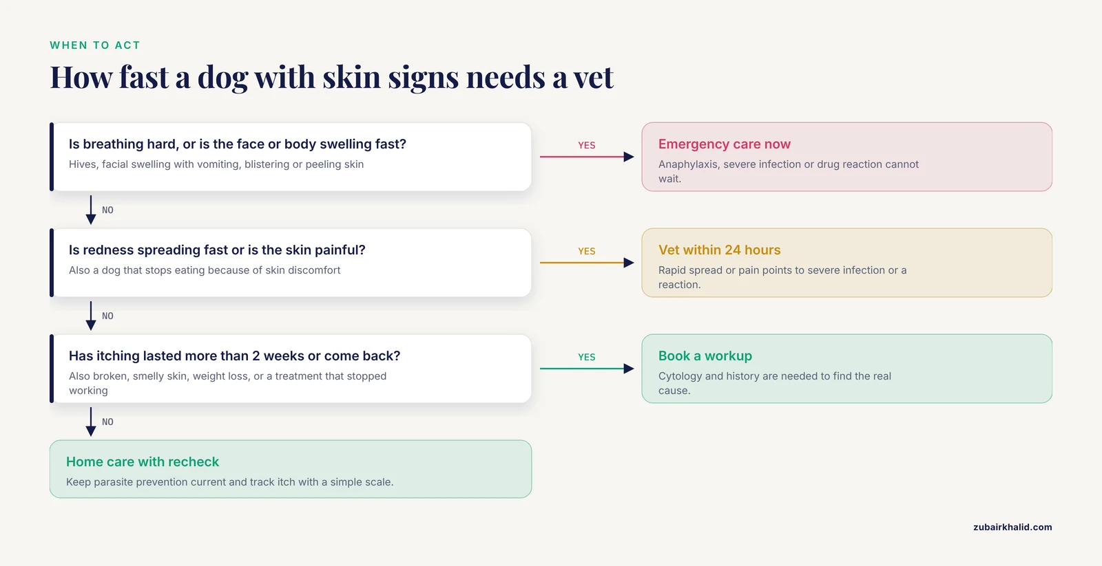
Call an emergency clinic immediately if your dog shows any of the following:
- Facial swelling, hives, or swelling around the eyes that develops quickly
- Difficulty breathing, noisy breathing, or collapse
- Vomiting, diarrhea, or lethargy alongside a skin reaction
- Extensive blistering, peeling, or sloughing skin
- A wound or hot, painful swelling that is spreading rapidly
- Sudden inability to walk or severe pain when touched
- Bleeding that does not stop after 5 minutes of firm pressure
These signs can indicate anaphylaxis, severe infection, or a drug reaction. They are not the typical presentation of ordinary skin disease, and they should not be managed at home.
Limitations and When to Contact a Veterinarian
This article covers the most common patterns, but individual dogs need a veterinarian who can examine the skin, run cytology, and review the whole history. Contact your veterinarian if itching persists beyond 2 weeks despite home care, if the skin is painful or smells strongly, if new lumps or masses appear, if your dog is losing weight, or if a previously effective treatment stops working. A dog with repeated ear infections, recurring pyoderma, or year-round paw licking deserves a full allergy workup rather than another course of the same medication.
Related Guides
- Dog Skin Rash on Legs: Causes and Treatment Options: Covers leg rashes that can mimic other canine skin conditions
- Keratin Buildup on Dog Skin and Paws: Causes and Treatment: Explains keratin buildup on paws, useful for chronic thickened skin
- Skin Necrosis in Dogs: Causes, Warning Signs, and Care: Highlights skin necrosis warning signs and when urgent care is needed
- Dark Patches on Dog Skin: Causes and When to Worry: Details dark skin patches and when they warrant veterinary concern
- What Causes Inflammation in Dogs? Gut, Skin, and Glands: Explores gut, skin, and gland inflammation links in dogs
- Heartworm Disease in Dogs: Prevention, Diagnosis, and Treatment: Covers heartworm prevention and diagnosis for dog parasite care
Frequently Asked Questions
What is the most common skin disease in dogs?
Canine atopic dermatitis is the most frequently diagnosed pruritic skin disease in dogs, accounting for 59.1 percent of cases in a large Swiss prevalence study [6].
How can I tell if my dog's skin problem is an allergy or an infection?
You cannot tell reliably by appearance alone. Infections can mimic allergies, and allergies frequently cause secondary infections. Cytology is the quickest way to find out.
Is dog skin disease contagious to humans?
Most common canine skin diseases are not contagious to people. Some parasites and fungal infections can affect humans, so ask your veterinarian about specific risks in your household.
How long does it take for dog skin disease to clear up?
Superficial bacterial infections often need 3 to 6 weeks of treatment. Allergic disease is managed long term rather than cured, and improvement in clinical trials is usually measured at 21 to 28 days [10][11].
Can I use human shampoo on my dog?
No. Human shampoos are formulated for a different skin pH and can worsen dryness and itching. Use a veterinary-recommended product.
Do probiotics really help dogs with itchy skin?
Evidence is encouraging but not conclusive. One randomized trial found faster improvement in owner-assessed signs with a probiotic and nutraceutical blend [17], while a smaller postbiotic trial found a mild, non-significant improvement in itch [18].
When should I see a veterinarian instead of treating at home?
See a veterinarian if itching lasts more than 2 weeks, if the skin is broken, painful, or smelly, if the dog is losing weight, or if signs keep coming back after treatment.
Can skin disease in dogs be cured?
Some causes, such as bacterial infection and parasites, can be cured. Allergic skin disease is lifelong and is managed rather than cured, usually with a combination of reactive and proactive therapy [15].
Sources
- An innovative three-dimensional skin model for advancing canine atopic dermatitis research.
- Comparison of human and canine atopic dermatitis transcriptome: A meta-analysis and review from a one health perspective.
- Preliminary Measurement of Faecal Short-Chain Fatty Acids in Dogs With Canine Atopic Dermatitis.
- A prospective study evaluating the correlation between local weather conditions, pollen counts and pruritus of dogs with atopic dermatitis.
- Canine atopic dermatitis clinical trials: study design and methodological considerations.
- A vertical study on the prevalence and clinical symptoms of canine pruritic dermatoses in Switzerland.
- Multimodal Diagnosis of Canine Leishmaniosis in a Dog Relocated from Italy to Romania: A Case Report.
- Retrospective Evaluation of Canine Cutaneous Periocular Idiopathic Histiocytic Disease.
- Topical cream with essential oils, zinc and salicylic acid reduces pruritus and skin lesion scores in pruritic dogs.
- Effect of a standardised Ophytrium-containing shampoo and a leave-on mousse protocol on dogs with irritated and pruritic skin.
- Efficacy and Field Safety of the Second-Generation, Highly Selective JAK1 Inhibitor Atinvicitinib for the Treatment of Canine Atopic Dermatitis: A Multicentre, Blinded, Randomised, Placebo-Controlled Clinical Trial in Client-Owned Dogs.
- Evaluation of oclacitinib maleate and prednisolone combined therapy for the control of atopic dermatitis in dogs: A controlled clinical trial.
- A blinded, non-inferiority randomised clinical trial comparing the efficacy and safety of low-dose methotrexate and ciclosporin for the control of moderate to severe canine atopic dermatitis.
- Efficacy and Safety of Allogeneic Mesenchymal Stromal Cells Versus Lokivetmab in Mild-To-Moderate Canine Atopic Dermatitis: A Single-Blind Randomised Study.
- Treatment of Canine Atopic Dermatitis: Updated Guidelines From the International Committee on Allergic Diseases of Animals (ICADA).
- Evaluation of One-Time Oral Oclacitinib Administration on Pruritic Behaviours After Intradermal Interleukin-31-Induced Pruritus Injections ("Reactive" Model) in Healthy Dogs: A Blinded, Randomised, Cross-Over Study.
- A Randomized Controlled Trial to Evaluate the Impact of a Novel Probiotic and Nutraceutical Supplement on Pruritic Dermatitis and the Gut Microbiota in Privately Owned Dogs.
- Efficacy of Heat-Killed Lactobacillus plantarum L-137 Supplementation in Treatment of Canine Atopic Dermatitis.
- Assessment of owner perceptions of caregiver burden, veterinarian-client relationship and satisfaction with the provider in canine pruritus: An experimental vignette study.
- Clinical observations during adjunctive Cannabis treatment and subsequent oclacitinib dose reduction in dogs with atopic dermatitis.