Hematocrit Low or High: Causes and Interpretation
By Dr. Zubair Khalid, DVM, MS, PhD ·
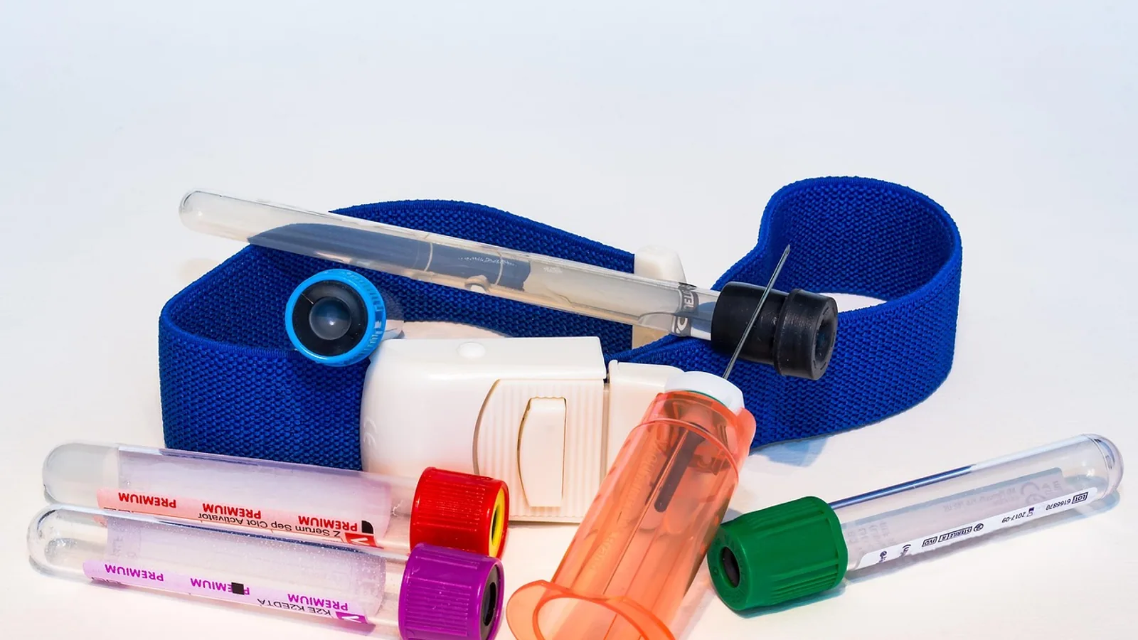
A low hematocrit means the percentage of blood volume occupied by red blood cells has fallen, and a high hematocrit means it has risen. The number itself is never the diagnosis. Interpretation depends on the species, the hydration status of the patient, the reticulocyte response, and the total protein, and those four pieces of information separate a mild lab quirk from a life-threatening bleed or a bone marrow failure.
This article is educational and is not a substitute for veterinary diagnosis or treatment.
What Hematocrit and PCV Actually Measure


Hematocrit (HCT) is the fraction of a blood sample occupied by erythrocytes, expressed as a percentage or as a decimal (a hematocrit of 45% is 0.45 L/L). Packed cell volume (PCV) is the same concept measured a different way. A PCV is produced by spinning a capillary tube of whole blood in a microhematocrit centrifuge until the cells pack at the bottom, then reading the height of the red cell column against the total column height. An analyzer HCT is calculated from the red blood cell count and the mean corpuscular volume (MCV), or measured by a conductivity or optical method depending on the instrument.
These two numbers are close but not identical. In a study of 45 dogs, 45 cats, and 33 horses comparing microhematocrit PCV with hematocrit from a Cobas b121 blood gas analyzer, the mean difference (bias) was -2.4% in dogs, -2.2% in cats, and -0.1% in horses, with no significant difference between methods [1]. In practical terms, a dog or cat PCV often reads a couple of points higher than the analyzer HCT on the same sample. That gap matters when you are tracking a patient across different machines, so always compare like with like. Automated analyzers have been validated against microhematocrit and reference instruments in dogs and cats with excellent correlation for red cell parameters, hematocrit, and PCV, with total observed error staying under the total allowable error of 10% for hematocrit and PCV [2]. Modern point-of-care and in-house analyzers are reliable, but the method still matters when a value sits right at a decision point.
Two other terms appear constantly in reports. Anemia is a low red cell mass, defined by a hematocrit or PCV below the reference interval. Polycythemia (also called erythrocytosis) is a high red cell mass. Relative polycythemia means the red cell mass is normal but the plasma volume is low, so the cells are concentrated. Absolute polycythemia means the total number of red cells has genuinely increased.
Species Reference Intervals for HCT and PCV
Reference intervals are method-dependent and laboratory-dependent. The ranges below are representative adult values in percentages. Use the interval printed on your own laboratory report whenever it is available, because a value flagged as abnormal by one analyzer may be normal by another.
| Species | Typical HCT/PCV range (%) | Most common cause of a low value | Most common cause of a high value |
|---|---|---|---|
| Dog | 37 to 55 | Blood loss, hemolysis, chronic inflammation | Dehydration, then splenic contraction and hypoxia |
| Cat | 30 to 45 (often cited up to 48) | Blood loss, hemolysis, chronic kidney disease | Dehydration, stress, then hypoxia |
| Horse | 32 to 52 | Blood loss, chronic disease, parasitism | Splenic contraction from excitement or exercise, dehydration |
| Cattle | 24 to 46 | Blood loss from parasitism, chronic disease | Dehydration, then hypoxia from cardiac or pulmonary disease |
| Ferret | 44 to 60 | Blood loss, hemolysis, bone marrow disease | Dehydration, splenic contraction |
Cats normally run higher than dogs in many published intervals, and healthy cats can sit in the low 40s without any disease. A cat with a PCV of 32% is at the bottom of normal, while a dog with the same number is mildly anemic. This species difference is one of the most common sources of misreading a lab report. In a multi-institutional study of 851 critically ill animals, the median admission PCV was 42% in dogs and 31% in cats, which reflects both species biology and the fact that sick cats present lower [3]. In a prospective study of anemic dogs and cats requiring transfusion, median pre-transfusion PCV was 14% in dogs and 12% in cats [4].
Horses have a large splenic reservoir of red cells. Excitement, exercise, or pain can push that reservoir into circulation and raise the PCV by several points within minutes, so a high PCV in a nervous horse is often physiologic rather than pathologic. Cattle tend to run lower than dogs, and a bovine PCV in the high 20s can be normal for a healthy adult.
Low Hematocrit: The Three Mechanisms
A low hematocrit always comes down to one of three processes: red cells are being lost, red cells are being destroyed, or red cells are not being produced fast enough. The reticulocyte count tells you which of the first two is happening and whether the bone marrow is responding.
Blood Loss
Acute hemorrhage lowers the PCV, but not immediately. In the first hours after a bleed, the patient loses whole blood, so the PCV and total protein fall together but the PCV may still look normal until fluid shifts into the vasculature. By 12 to 24 hours, the PCV drops as plasma volume is restored. Chronic blood loss, such as from a bleeding gastrointestinal lesion or a heavy parasite burden, produces a low PCV with a low or normal total protein and often microcytic, hypochromic red cells as iron stores are depleted.
Blood loss is the most common cause of anemia in cats receiving xenotransfusion in one large case series, where surgical blood loss was the leading indication in 17 of 49 cats [5]. In a separate study of 105 cats given canine blood, hemorrhage was a more frequent indication than hemolysis or hypoproliferative anemia [6]. These patterns reflect real-world emergency practice, where trauma and surgical bleeding drive transfusion decisions.
Hemolysis
Hemolysis is destruction of red cells before the end of their normal lifespan. It produces a low PCV with a normal or high total protein, and often with icterus, hemoglobinuria, or a regenerative reticulocyte response. Immune-mediated hemolytic anemia is a classic cause in dogs and cats. Infectious causes vary by region and species, and vector-borne pathogens such as Babesia and Ehrlichia species are recognized in dogs in some parts of the world [7].
Hemolysis also occurs as a consequence of transfusion. Cats given canine packed red blood cells can develop acute hemolysis, and in one study of 53 cats, 25% had acute hemolysis within 24 hours, though only 8% met the full definition of an acute hemolytic transfusion reaction [8]. Delayed hemolytic reactions are even more common after xenotransfusion, occurring in 25 of 39 cats in one series, with icterus appearing at a median of 1.9 days and hemolytic serum at a median of 2 days [5]. This is a strong argument for using species-matched blood whenever it is available.
Non-Regenerative Anemia
When the bone marrow cannot produce enough red cells, the reticulocyte count stays low and the anemia is non-regenerative. Causes include chronic kidney disease (lack of erythropoietin), chronic inflammatory disease (anemia of inflammation), endocrine disease, iron deficiency, and primary bone marrow disease such as aplastic anemia or marrow infiltration by neoplasia. These patients often have a normal total protein and no evidence of blood loss or hemolysis.
A fourth category deserves mention because it is iatrogenic. Hospital-acquired anemia develops during hospitalization, driven largely by repeated blood sampling. In a prospective study of 56 hospitalized dogs and cats, the incidence of hospital-acquired anemia was 34% by 24 hours, 43% by 48 hours, and 48% by 72 hours, and cumulative phlebotomy volumes greater than 3% of total blood volume significantly increased the risk [9]. A larger multi-institutional study of 851 critically ill animals found an admission anemia prevalence of 32% and an overall prevalence during hospitalization of 56%, with the last recorded PCV significantly lower than the admission PCV in both dogs and cats [3]. If your pet is in the hospital for several days and the PCV keeps drifting down, ask how much blood has been drawn.
High Hematocrit: Relative Versus Absolute
A high hematocrit is either relative or absolute, and the distinction changes the entire diagnostic path.
Relative Polycythemia from Dehydration
Relative polycythemia means the red cell mass is normal but plasma volume is reduced. Dehydration from vomiting, diarrhea, poor water intake, or third-space fluid loss concentrates the red cells. The clues are a high total protein, a high blood urea nitrogen and creatinine, and a patient that looks volume-depleted. Splenic contraction is the other relative cause. In horses, excitement or exercise can raise the PCV several points within minutes. In dogs and cats, fear and restraint can do the same on a smaller scale.
The treatment for relative polycythemia is fluid, not phlebotomy. Once the patient is rehydrated, the PCV normalizes. This is the single most common reason a high hematocrit turns out to be benign.
Absolute Polycythemia from Hypoxia or Erythropoietin Excess
Absolute polycythemia means the red cell mass has genuinely increased. It has two broad causes. The first is chronic hypoxia, which drives erythropoietin production. Chronic cardiac disease, chronic pulmonary disease, and right-to-left shunts can all cause this. The second is inappropriate erythropoietin production or an erythropoietin-independent marrow disorder, sometimes called polycythemia vera. These patients have a high PCV with a normal or low total protein and no evidence of dehydration.
The distinction matters because the treatments differ. Hypoxic polycythemia is managed by addressing the underlying cardiac or pulmonary disease. Polycythemia vera may require phlebotomy or myelosuppressive therapy. Lowering hematocrit in a dehydrated patient by removing blood would be harmful, while lowering it in a patient with true polycythemia and hyperviscosity can be therapeutic.
Stepwise Interpretation Flowchart
The following flowchart shows the main decision path from an abnormal hematocrit to a working diagnosis.
flowchart TD
    A[Abnormal hematocrit] --> B{High or low}
    B -->|High| C{Total protein high}
    C -->|Yes| D[Relative from dehydration]
    C -->|No| E{Evidence of hypoxia}
    E -->|Yes| F[Absolute from chronic hypoxia]
    E -->|No| G[Consider erythropoietin excess]
    B -->|Low| H{Reticulocyte count}
    H -->|High| I[Regenerative anemia]
    H -->|Low| J[Non regenerative anemia]
    I --> K{Total protein}
    K -->|Low| L[Blood loss]
    K -->|Normal or high| M[Hemolysis]
    J --> N[Consider kidney disease marrow disease or inflammation]
The Biology in Plain Language
Red blood cells are produced in the bone marrow under the control of erythropoietin, a hormone made mainly by the kidneys in response to low oxygen delivery. A red cell circulates for roughly 110 to 120 days in dogs, somewhat less in cats, and the marrow replaces cells continuously. When the PCV falls, the kidney senses reduced oxygen delivery and increases erythropoietin, which stimulates the marrow to release reticulocytes, the immature red cells that still contain residual RNA. A high reticulocyte count means the marrow is working. A low reticulocyte count in an anemic patient means the marrow is not responding, and the problem is either outside the marrow (lack of erythropoietin, lack of iron, chronic inflammation) or inside it (marrow disease).
Reticulocytes are counted as a percentage of red cells, but a percentage can mislead. A cat with a PCV of 12% and 5% reticulocytes has far fewer absolute reticulocytes than a cat with a PCV of 35% and 5% reticulocytes. The corrected reticulocyte percentage and the absolute reticulocyte count adjust for this, and most laboratories report an absolute count. In dogs, an absolute reticulocyte count above roughly 60,000 to 80,000 per microliter is generally regenerative, though the exact threshold varies by laboratory. Cats are different. Cats normally have very low reticulocyte counts and can take several days longer to mount a response than dogs, so a cat with a modest reticulocyte count may still be regenerative. Aggregated reticulocytes are the most immature form and their presence signals an active marrow response.
Total protein is the other half of the picture. In blood loss, red cells and protein are lost together, so both fall. In hemolysis, red cells are destroyed but protein is retained, so the PCV falls while protein stays normal or rises. In non-regenerative anemia, protein is usually normal. This single pairing of PCV and total protein resolves most cases before any advanced testing.
Practical Implications for Owners and Keepers
If your pet's hematocrit is flagged as low, the first question is whether the pet is stable. A PCV below roughly 15% in a dog or cat, or a rapidly falling PCV, is an emergency. Signs of severe anemia include pale or white gums, weakness, collapse, rapid breathing, and a fast heart rate. These animals need immediate veterinary care, and many need a transfusion.
If the hematocrit is high, the first question is whether the pet is dehydrated. A high PCV with a high total protein in a pet that has been vomiting or not drinking usually means dehydration, and fluid therapy is the treatment. A high PCV with a normal total protein in a pet with no access to water loss and no evidence of dehydration warrants a search for cardiac or pulmonary disease.
Several practical points help owners understand what they are seeing. A single abnormal value is a data point, not a diagnosis. Trends matter more than isolated numbers. A PCV that falls from 45% to 30% over 48 hours is more concerning than a stable 30%. Ask for the reticulocyte count when anemia is found, because it changes the entire differential list. Ask whether the total protein was measured at the same time, because the pairing is what separates blood loss from hemolysis. And if your pet is hospitalized, ask how much blood has been drawn, because hospital-acquired anemia is common and largely preventable [9][3].
Clinical Relevance, Limitations and Common Mistakes
The most common mistake is comparing a dog's PCV to a cat's. Cats normally run higher than dogs, and a value that is normal in a cat may be anemic in a dog. The second most common mistake is treating a high PCV caused by dehydration with phlebotomy. Removing blood from a dehydrated patient worsens the problem. The third is ignoring the total protein. A low PCV with a low total protein points to blood loss, while a low PCV with a normal total protein points to hemolysis or non-regenerative anemia, and that distinction drives the diagnostic plan.
Method differences are a real source of confusion. A PCV from a spun microhematocrit and an HCT from an analyzer can differ by a couple of percentage points in dogs and cats [1]. If a patient is being monitored across different machines, the trend may look like a change when it is really a method difference. Stick to one method when tracking a patient over time.
Transfusion practice adds another layer. Cats given canine blood can develop acute hemolysis and delayed hemolytic reactions at high rates [8][5]. Species-matched blood is always preferred when available. When it is not, the risks and benefits must be weighed carefully, and the patient monitored closely for icterus and hemoglobinuria in the days after transfusion.
Finally, hospital-acquired anemia is underrecognized. Cumulative phlebotomy volumes above 3% of total blood volume significantly increase the risk, and the incidence climbs with each day of hospitalization [9]. Clinicians should group blood draws, use the smallest volume necessary, and consider the cumulative effect when ordering daily panels.
Individual cases need a veterinarian who can examine the patient, review the full blood work, and account for the species, the hydration status, and the clinical picture. This article is educational and is not a substitute for veterinary diagnosis or treatment.
Frequently Asked Questions
What is a normal hematocrit for a dog?
Most adult dogs run between 37% and 55%, though the exact interval depends on the laboratory and method. Values below the low end suggest anemia, and values above the high end suggest dehydration or another cause of concentration.
What is a normal hematocrit for a cat?
Cats typically run 30% to 45%, and many healthy cats sit in the low 40s. Cats normally run higher than dogs, so a value that looks normal in a dog may be low for a cat.
Can dehydration cause a high hematocrit?
Yes. Dehydration reduces plasma volume, which concentrates the red cells and raises the PCV. The total protein is usually high as well, and the PCV normalizes with fluid therapy.
Does a high hematocrit always mean polycythemia?
No. Relative polycythemia from dehydration or splenic contraction is far more common than true absolute polycythemia. The total protein and the clinical picture separate the two.
What is the difference between PCV and HCT?
PCV is measured by spinning a blood sample in a microhematocrit tube and reading the packed cell layer. HCT is calculated or measured by an automated analyzer. The two are close but can differ by a couple of percentage points in dogs and cats.
Why does my cat's hematocrit keep dropping in the hospital?
Repeated blood sampling can cause hospital-acquired anemia. Cumulative phlebotomy volumes above 3% of total blood volume significantly increase the risk, and the incidence rises with each day of hospitalization.
What does a low reticulocyte count mean in an anemic pet?
A low reticulocyte count means the bone marrow is not responding to the anemia. Causes include chronic kidney disease, chronic inflammation, iron deficiency, and primary bone marrow disease.
Can a high hematocrit be dangerous?
Yes. Severe absolute polycythemia increases blood viscosity and can impair circulation. Relative polycythemia from dehydration is dangerous for a different reason, because the underlying fluid loss can cause organ dysfunction if it is not corrected.
Related Articles
- Low ligation efficiency causes
- Lymphoma in Cats: High-Grade vs Low-Grade GI Alimentary Lymphoma
- Sandwich ELISA Troubleshooting
- Diabetes Diet for Cats: High-Protein Low-Carb Remission Diet
- GC Content and Primer Design: How to Handle High and Low GC Templates
- ELISA Troubleshooting: High Background, Low Signal, and Poor Reproducibility
- High BUN Blood Test: Causes and What to Do Next
Sources
- Agreement between hematocrit values determined by the Cobas b121 blood gas analyzer and the microhematocrit method in dogs, cats, and horses.
- Evaluation of the scil vCell 5, a novel laser- and impedance-based point-of-care hematology analyzer, for use in dogs and cats.
- Hospital-acquired Anemia in Critically Ill Dogs and Cats: A Multi-Institutional Study.
- Left heart dimensions in anemic cats and dogs before and after blood transfusion.
- Xenotransfusion of canine blood to cats: a review of 49 cases and their outcome.
- Retrospective study of canine blood xenotransfusion compared with type-matched feline blood allotransfusion to cats: indications, effectiveness, limitations and adverse effects.
- First molecular confirmation of multiple zoonotic vector-borne diseases in pet dogs and cats of Hong Kong SAR.
- Incidence of acute haemolysis in cats receiving canine packed red blood cells (xenotransfusions).
- Incidence of hospital-acquired anemia in hospitalized dogs and cats.