Coombs Test: Direct vs Indirect Antiglobulin Testing
By Dr. Zubair Khalid, DVM, MS, PhD ·
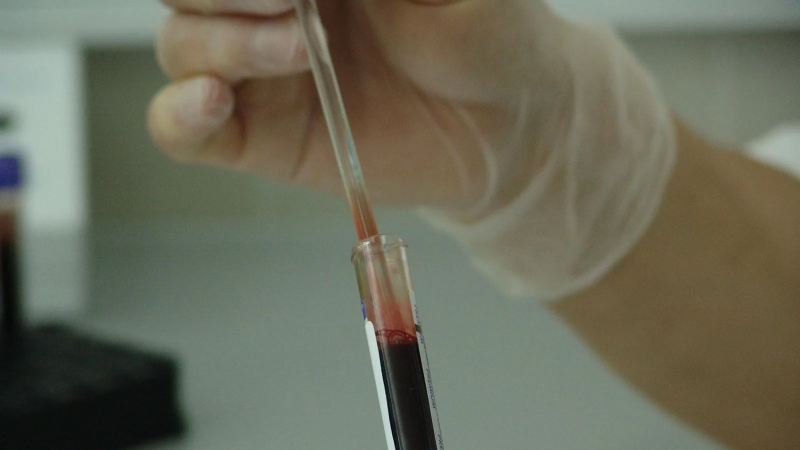
The Coombs test, also called the antiglobulin test, detects antibodies or complement proteins stuck to the surface of red blood cells. The direct antiglobulin test (DAT) uses the patient's own red cells to detect antibody already bound in the body, while the indirect antiglobulin test (IAT) uses the patient's serum mixed with reagent red cells to detect circulating antibodies that can bind red cells in the laboratory.
Both tests share the same core trick. Antibodies against red cells are often too sparse or too small to clump cells on their own, so a bridging reagent called antihuman globulin is added to link antibody-coated cells together into a visible lattice. That visible clumping, called agglutination, is the positive endpoint.
This article explains the biology behind the Coombs antiglobulin test, walks through the stepwise protocol, compares DAT and IAT side by side, and covers how the test is used in dogs and cats with suspected immune-mediated hemolytic anemia.
This article is educational and is not a substitute for veterinary diagnosis or treatment.
What the Coombs Test Actually Detects
Red blood cells carry surface antigens. When an animal or person makes antibodies against those antigens, the antibodies can attach to red cell membranes. Some antibodies also activate complement, a cascade of plasma proteins that coats the cell with fragments such as C3d. Once a red cell is coated with immunoglobulin G (IgG), immunoglobulin M (IgM), or complement, it becomes a target for destruction in the spleen, liver, and bloodstream.
The problem for the laboratory is that a coated red cell does not look different under a microscope. The antibody layer is invisible. The Coombs test makes that layer visible by adding a second antibody, antihuman globulin, which is produced in another species and directed against the patient's immunoglobulins or complement proteins. Antihuman globulin binds the Fc portion of IgG or the complement fragment on adjacent red cells and cross-links them. If enough cells are cross-linked, they form visible aggregates.
Two variables decide whether you get agglutination:
- Whether red cells in the sample are already coated with antibody or complement in vivo.
- Whether the serum contains free antibody that can coat reagent red cells in vitro.
The DAT answers the first question. The IAT answers the second.
A useful way to remember the difference is that the DAT asks "are the patient's cells already coated?" and the IAT asks "can the patient's serum coat red cells?" The DAT is a snapshot of what happened inside the body. The IAT is a challenge test performed in a tube, gel card, or microplate.
Direct Antiglobulin Test (DAT): Detecting In Vivo Sensitization
The DAT is performed on a sample of the patient's anticoagulated blood, typically collected in EDTA to prevent complement activation during storage. The red cells are washed to remove plasma proteins, especially free IgG, that would otherwise neutralize the antihuman globulin reagent. After washing, antihuman globulin is added, the tube is centrifuged, and the cell button is examined for agglutination.
A positive DAT means the patient's red cells were coated with antibody or complement at the moment of sampling. This is called in vivo sensitization. It does not by itself prove that hemolysis is occurring. A positive DAT is a laboratory finding, and the diagnosis of immune-mediated hemolytic anemia requires that finding to be combined with evidence of red cell destruction such as anemia, elevated bilirubin, elevated lactate dehydrogenase, low haptoglobin, or spherocytosis on a blood film [1].
That distinction matters clinically. In a study of 92 patients with systemic lupus erythematosus and no hemolytic anemia, 33.6% had a positive direct Coombs test, likely because immune complexes were binding to red cells through complement receptor 1 [2]. A positive DAT in that setting reflects immune complex load, not active hemolysis. The same principle applies in veterinary medicine: a positive DAT in a dog with a normal packed cell volume and no evidence of regeneration or hemolysis should not be treated as immune-mediated hemolytic anemia.
What a Positive DAT Means in Practice
A positive DAT narrows the differential diagnosis. It tells the clinician that an immune mechanism is involved. The next steps are to determine the specificity of the coating (IgG alone, complement alone, or both) and to correlate the result with the clinical picture.
In a study of 501 anemic patients in eastern India, DAT was positive in 64 patients. IgG was found in 86% of positive samples, and C3d alone was found in 14%. Warm autoimmune hemolytic anemia accounted for 93.7% of the DAT-positive cases, and the strength of the DAT reaction was directly proportional to the degree of hemolysis [3]. That last point is clinically useful: a stronger reaction generally correlates with more active disease, though the correlation is not perfect and should not be used as the sole measure of severity.
What a Negative DAT Means
A negative DAT does not exclude immune-mediated hemolysis. So-called "Coombs-negative" or DAT-negative hemolytic anemia is a recognized entity [1]. Possible explanations include:
- The density of antibody on the red cell surface is below the detection threshold of the assay.
- The antibody is of a class or subclass that the antihuman globulin reagent does not detect well, such as IgA or IgM in low concentration.
- The antibody dissociates from the red cell during sample storage or washing.
- The hemolysis is complement-mediated and the complement fragments are lost from the cell surface before testing.
In these cases, the clinician must rely on other evidence of immune-mediated destruction, including response to immunosuppressive therapy, exclusion of other causes, and sometimes more sensitive techniques such as flow cytometry or enzyme-linked antiglobulin testing [4].
Indirect Antiglobulin Test (IAT): Detecting Circulating Antibodies
The IAT uses the patient's serum or plasma, not the patient's red cells. The serum is incubated with reagent red cells of known antigen type. If the serum contains antibodies against those antigens, the antibodies bind to the reagent cells. After incubation, the cells are washed and antihuman globulin is added. Agglutination indicates that the serum contained antibodies capable of binding red cells.
The IAT is the test used in crossmatching and in antibody screening, but its clinical role extends beyond transfusion medicine. In neonatal hemolytic disease, the IAT is used to test the mother's serum for antibodies against the baby's red cell antigens. A study of 2,769 babies and their mothers found that DAT positivity in newborns was 3.3%, and the need for phototherapy was significantly higher in DAT-positive infants [5]. The IAT in the mother helped identify the specific antibody responsible, which guided monitoring and treatment.
In veterinary medicine, the IAT is used less commonly than the DAT, but it has a role in investigating suspected immune-mediated hemolytic anemia when the DAT is negative. If the patient's serum contains antibodies that bind reagent dog or cat red cells, that supports an immune-mediated process even when the direct test is negative.
Why the IAT Matters When the DAT Is Negative
The DAT and IAT are complementary. A patient can have a negative DAT and a positive IAT if the antibodies are present in circulation but have not yet coated a significant fraction of the patient's own red cells. This pattern can occur early in the disease course or when the antibody has low affinity for the patient's own cells but high affinity for reagent cells.
Conversely, a positive DAT with a negative IAT can occur when the antibody is entirely cell-bound and no free antibody remains in the serum. This is common in warm autoimmune hemolytic anemia, where the autoantibody is often completely absorbed onto the patient's red cells.
Stepwise Protocol for the Antiglobulin Coombs Test
The tube method remains the reference technique, though gel column technology and flow cytometry are increasingly used. The gel test, developed by Lapierre in 1984, was designed to standardize antiglobulin testing and improve sensitivity and specificity [6]. In a blinded retrospective study of 40 patient sera, the gel test detected and identified 18 known antibodies, while the tube method detected 16 [6]. The gel test was also easier to read and more reproducible according to the technologists who performed it [6].
Regardless of platform, the core steps are the same. The following protocol describes the tube method for the DAT, with notes on how the IAT differs.
Step 1: Sample Collection and Preparation
For the DAT, collect blood in EDTA. For the IAT, collect blood in a plain tube or a tube without anticoagulant to obtain serum, or use plasma if the assay is validated for it. Label the tube with the patient identifier and the date and time of collection.
Step 2: Wash the Red Cells
Wash the red cells three to four times with isotonic saline. Washing removes plasma proteins, especially free IgG, that would otherwise bind the antihuman globulin reagent and cause a false negative result. Inadequate washing is one of the most common causes of false-negative DAT results.
The washing step is also time-sensitive. A study of 25 samples with positive IgG DATs found that delays after preparation of the red cell suspension or after the last wash decant could weaken the reaction. Three samples were weakened by delays after red cell suspension preparation, and one sample was weakened when the addition of anti-IgG was delayed [7]. The practical lesson is to proceed without interruption once the cells are washed and the suspension is prepared.
Step 3: Prepare the Cell Suspension
Resuspend the washed red cells in saline to make a 2% to 5% suspension. The exact concentration depends on the assay and the laboratory's standard operating procedure. A suspension that is too concentrated can cause false-positive results from rouleaux or nonspecific clumping. A suspension that is too dilute can cause false-negative results because there are too few cells to form visible agglutinates.
Step 4: Add Antihuman Globulin
Add the antihuman globulin reagent to the tube. The reagent may be polyspecific (detecting IgG and complement) or monospecific (detecting IgG alone or complement alone). Monospecific reagents are used when the laboratory needs to determine whether the coating is IgG, complement, or both.
Step 5: Centrifuge
Centrifuge the tube under conditions specified by the reagent manufacturer. Centrifugation packs the cells at the bottom of the tube and brings antibody-coated cells into close contact with the antihuman globulin.
Step 6: Read for Agglutination
Gently resuspend the cell button and examine it for agglutination. A positive result appears as visible clumps of red cells. A negative result appears as a smooth suspension with no clumps. Grade the reaction from weak (1+) to strong (4+) according to the laboratory's grading scheme.
Step 7: Add Control Cells (Check Cells)
If the result is negative, add IgG-coated control cells to the tube and centrifuge again. If the control cells agglutinate, the antihuman globulin reagent was active and the negative result is valid. If the control cells do not agglutinate, the reagent was neutralized or the washing was inadequate, and the test must be repeated. This step is called the check cell or Coombs control step.
How the IAT Differs
For the IAT, the steps are similar but the order changes. Incubate the patient's serum with reagent red cells first, allowing antibodies in the serum to bind the reagent cells. Then wash the cells to remove unbound serum proteins, add antihuman globulin, centrifuge, and read for agglutination. The check cell step is also performed for negative IAT results.
The following flowchart summarizes the decision path for choosing between the DAT and IAT and interpreting the result.
flowchart TD
    A[Suspected immune hemolysis] --> B{Are patient red cells coated}
    B -->|Yes| C[Perform DAT on patient red cells]
    B -->|No| D[Perform IAT on patient serum]
    C --> E[Wash cells add antihuman globulin centrifuge]
    D --> F[Incubate serum with reagent cells wash add antihuman globulin centrifuge]
    E --> G{Agglutination}
    F --> G
    G -->|Positive| H[Antibody or complement detected]
    G -->|Negative| I[Add control cells to validate]
    I --> J{Control cells agglutinate}
    J -->|Yes| K[Negative result valid]
    J -->|No| L[Repeat test with fresh sample]
Direct vs Indirect Coombs Test: Comparison Table
| Feature | Direct Antiglobulin Test (DAT) | Indirect Antiglobulin Test (IAT) |
|---|---|---|
| Sample used | Patient's red cells (EDTA whole blood) | Patient's serum or plasma plus reagent red cells |
| What is detected | Antibody or complement already bound to red cells in vivo | Circulating antibodies that bind red cells in vitro |
| Timing of sensitization | In vivo | In vitro |
| Primary clinical use | Diagnosis of immune-mediated hemolytic anemia, transfusion reaction, hemolytic disease of the newborn | Antibody screening, crossmatching, investigation of DAT-negative hemolysis, maternal antibody detection in hemolytic disease of the newborn |
| Positive result means | Red cells are coated with IgG, complement, or both | Serum contains antibodies against red cell antigens |
| Negative result means | No detectable coating on red cells at the time of sampling | No detectable circulating antibodies against the reagent cells used |
| Common causes of false negative | Inadequate washing, delayed testing after washing, low antibody density, prozone effect | Inadequate washing, low antibody titer, weak antibody affinity, improper incubation |
| Common causes of false positive | Prior transfusion, recent drugs, nonspecific IgG binding, rouleaux | Contaminated reagents, nonspecific agglutination, cold agglutinins |
| Veterinary application | Confirms immune-mediated hemolytic anemia in dogs and cats | Less commonly used, but helpful when DAT is negative and immune hemolysis is still suspected |
Clinical Relevance, Limitations and Common Mistakes
The Coombs test is a tool, not a diagnosis. A positive DAT in a dog with anemia, icterus, and spherocytosis strongly supports immune-mediated hemolytic anemia. A positive DAT in a dog with a normal hematocrit and no evidence of hemolysis may be an incidental finding.
In a study of 1,127 thrombocytopenic dogs, 39.3% were DAT-positive for platelet-bound IgG using a flow cytometry-based assay. DAT-positive dogs had significantly lower platelet counts than DAT-negative dogs, and a weak inverse correlation was found between the percentage of IgG-bound platelets and platelet count [8]. This shows that the DAT principle extends beyond red cells to platelets, and that the strength of positivity can carry prognostic information.
False Negatives from Inadequate Washing
Inadequate washing is the most common technical cause of a false-negative DAT. Residual plasma IgG binds the antihuman globulin reagent before it can reach the red cells. The check cell step is designed to catch this error. If the check cells fail to agglutinate, the test is invalid and must be repeated with more thorough washing.
Delays after washing can also weaken the result. The study of 25 samples found that delays of as little as one minute after the last wash decant could weaken some reactions [7]. This does not mean every delay causes a false negative, but it does mean that the assay should be performed without unnecessary interruption.
False Positives from Transfusion and Drugs
A positive DAT can occur after transfusion because donor red cells coated with antibody or complement are present in the recipient's circulation. It can also occur with certain drugs. Drug-induced immune hemolytic anemia is rare but potentially life-threatening. A case report of penicillin-induced immune hemolysis described a patient who presented with gross hematuria and a strongly positive direct Coombs test after a 10-day course of oral penicillin [9]. Another case report described alectinib-induced hemolytic anemia with a positive DAT that turned negative after the drug was discontinued and recurred when the drug was resumed [10].
Ceftriaxone is another drug associated with immune hemolysis. A case report described a patient who developed dark brown urine and shock one hour after receiving ceftriaxone, with a hemoglobin drop to 26 g/L and a positive direct Coombs test. The patient was found to have ceftriaxone antibodies of both IgG and IgM classes [11].
These cases illustrate a key point: a positive DAT in a patient receiving a new medication should prompt consideration of drug-induced immune hemolysis, especially if the anemia is abrupt and the clinical picture is severe.
The DAT in Hemolytic Disease of the Newborn
In newborns, the DAT is performed on cord blood. A positive DAT in a newborn with jaundice or anemia supports immune-mediated hemolysis. The IAT is performed on the mother's serum to identify the specific antibody. In a study of 2,769 babies, the prevalence of DAT positivity was 3.3%. ABO incompatibility accounted for 45.9% of DAT-positive cases, RhD incompatibility for 5.7%, and combined RhD and ABO incompatibility for 10.3% [5]. The need for phototherapy was significantly higher in DAT-positive infants [5].
The DAT in Other Conditions
A positive DAT is not limited to classical autoimmune hemolytic anemia. It has been reported in post-artesunate delayed hemolysis in severe malaria, where DAT positivity was found in 54.3% of patients tested but was not associated with the occurrence of delayed hemolysis [12]. It has also been reported in systemic loxoscelism from brown spider envenomation, where a patient developed a strongly positive DAT (4+) with immune-mediated hemolysis [13].
These examples show that a positive DAT can be a marker of immune activation rather than a specific diagnosis. The clinical context determines what the result means.
What the Coombs Test Cannot Do
The Coombs test cannot distinguish between autoantibodies and alloantibodies on its own. It cannot predict the severity of hemolysis. It cannot replace a complete blood count, blood film review, or biochemical panel. A negative DAT does not exclude immune-mediated hemolysis, and a positive DAT does not confirm it.
Individual cases require veterinary assessment. A veterinarian will interpret the Coombs test alongside the physical examination, history, and other laboratory findings.
Veterinary Use in Dogs and Cats
In veterinary medicine, the DAT is most commonly used to support a diagnosis of immune-mediated hemolytic anemia (IMHA) in dogs and cats. IMHA is a common cause of severe anemia in dogs, and the DAT can help confirm an immune cause when the clinical picture is ambiguous.
The principle is the same as in human medicine. The DAT detects IgG or complement on the surface of the patient's red cells. In dogs, the test is typically performed with species-specific antihuman globulin reagents that cross-react with canine immunoglobulins and complement. In cats, similar reagents are used, though the test is less commonly performed.
A positive DAT in a dog with regenerative anemia, spherocytosis, and icterus supports IMHA. A negative DAT does not exclude IMHA, and many dogs with clinically diagnosed IMHA have negative DAT results. The diagnosis in those cases rests on exclusion of other causes and response to immunosuppressive therapy.
The IAT is used less frequently in veterinary medicine, but it can be helpful when the DAT is negative and immune-mediated hemolysis is still suspected. The IAT can detect circulating antibodies that have not yet coated a sufficient number of red cells to produce a positive DAT.
Flow cytometry is an emerging platform for the DAT in veterinary medicine. A study of 1,127 thrombocytopenic dogs used flow cytometry to detect platelet-bound IgG and established a diagnostic cutoff of 10% IgG-bound platelets [8]. This approach is more sensitive than traditional tube methods and can be applied to red cells as well as platelets.
Veterinarians should be aware that prior transfusion can cause a positive DAT in dogs and cats, just as in humans. Recent drug administration can also cause a positive DAT. These factors should be considered when interpreting the result.
Frequently Asked Questions
What is the difference between a direct and indirect Coombs test?
The direct Coombs test (DAT) uses the patient's red cells and detects antibodies or complement already bound to those cells in the body. The indirect Coombs test (IAT) uses the patient's serum mixed with reagent red cells and detects circulating antibodies that can bind red cells in the laboratory.
What does a positive Coombs test mean?
A positive Coombs test means that antibodies or complement were detected on red cells (DAT) or that circulating antibodies against red cells were detected in serum (IAT). A positive result supports an immune-mediated process but does not confirm active hemolysis on its own.
Can a Coombs test be negative in immune-mediated hemolytic anemia?
Yes. A negative DAT does not exclude immune-mediated hemolytic anemia. Antibody density may be below the detection threshold, the antibody may be of a class not detected by the reagent, or the antibody may dissociate during sample handling.
Why is washing important in the Coombs test?
Washing removes unbound plasma proteins, especially free IgG, that would otherwise neutralize the antihuman globulin reagent and cause a false-negative result. Inadequate washing is a common cause of false-negative DAT results.
What causes a false-positive Coombs test?
Prior transfusion, recent drug administration, nonspecific IgG binding, and rouleaux formation can all cause a false-positive DAT. Drug-induced immune hemolysis is a rare but important cause of a positive DAT.
Is the Coombs test used in dogs and cats?
Yes. The DAT is used in dogs and cats to support a diagnosis of immune-mediated hemolytic anemia. The IAT is used less commonly but can help when the DAT is negative and immune hemolysis is still suspected.
How long does a Coombs test take?
The tube method for the DAT can be completed in under an hour once the sample is received. The IAT requires an incubation step and may take longer. Gel and flow cytometry platforms have different turnaround times depending on the laboratory.
Can a Coombs test be positive without anemia?
Yes. A positive DAT can occur without hemolytic anemia, as seen in some patients with systemic lupus erythematosus and in some patients receiving certain drugs. The result must be interpreted alongside the clinical picture and other laboratory findings.
Related Articles
- Direct vs Indirect Immunofluorescence in Viral Diagnostics
- Feed Testing for Mycotoxins: When and How to Test
- Immunoassay Drug Testing: Principles and Limitations
- Pilot Testing Your Research Question
- Thyroid Function Testing in Veterinary Medicine
- Motility Testing in Microbiology: Semisolid Agar Method
- Indirect Fluorescent Antibody Test: How It Works
- How to Tell If an Egg Is Still Good: 4 Safe Tests
Sources
- Dissecting the direct antiglobulin test in warm autoimmune hemolytic anemia: clinical and laboratory correlations.
- Disease activity in systemic lupus erythematosus in relation to direct Coombs test positivity without haemolytic anaemia: a single-centre cross-sectional study.
- Role of direct antiglobulin test in anemia under evaluation: Its prevalence, laboratory workup, and significance in further patient management-A study from eastern India.
- Estimation of transfused red cell survival using an enzyme-linked antiglobulin test.
- Neonatal hemolytic disease: How should we use indirect and direct antiglobulin tests?
- Evaluation and implementation of the gel test for indirect antiglobulin testing in a community hospital laboratory.
- How slow can you be when performing a direct antiglobulin test?
- Diagnostic evaluation of a flow cytometry-based direct antiglobulin test and demographic analysis in dogs with suspected immune thrombocytopenia.
- [[In macrohematuria, medication history also needs to be considered : Penicillin-induced immune hemolysis: a case report and review of literature].](https://pubmed.ncbi.nlm.nih.gov/37493757/)
- Alectinib-induced Hemolytic Anemia with Positive Direct Antiglobulin Test in a Patient with Lung Adenocarcinoma: A Possible Drug-drug Interaction Effect.
- First application of whole blood exchange-lymphoplasmapheresis combined transfusion for restoring immune homeostasis of ceftriaxone-induced hemolytic crisis: a case report.
- Favorable outcome without corticosteroids during post-artesunate delayed hemolysis with positive direct antiglobulin test in severe imported Plasmodium falciparum malaria, France.
- Systemic loxoscelism with hemolysis and positive Coombs: diagnostic and physiopathological implications.