
Protostrongylus rufescens (Small Lungworm) in Sheep and Goats: Diagnosis and Clinical Relevance
Etiology and Taxonomy
Protostrongylus rufescens (Leuckart, 1895) is a metastrongyloid nematode belonging to the family Protostrongylidae. It is one of the principal small lungworms infecting domestic and wild small ruminants, including sheep (Ovis aries) and goats (Capra hircus). The adult parasites reside in the bronchioles and alveolar ducts, where they induce chronic proliferative and inflammatory responses. The taxonomic position within the Protostrongylidae is supported by morphological features of the bursa and spicules in males, as well as by molecular phylogenetic analyses of the mitochondrial genome [1]. Cytogenetic studies have demonstrated a diploid chromosome number of 2n = 12 in P. rufescens, with a distinctive karyotype that aids in species discrimination from other protostrongylids such as Muellerius capillaris and Cystocaulus ocreatus [2].
The species is often co-endemic with other protostrongylid nematodes, and mixed infections are common in many regions [3, 4]. Biochemical characterization using malic dehydrogenase isoenzyme patterns has provided additional discriminative tools for distinguishing P. rufescens from other lungworm species in livestock [5, 6].
Epidemiology
Protostrongylus rufescens has a global distribution but is particularly prevalent in temperate, Mediterranean, and subtropical climates where intermediate gastropod hosts are abundant. Prevalence studies in small ruminants have reported widely varying infection rates depending on geographic location, management system, and diagnostic method.
A postmortem study in the central highlands of Ethiopia (North Shewa) identified P. rufescens in a substantial proportion of sheep and cattle examined, emphasizing the role of necropsy for accurate prevalence estimation [7]. In the Wolaita zone of southern Ethiopia, lungworm infection in small ruminants was associated with several risk factors including grazing on communal pastures, lack of anthelmintic treatment, and high rainfall [8]. Similarly, surveys in northeastern Iran have documented infection rates exceeding 30% in sheep and goats, with P. rufescens identified as one of the dominant protostrongylid species [9].
Breed-related susceptibility has been observed; comparative studies involving Romanov and Lacaune sheep in France showed differences in the intensity of P. rufescens infection, suggesting a genetic component to host resistance [10]. Work in Mediterranean climates further demonstrated that meat sheep breeds under certain anthelmintic regimens exhibited variable infection levels, with implications for targeted selective treatment strategies [11].
In Morocco, epidemiological surveys of goats revealed that P. rufescens infections were influenced by altitude, rainfall, and goat age, with younger animals harboring higher larval counts [12]. Likewise, a retrospective study from Brazil spanning four decades confirmed the presence of P. rufescens in ruminants across multiple states, with a greater proportion of cases in the southern region where temperate conditions favor gastropod survival [13]. Reports from Uzbekistan have quantified infection levels in definitive caprine hosts and intermediate gastropod populations, highlighting the ecological coupling between pasture contamination and snail density [14]. Wildlife reservoirs, including mouflon (Ovis musimon), markhor (Capra falconeri), urial (Ovis vignei), and Chiltan wild goat (Capra aegagrus chiltanensis), contribute to the sylvatic cycle and may serve as sources of reinfection for domestic flocks [15, 16].
Life Cycle and Transmission
The life cycle of P. rufescens is indirect and requires a terrestrial gastropod intermediate host. First-stage larvae (L1) are passed in the feces of infected sheep or goats. These larvae are motile and must penetrate the foot or mantle of a suitable snail or slug species to continue development. Within the gastropod, the larvae undergo two molts to reach the third-stage (L3) infective form. The prepatent period in the definitive host is approximately 4 to 6 weeks.
Environmental factors critically influence larval survival. Experimental studies have shown that first-stage protostrongylid larvae can survive for several weeks under cool, humid conditions but are rapidly killed by desiccation and high temperatures [17]. Grazing management therefore plays a major role in transmission dynamics. Pasture contamination is highest during periods of moderate temperature and adequate moisture, conditions that also favor gastropod activity.
After ingestion of infected gastropods by the grazing ruminant, the L3 larvae penetrate the intestinal wall and migrate via the lymphatic and circulatory systems to the lungs. The final molt occurs in the bronchioles, and adults become established in the small airways. Grazing animals cannot become infected by ingestion of free L1 alone; the obligate gastropod host is required.
Clinical Signs and Pathology
In most cases, infections with P. rufescens are subclinical or produce only mild respiratory signs, especially in adult animals. However, heavy burdens in lambs and kids can lead to chronic coughing, reduced growth rates, dyspnea, and secondary bacterial pneumonia. Disease is more pronounced under conditions of poor nutrition, concurrent parasitism, or high stocking density.
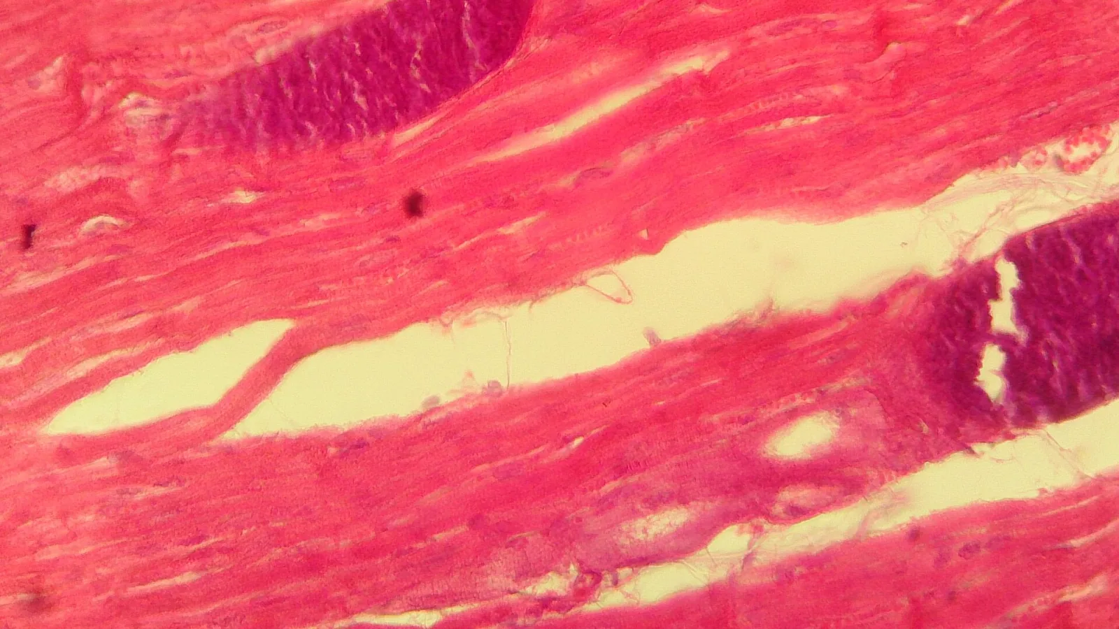
Pathologically, the verminous pneumonia induced by P. rufescens is characterized by a nodular, interstitial pattern. Grossly, raised, grayish-green nodules (1 to 3 mm) are scattered throughout the diaphragmatic lung lobes. Histologically, these nodules consist of aggregates of eosinophils, macrophages, and multinucleated giant cells surrounding adult worms and eggs within the alveolar septa [4]. Alveolar mastocytosis and eosinophilia have been documented in lambs concurrently infected with P. rufescens and Haemonchus contortus, indicating a mixed Th2 response [18]. In mixed infections with other protostrongylids (e.g., Muellerius capillaris), the pathological changes are more extensive and may include coalescing granulomas [4].
Sheep and goats with P. rufescens infection may also harbor Dictyocaulus filaria (large lungworm), necessitating differential diagnosis. Comparative pathology of lungworm infections in Moroccan sheep highlighted that P. rufescens lesions are predominantly alveolar and bronchiolar, whereas D. filaria causes more conspicuous bronchitis [19]. Other studies have noted that P. rufescens infection can present with no overt clinical signs despite moderate worm burdens, making reliance on clinical examination alone unreliable for diagnosis [20].
Diagnostic Approaches
Accurate diagnosis of P. rufescens infection is essential for implementing appropriate control measures and avoiding unnecessary anthelmintic use. The following diagnostic modalities are employed:
Fecal Examination: Baermann Technique
The Baermann technique remains the cornerstone of antemortem diagnosis. This method relies on the active migration of first-stage larvae (L1) from fecal material into warm water. Fresh feces (10 to 15 g) are placed on a mesh screen lined with gauze, suspended in a funnel filled with tap water, and incubated at 25 to 30 degrees Celsius for 12 to 24 hours. Larvae sediment at the bottom of the funnel and are collected for microscopic examination.
Protostrongylus rufescens L1 are characterized by a blunt, slightly curved tail with a distinct dorsal spine; their length ranges from 250 to 350 micrometres. Morphometric differentiation from Muellerius capillaris (which has a kinked tail) and Cystocaulus ocreatus (which has a terminal filament) is critical. In mixed infections, species-level identification may be challenging and requires expertise.
The relationship between fecal larval count and adult worm burden has been evaluated; a significant but variable correlation exists, indicating that quantitative larval counts can provide a reasonable estimate of infection intensity at the population level [20].
Molecular Diagnostics
DNA-based methods offer superior sensitivity and specificity for species identification. PCR amplification of the internal transcribed spacer 2 (ITS-2) region or the mitochondrial cytochrome c oxidase subunit I (cox1) gene can reliably differentiate P. rufescens from other lungworms [1]. These assays are particularly useful when morphological differentiation is ambiguous or when low larval numbers are present.
High-throughput sequencing approaches (e.g., amplicon-based metabarcoding) have been applied to pooled fecal samples for herd-level surveillance, though such methods remain primarily research tools.
Postmortem Examination
Necropsy remains the gold standard for definitive diagnosis and burden quantification. The lungs should be removed and examined for nodules. The Baermann technique can be applied to lung tissue homogenates to recover larvae. Histopathology confirms the presence of intralesional nematodes and associated granulomatous inflammation.
Serology and Imaging
No commercial serological tests are widely available for P. rufescens. Thoracic ultrasonography and radiography may reveal interstitial patterns but are not specific. These imaging modalities are rarely used in routine field diagnosis.
Diagnostic Workflow
The following decision tree summarizes the recommended diagnostic approach for suspected lungworm infection in sheep and goats.
flowchart TD
 A[Sheep or goat with chronic cough, reduced growth, or respiratory signs] --> B{History and grazing management suggest lungworm risk?}
 B -->|Yes| C[Collect fresh feces for Baermann technique]
 B -->|No| D["Consider other respiratory pathogens: bacterial, viral, Dictyocaulus"]
 C --> E{Larvae detected?}
 E -->|Yes| F["Identify L1 morphology: tail spine, size, shape"]
 F --> G["Species-level identification: P. rufescens vs others"]
 G --> H["Molecular confirmation if needed: PCR of ITS-2 or cox1"]
 H --> I[Quantify larval count to estimate burden]
 I --> J["Treat with appropriate anthelmintic: eprinomectin, ivermectin"]
 E -->|No| K[Repeat Baermann on pooled samples or consider necropsy]
 K --> L[Postmortem lung examination for nodules and adult worms]
 L --> M[Confirm with histopathology or larval recovery from lung]
 M --> N[If negative, investigate other respiratory diseases]
Treatment and Control
Effective anthelmintic options for P. rufescens include macrocyclic lactones and certain benzimidazoles. Topical eprinomectin (5 mg/mL solution) administered at 1 mg/kg bodyweight has demonstrated high efficacy against natural infections in sheep, with reduction in larval counts exceeding 95% [21]. Ivermectin delivered via a controlled-release capsule (designed for sustained gut release) has also shown excellent activity against small lungworms including P. rufescens, likely due to prolonged drug exposure in the pulmonary tissues [22, 23]. In earlier studies, ivermectin capsules achieved near-complete elimination of protostrongylid larvae [22]. Netobimin, a probenzimidazole, has been used in mouflon with variable success [16], and luxabendazole has demonstrated efficacy against lungworms in naturally infected sheep [24].
Anthelmintic resistance in protostrongylids has not been widely documented, but prudent use is advised to delay its emergence. Targeted selective treatment based on larval counts, clinical signs, and risk factors may help reduce selection pressure [11].
Control strategies focus on reducing exposure to infected gastropods. Pasture rotation, avoidance of heavy grazing during cool wet seasons, and drainage of snail habitats can lower transmission. Because gastropod populations are difficult to eliminate, strategic anthelmintic treatments timed to coincide with periods of high larval pickup are recommended. For flocks with persistent problems, treating all animals before turnout to clean pasture may be beneficial.
No vaccine is commercially available. While studies have explored cross-protection using Dictyocaulus filaria vaccine in goats [25], this approach does not target P. rufescens specifically.
Conclusion
Protostrongylus rufescens is a globally distributed small lungworm of small ruminants with significant subclinical impacts on productivity. Its indirect life cycle involving gastropod intermediate hosts makes control reliant on integrated management combining strategic anthelmintic use and grazing management. Antemortem diagnosis relies on the Baermann technique, with molecular methods providing species-level confirmation. Postmortem examination remains essential for quantifying burdens and differentiating mixed infections. Continued surveillance, particularly in regions with expanding small ruminant production, is needed to understand epidemiological trends and to guide evidence-based control programs.
References
[1] Jabbar A, Mohandas N, Jex AR et al. The mitochondrial genome of Protostrongylus rufescens, implications for population and systematic studies. Parasit Vectors. 2013. https://pubmed.ncbi.nlm.nih.gov/24025317/
[2] Cutillas C, Guevara DC, Valero A et al. Protostrongylus rufescens: a cytogenetic study. J Helminthol. 1987. https://pubmed.ncbi.nlm.nih.gov/3571924/
[3] Panayotova-Pencheva MS. Species composition and morphology of protostrongylids (Nematoda: Protostrongylidae) in ruminants from Bulgaria. Parasitol Res. 2011. https://pubmed.ncbi.nlm.nih.gov/21461727/
[4] Panayotova-Pencheva MS, Alexandrov MT. Some pathological features of lungs from domestic and wild ruminants with single and mixed protostrongylid infections. Vet Med Int. 2010. https://pubmed.ncbi.nlm.nih.gov/20445790/
[5] Cutillas C, Arias P, Spakulova M. Malic dehydrogenase isoenzymatic pattern in lung-nematode parasite species. Parasitol Res. 1996. https://pubmed.ncbi.nlm.nih.gov/8825454/
[6] Cutillas C, Espina C, Spakulová M et al. Differential diagnosis of lung nematode parasites from livestock by electrophoretic techniques. Int J Parasitol. 1995. https://pubmed.ncbi.nlm.nih.gov/7622328/
[7] Engdawork A, Kumsa B. Prevalence and Species Identification of Lungworms in Sheep and Cattle: A Postmortem Study in North Shewa, Central Highlands of Ethiopia. Vet Med Int. 2025. https://pubmed.ncbi.nlm.nih.gov/41234707/
[8] Tessema W, Getachew M, Tora E. Prevalence and Risk Factors of Lungworm Infection in Small Ruminants in Selected Districts of Wolaita Zone, Southern Ethiopia. J Parasitol Res. 2024. https://pubmed.ncbi.nlm.nih.gov/38633028/
[9] Borji H, Azizzadeh M, Ebrahimi M et al. Study on small ruminant lungworms and associated risk factors in northeastern Iran. Asian Pac J Trop Med. 2012. https://pubmed.ncbi.nlm.nih.gov/23146797/
[10] Gruner L, Cabaret J, Sauve C et al. Comparative susceptibility of Romanov and Lacaune sheep to gastrointestinal nematodes and small lungworms. Vet Parasitol. 1986. https://pubmed.ncbi.nlm.nih.gov/3962166/
[11] Bentounsi B, Cabaret J. Small-Lungworm (Protostrongylidae) Infections in Relation to Meat Sheep Breeds, Mediterranean Climates, and Anthelmintic Regimens. Vet Sci. 2025. https://pubmed.ncbi.nlm.nih.gov/40431564/
[12] Berrag B, Urquhart GM. Epidemiological aspects of lungworm infections of goats in Morocco. Vet Parasitol. 1996. https://pubmed.ncbi.nlm.nih.gov/8750686/
[13] de Macedo LO, Lima TARF, Verocai GG et al. Lungworms in ruminants from Brazil: A retrospective epidemiological study over four decades. Vet Parasitol Reg Stud Reports. 2021. https://pubmed.ncbi.nlm.nih.gov/34879956/
[14] Kuchboev AE, Krücken J, Karimova RR et al. Infection levels of protostrongylid nematodes in definitive caprine and intermediate gastropod hosts from Uzbekistan. J Helminthol. 2017. https://pubmed.ncbi.nlm.nih.gov/27018914/
[15] Ahmed S. Parasites of markhor, urial and Chiltan wild goat in Pakistan. Ann Parasitol. 2020. https://pubmed.ncbi.nlm.nih.gov/32198990/
[16] Meana A, Luzón-Peña M, Santiago-Moreno J et al. Natural infection by gastrointestinal and bronchopulmonary nematodes in mouflons (Ovis musimon) and their response to netobimin treatment. J Wildl Dis. 1996. https://pubmed.ncbi.nlm.nih.gov/8627934/
[17] Cabaret J, Riseani SR, Baeza E. Survival of sheep and goat first stage protostrongylid larvae in experimental conditions: influence of humidity and temperature. J Helminthol. 1991. https://pubmed.ncbi.nlm.nih.gov/1940250/
[18] Mansfield LS, Gamble HR. Alveolar mastocytosis and eosinophilia in lambs with naturally acquired nematode infections of Protostrongylus rufescens and Haemonchus contortus. Vet Immunol Immunopathol. 1995. https://pubmed.ncbi.nlm.nih.gov/8746699/
[19] Bouljihad M, Berrag B, Leipold HW. Gross and light-microscopic features of ovine pulmonary hydatidosis and verminous pneumonias in Morocco. Zentralbl Veterinarmed B. 1995. https://pubmed.ncbi.nlm.nih.gov/8592907/
[20] Rehbein S, Hamel D. A note on the relationship between fecal larval excretion and Protostrongylus rufescens lungworm burden in sheep. Parasitol Res. 2022. https://pubmed.ncbi.nlm.nih.gov/35290504/
[21] Rehbein S, Knaus M, Li J et al. Treatment of natural Protostrongylus rufescens lungworm infection in sheep with eprinomectin 5 mg/mL topical solution. Vet Parasitol. 2022. https://pubmed.ncbi.nlm.nih.gov/34959085/
[22] Rehbein S, Visser M. Efficacy of ivermectin delivered via a controlled-release capsule against small lungworms (Protostrongylidae) in sheep. J Vet Med B Infect Dis Vet Public Health. 2002. https://pubmed.ncbi.nlm.nih.gov/12420864/
[23] Rehbein S, Batty AF, Barth D et al. Efficacy of an ivermectin controlled-release capsule against nematode and arthropod endoparasites in sheep. Vet Rec. 1998. https://pubmed.ncbi.nlm.nih.gov/9571756/
[24] Kassai T, Takáts C, Fok E et al. Activity of luxabendazole against liver flukes, gastrointestinal roundworms, and lungworms in naturally infected sheep. Parasitol Res. 1988. https://pubmed.ncbi.nlm.nih.gov/2974592/
[25] Sharma RL. Parasitic bronchitis in goats and the possible use of Dictyocaulus filaria vaccine for its control. Vet Parasitol. 1994. https://pubmed.ncbi.nlm.nih.gov/8171828/
[26] Zafari S, Mohtasebi S, Sazmand A et al. The Prevalence and Control of Lungworms of Pastoral Ruminants in Iran. Pathogens. 2022. https://pubmed.ncbi.nlm.nih.gov/36558726/
[27] Kuchboev AE, Kazakov I, Asrarov MI et al. [The effect of the homogenates from different developmental stages of the nematode Protostrongylus rufescens (Leuckart, 1895) on mitochondrial and lipid bilayer membranes]. Parazitologiia. 2007. https://pubmed.ncbi.nlm.nih.gov/17460939/
[28] Rehbein S, Visser M, Winter R. [Endoparasitic infections in sheep from the Swabian Alb]. Dtsch Tierarztl Wochenschr. 1998. https://pubmed.ncbi.nlm.nih.gov/9857565/
[29] Reguera-Feo A, Castañón-Ordóñez L, del Campillo MC. Ecological relations among first-stage larvae of four species of Protostrongylidae (Nematoda) within their ovine host. Appl Parasitol. 1996. https://pubmed.ncbi.nlm.nih.gov/8574249/
[30] Mansfield LS, Gamble HR, Baker JS et al. Lungworm infection in a sheep flock in Maryland. J Am Vet Med Assoc. 1993. https://pubmed.ncbi.nlm.nih.gov/8449799/
Disclaimer: This article is for educational and informational purposes only. It is not intended to substitute for professional veterinary advice, diagnosis, treatment, or regulatory guidance. Always consult a licensed veterinarian or qualified specialist regarding animal health, disease diagnosis, and therapeutic decisions.